. 2009 Dec;62(6):1368-78.
doi: 10.1002/mrm.22140.

Hybrid adiabatic-rectangular pulse train for effective saturation of magnetization within the whole heart at 3 T

Affiliations

Hybrid adiabatic-rectangular pulse train for effective saturation of magnetization within the whole heart at 3 T

Daniel Kim et al. Magn Reson Med. 2009 Dec.

Abstract

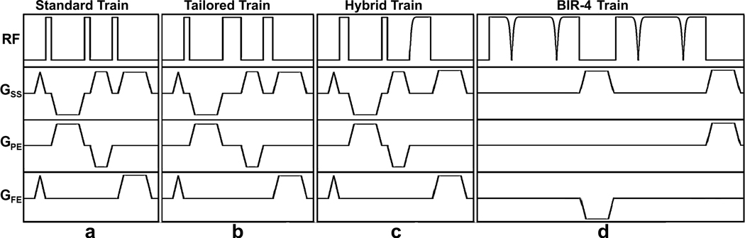
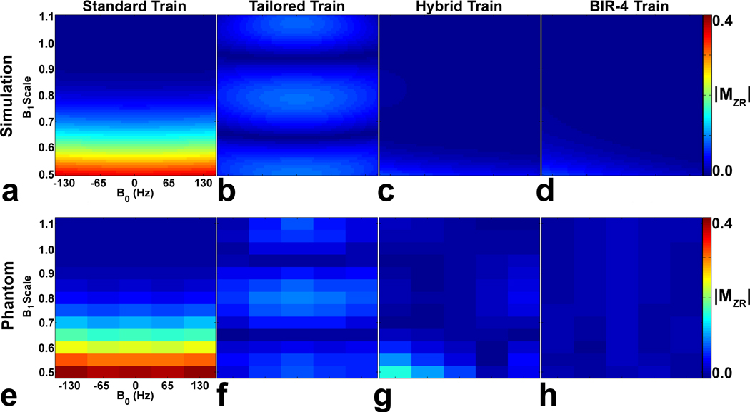
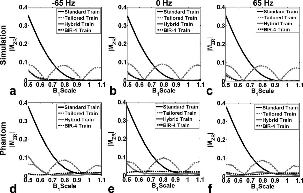
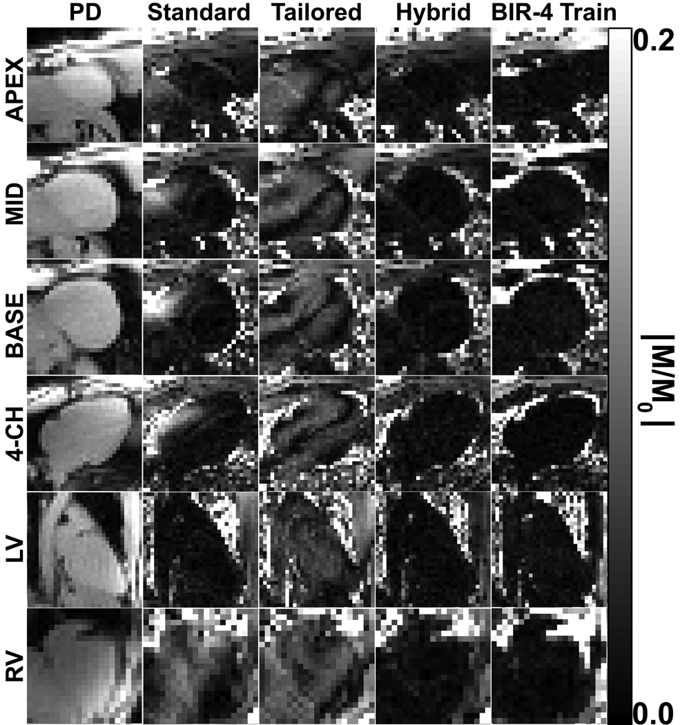
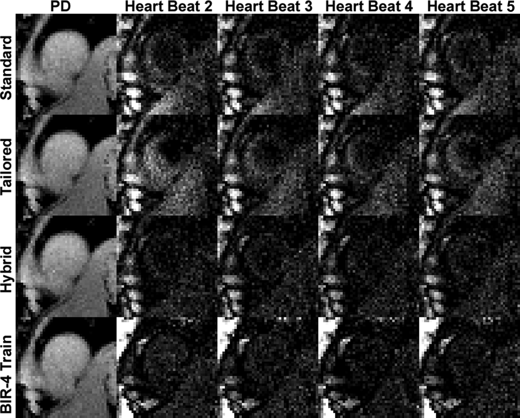
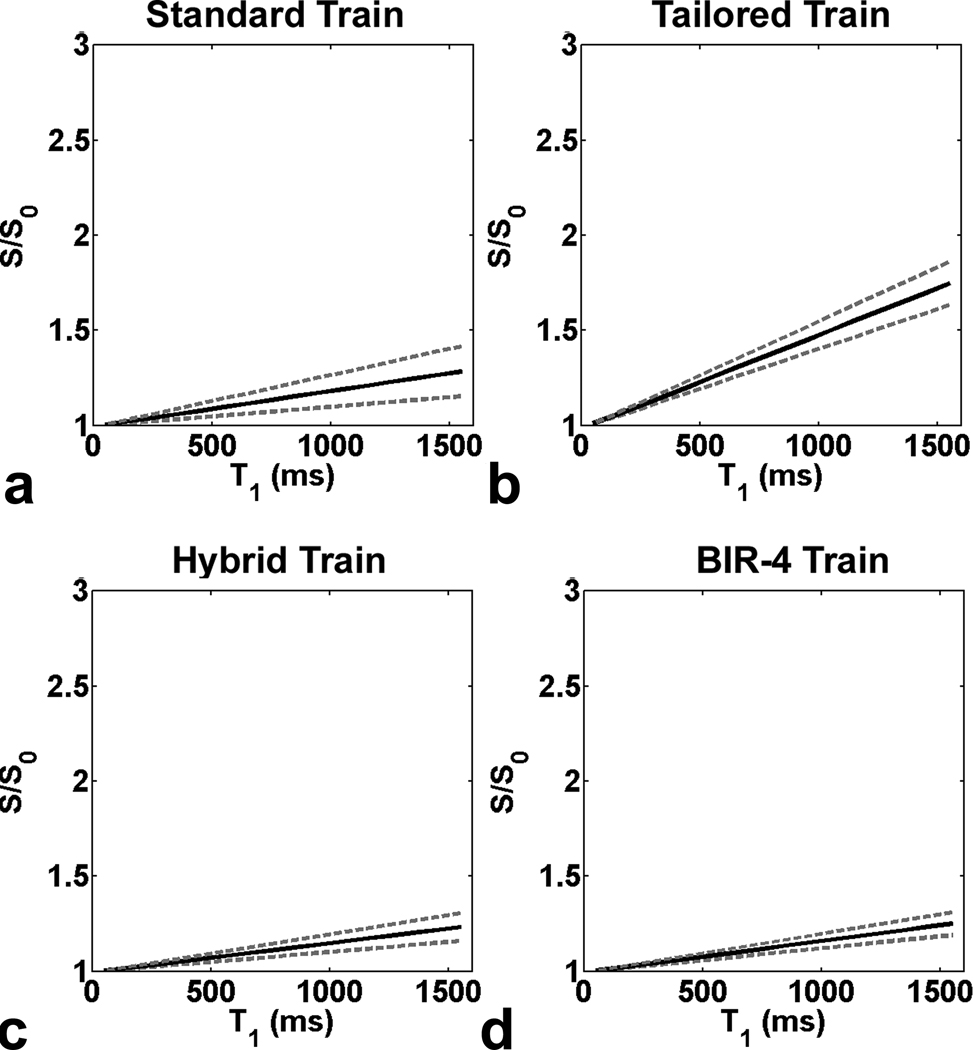
Uniform T(1)-weighting is a major challenge for first-pass cardiac perfusion MRI at 3 T. Previously proposed adiabatic amplitude of radiofrequency field (B(1))-insensitive rotation (BIR-4) pulse and standard and tailored pulse trains of three nonselective pulses have been important developments but each pulse has limitations at 3 T. As an extension of the tailored pulse train, we developed a hybrid pulse train by synergistically combining two nonselective rectangular radiofrequency pulses and an adiabatic half-passage pulse, in order to achieve effective saturation of magnetization within the heart, while remaining within clinically acceptable specific absorption rate limits. The standard pulse train, tailored pulse train, hybrid pulse train, and BIR-4 pulse train were evaluated through numerical, phantom, and in vivo experiments. Among the four saturation pulses, only the hybrid pulse train yielded residual magnetization <2% of equilibrium magnetization in the heart while remaining within clinically acceptable specific absorption rate limits for multislice first-pass cardiac perfusion MRI at 3 T.


Figures

Figure 1
Figure 1
Pulse sequence diagrams: (a) standard pulse train, (b) tailored pulse train, (c) hybrid pulse train, and (d) BIR-4 pulse train. The spoiler gradients are applied before and after each saturation pulse to dephase the transverse magnetization. The crusher gradients in between RF pulses are cycled to eliminate stimulated echoes. These diagrams are drawn to approximate proportions but not to exact scale. GSS: slice-select gradient; GPE: phase-encoding gradient; GFE: frequency-encoding gradient. The RF pulse characteristics are summarized in Table 1.
Figure 2
Figure 2
(Top row) Simulated MZR maps as a function of B0 and B1+ scales. Compared with the (a) standard pulse train and (b) tailored pulse train, both the (c) hybrid pulse train and the (d) BIR-4 pulse train yielded considerably lower MZR maps. The resulting RMS MZR measurements were 0.136, 0.055, 0.013, and 0.012 for the standard train, tailored train, hybrid train, and BIR-4 pulse train, respectively. (Bottom row) MZR maps of the phantom as a function of B0 and B1+ scales. Consistent with the simulation results, the corresponding phantom MZR maps were in good agreement, and both the (g) hybrid pulse train and (h) BIR-4 pulse train produced considerably lower MZR maps than the (d) standard and (e) tailored pulse trains. The resulting RMS MZR measurements were 0.164, 0.053, 0.035, and 0.016 for the standard train, tailored train, hybrid train, and BIR-4 pulse train, respectively. The MZR maps are displayed with identically narrow grayscales (0 – 0.4 in dimensionless units) to bring out regional variations.
Figure 3
Figure 3
(Top row) Numerically simulated profiles of MZR as a function of B1+ scale: (a) −65 Hz, (b) 0 Hz, and (c) 65 Hz off-resonance. (Bottom row) Corresponding profiles of MZR in the phantom as a function of B1+ scale: (d) −65 Hz, (e) 0 Hz, and (f) 65 Hz off-resonance. Note that the standard pulse train yielded lower MZR than the tailored pulse train for B1+ scales > 0.72, and vice versa for B1+ scales < 0.72. In contrast, the hybrid pulse train and BIR-4 pulse train yielded considerably lower MZR throughout the B1+ scale.
Figure 4
Figure 4
Representative set of normalized SR images from one volunteer that compares the performance of four saturation pulses. The standard pulse train performed well within the LV (dark regions) but poorly within the RV (bright regions). The tailored pulse train performed similarly within the whole heart, but at the expense of producing medium level of MZR values (gray regions and dark bands) within both ventricles. In contrast, both the hybrid RF pulse train and BIR-4 pulse train yielded uniformly minimal MZR within both ventricles (dark regions). The normalized images are displayed with identically narrow grayscales (0 – 0.2 in dimensionless units) to bring out regional variations.
Figure 5
Figure 5
Representative repeated acquisitions of non-contrast T1-weighted images over multiple heart beats: (first row) standard train, (second row) tailored train, (third row) hybrid train, and (fourth row) BIR-4 train. Both the standard and tailored pulse trains yielded residual signal compared with the hybrid and BIR-4 pulse trains. The T1-weighted images are displayed with identically grayscales (200 – 1000 in arbitrary units) to bring out regional variations.
Figure 6
Figure 6
Representative first-pass cardiac perfusion images at (first column) pre-contrast, (second column) peak blood enhancement, and (third column) peak myocardial wall enhancement, using the hybrid pulse train as the saturation pulse: (first row) apex, (second row) mid-ventricular, and (third row) base. (Fourth column) The corresponding plots of normalized T1-weighted signal for each of 16 segments as a function of cardiac cycle. These normalized signal-time curves show good agreement between segments, suggesting that the hybrid pulse train had produced uniform T1-weighted signal. The T1-weighted images are displayed with identically grayscales (200 – 1500 in arbitrary units).
Figure 7
Figure 7
Simulated plots of S/S0 as a function of T1 for the LV: (a) standard pulse train, (b) tailored pulse train, (c) hybrid pulse train, and (d) BIR-4 pulse train. The standard pulse train yielded relatively accurate S/S0, whereas the tailored pulse train yielded less accurate S/S0. Compared with the standard and tailored pulse trains, both the hybrid pulse train and BIR-4 pulse train produced more accurate S/S0. Solid line represents S/S0 calculated with mean in vivo MZR. Upper and lower dotted lines represent S/S0 calculated with mean+SD and mean−SD in vivo MZR, respectively.

References

    1. Cheng AS, Pegg TJ, Karamitsos TD, Searle N, Jerosch-Herold M, Choudhury RP, Banning AP, Neubauer S, Robson MD, Selvanayagam JB. Cardiovascular magnetic resonance perfusion imaging at 3-tesla for the detection of coronary artery disease: a comparison with 1.5-tesla. Journal of the American College of Cardiology. 2007;49(25):2440–2449. - PubMed
    1. Greenman RL, Shirosky JE, Mulkern RV, Rofsky NM. Double inversion black-blood fast spin-echo imaging of the human heart: a comparison between 1.5T and 3.0T. Journal of Magnetic Resonance Imaging. 2003;17(6):648–655. - PubMed
    1. Noeske R, Seifert F, Rhein KH, Rinneberg H. Human cardiac imaging at 3 T using phased array coils. Magnetic Resonance in Medicine. 2000;44(6):978–982. - PubMed
    1. Singerman RW, Denison TJ, Wen H, Balaban RS. Simulation of B1 field distribution and intrinsic signal-to-noise in cardiac MRI as a function of static magnetic field. Journal of Magnetic Resonance. 1997;125(1):72–83. - PubMed
    1. Schar M, Kozerke S, Fischer SE, Boesiger P. Cardiac SSFP imaging at 3 Tesla. Magnetic Resonance in Medicine. 2004;51(4):799–806. - PubMed

Publication types