. 2009 Nov;158(5):1236-47.
doi: 10.1111/j.1476-5381.2009.00401.x. Epub 2009 Sep 28.

alpha-Fetoprotein as a modulator of the pro-inflammatory response of human keratinocytes

Affiliations

alpha-Fetoprotein as a modulator of the pro-inflammatory response of human keratinocytes

A I Potapovich et al. Br J Pharmacol. 2009 Nov.

Abstract

Background and purpose: The immunomodulatory effects of alpha-fetoprotein (AFP) on lymphocytes and macrophages have been described in vitro and in vivo. Recombinant forms of human AFP have been proposed as potential therapeutic entities for the treatment of autoimmune diseases. We examined the effects of embryonic and recombinant human AFP on the spontaneous, UVA- and cytokine-induced pro-inflammatory responses of human keratinocytes.

Experimental approach: Cultures of primary and immortalized human keratinocytes (HaCaT) and human blood T lymphocytes were used. The effects of AFP on cytokine expression were studied by bioplexed elisa and quantitative reverse transcriptase polymerase chain reaction assay. Kinase and nuclear factor kappa B (NFkappaB) phosphorylation were quantified by intracellular elisa. Nuclear activator protein 1 and NFkappaB DNA binding activity was measured by specific assays. Nitric oxide and H(2)O(2) production and redox status were assessed by fluorescent probe and biochemical methods.

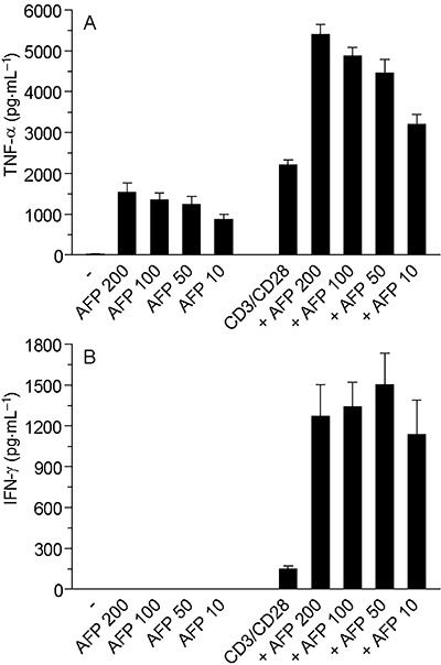
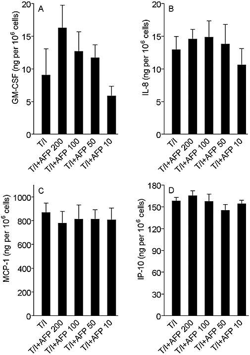
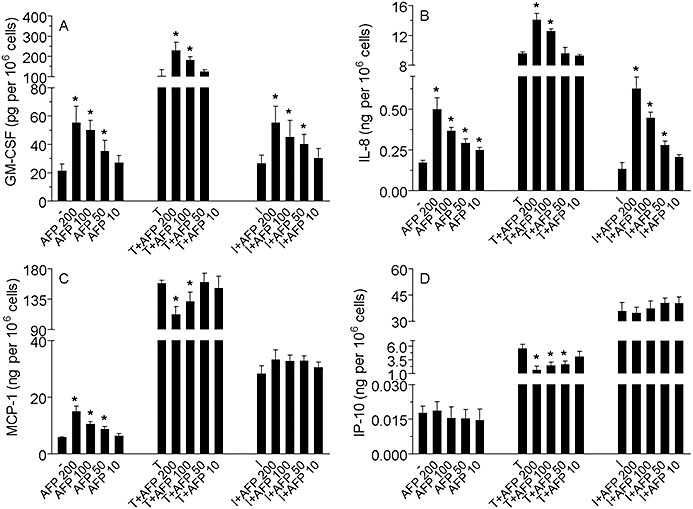
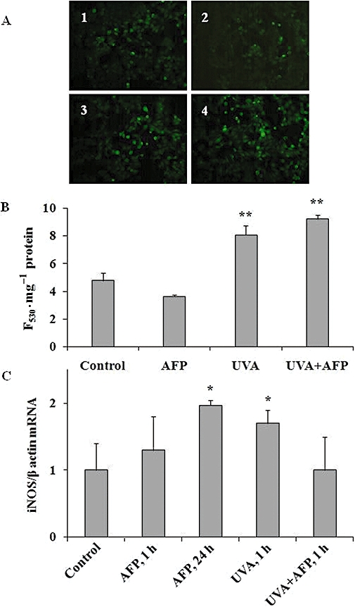
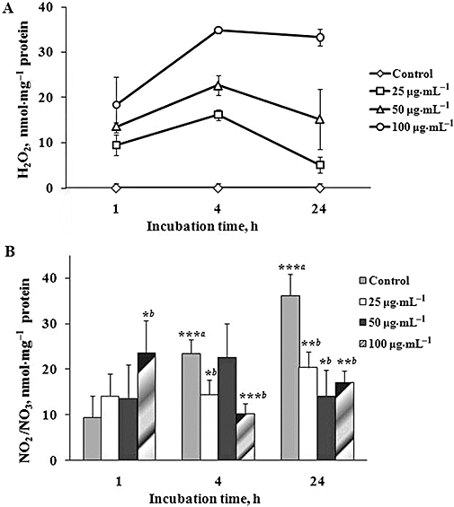
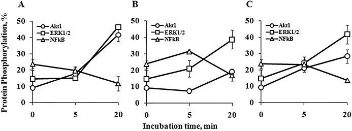
Key results: All forms of AFP enhanced baseline expression of cytokines, chemokines and growth factors. AFP dose-dependently increased tumour necrosis factor alpha-stimulated granulocyte macrophage colony stimulating factor and interleukin 8 expression and decreased tumour necrosis factor alpha-induced monocyte chemotactic protein 1 and IP-10 (interferon gamma-produced protein of 10 kDa) expression. AFP induced a marked activator protein 1 activation in human keratinocytes. AFP also increased H(2)O(2) and modulated nitrite/nitrate levels in non-stimulated keratinocytes whereas it did not affect these parameters or cytokine release from UVA-stimulated cells. Phosphorylation of extracellular signal-regulated kinase (ERK1/2) and Akt1 but not NFkappaB was activated by AFP alone or by its combination with UVA.

Conclusions and implications: Exogenous AFP induces activation of human keratinocytes, with de novo expression of a number of pro-inflammatory mediators and modulation of their pro-inflammatory response to cytokines or UVA. AFP may modulate inflammatory events in human skin.


Figures

Figure 3
Figure 3
AFP alone induces TNF-α in purified CD4 T cells and boosts the release of IFN-γ (A) and TNF-α (B) due to CD3/CD28 stimulation. T lymphocytes were cultured in 24-well clusters and treated with rhAFP, in the presence or absence of CD3/CD28 stimulation. The results are representative of data obtained with CD4-positive T cells from six distinct blood donors. AFP, α-fetoprotein; IFN-γ, interferon gamma; rhAFP, recombinant human alpha fetoprotein; TNF-α, tumour necrosis factor alpha.
Figure 2
Figure 2
Effect of gly-rhAFP on the release of GM-CSF (A), IL-8 (B), MCP-1 (C) and IP-10 (D) by normal human keratinocytes, used in combination with 100 ng·mL−1 TNF-α and 100 U·mL−1 IFN-γ (T/I). The results are expressed as the mean ± SD of three independent experiments. AFP, α-fetoprotein; gly-rhAFP, glycosylated recombinant human alpha fetoprotein; GM-CSF, granulocyte macrophage colony stimulating factor; IFN-γ, interferon gamma; IL, interleukin; IP-10, interferon gamma-produced protein of 10 kDa; MCP-1, monocyte chemotactic protein 1; TNF-α, tumour necrosis factor alpha.
Figure 1
Figure 1
Effect of gly-rhAFP on the release of GM-CSF (A), IL-8 (B), MCP-1 (C) and IP-10 (D) by normal human keratinocytes, used alone in the dose range 10–200 µg·mL−1, or in combination with 100 ng·mL−1 TNF-α (T) or 100 U·mL−1 IFN-γ (I). The results are expressed as the mean ± SD of three independent experiments. *P < 0.05 versus untreated controls (-) or versus stimulated controls (T or I). AFP, α-fetoprotein; gly-rhAFP, glycosylated recombinant human alpha fetoprotein; GM-CSF, granulocyte macrophage colony stimulating factor; IFN-γ, interferon gamma; IL, interleukin; IP-10, interferon gamma-produced protein of 10 kDa; MCP-1, monocyte chemotactic protein 1; TNF-α, tumour necrosis factor alpha.
Figure 5
Figure 5
Effects of gly-rhAFP (100 µg·mL−1) and UVA (2.5 J·cm−2) on intracellular NO levels and iNOS gene expression in HaCaT cells (mean ± SD). (A) Typical fluorescence micrographs of HaCaT cells incubated for 1 h with fluorescent probe DAF-2DA: 1 – control cells; 2 – cells exposed to gly-rhAFP; 3 – cells exposed to UVA, 4 – cells exposed to the combination of UVA and gly-rhAFP. (B) Fluorescence intensity (F530·mg−1 protein) in the lysates of DAF-2DA-loaded HaCaT cells. (C) iNOS gene expression expressed as fold induction related to control. *P < 0.05; **P < 0.01 versus control. AFP, α-fetoprotein; DAF-DA, 4,5-diaminofluorescein diacetate; gly-rhAFP, glycosylated recombinant human alpha fetoprotein; iNOS, inducible nitric oxide synthase.
Figure 4
Figure 4
Effects of glycosylated recombinant human alpha fetoprotein (gly-rhAFP) on H2O2 (A) and NO2/NO3 (B) levels in conditioned medium of HaCaT cells (mean ± SD). Cells were treated with various doses of gly-rhAFP (25, 50 and 100 µg·mL−1), and the conditioned medium was analysed at different time points (1, 4 and 24 h). ***aP < 0.005 versus control at 1 h; *bP < 0.05; **bP < 0.01; ***bP < 0.005 versus respective control.
Figure 6
Figure 6
Effects of gly-rhAFP and UVA on phosphorylation of NFκB, Akt1 and ERK1/2 in HaCaT cells (mean ± SD). 0 min – control cells without treatment; 5 and 20 min – time after treatment. (A) cells exposed to 100 µg·mL−1 gly-rhAFP; (B) cells exposed to 2.5 J·cm−2 UVA; (C) cells exposed to UVA + gly-rhAFP. Akt, protein kinase B; ERK, extracellular signal-regulated kinase; gly-rhAFP, glycosylated recombinant human alpha fetoprotein; NFκB, nuclear factor kappa B.
Figure 7
Figure 7
Gly-rhAFP activates AP-1 in normal human keratinocytes. (A) Western blot analysis of c-Fos (AP-1 subunit) and p65 (RelA, NFκB subunit) in nuclear lysates (10 µg per lane) from untreated cultures (-), incubated for 20 min with 50 or 10 µg·mL−1 rhAFP alone, or in the presence of 50 ng·mL−1 TNF-α or 50 U·mL−1 IFN-γ. Lamin B was used to assess equal loading. These results are representative of three independent experiments. (B) AP-1- and NFκB-specific DNA binding activity in keratinocytes following 2 h treatments. **P < 0.01 versus untreated keratinocyte cultures (-).*P < 0.05 versus untreated keratinocyte cultures. AFP, α-fetoprotein; AP-1, activator protein 1; gly-rhAFP, glycosylated recombinant human alpha fetoprotein; IFN-γ, interferon gamma; NFκB, nuclear factor kappa B; TNF-α, tumour necrosis factor alpha.

References

    1. Aebi H. Catalase in vitro. Methods Enzymol. 1984;105:121–126. - PubMed
    1. Alisa A, Boswell S, Pathan AA, Ayaru L, Williams R, Behboudi S. Human CD4(+) T cells recognize an epitope within alpha-fetoprotein sequence and develop into TGF-beta-producing CD4(+) T cells. J Immunol. 2008;180:5109–5117. - PubMed
    1. Bachelor MA, Bowden GT. UVA-mediated activation of signalling pathways involved in skin tumor promotion and progression. Semin Cancer Biol. 2004;14:131–138. - PubMed
    1. Bashir MM, Sharma MR, Werth VP. TNF-alpha production in the skin. Arch Dermatol Res. 2009;301:87–91. - PubMed
    1. Benevolensky SV, Marchenko AN, Kozlov DG, Zatsepin SS, Shingarova LN, Dudich IV, et al. Recombinant α-fetoprotein, method and means for preparation thereof, compositions on the base thereof and use thereof. WORLD PATENT WO-2006009492: 2006 January 26.

Publication types

MeSH terms