. 2009 Sep 29;2(1):28.
doi: 10.1186/1755-7682-2-28.

Regulation of the epithelial sodium channel [ENaC] in kidneys of salt-sensitive Dahl rats: insights on alternative splicing

Affiliations

Regulation of the epithelial sodium channel [ENaC] in kidneys of salt-sensitive Dahl rats: insights on alternative splicing

Marlene F Shehata. Int Arch Med. .

Abstract

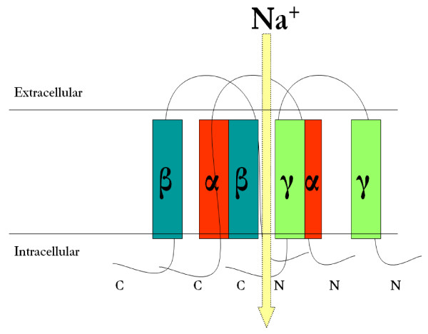
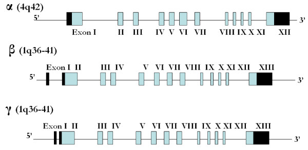
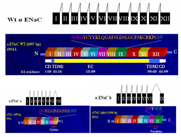
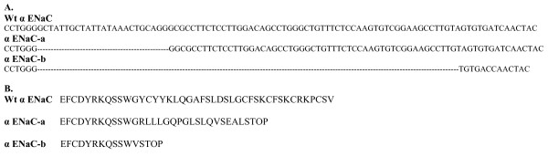
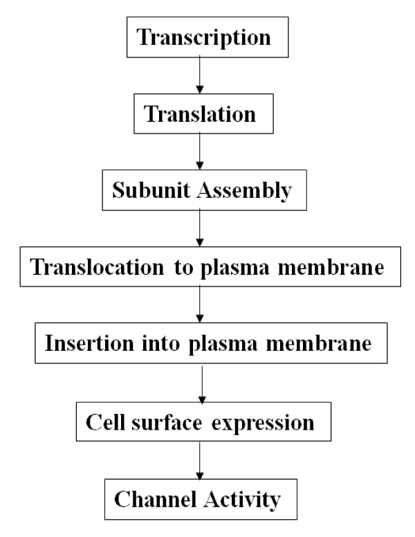
The epithelial sodium channel [ENaC] is critical for the maintenance of sodium balance, extracellular fluid volume and long term blood pressure control. Monogenic disorders causing ENaC hyperactivity have led to a severe form of hereditary hypertension in humans, known as Liddle's syndrome. Similarly, in animal models, ENaC hyperactivity has been well documented in kidneys of salt-sensitive [S] Dahl rats [a genetic model of salt-sensitive hypertension] versus their normotensive control [Dahl salt-resistant [R] rats]. The purpose of the present review is to highlight the differential regulation of ENaC in kidneys of Dahl S versus R rats. A systematic overview of the putative role of alternative splicing of the main alpha subunit of ENaC [alpha ENaC] in modulating ENaC expression in kidneys of Dahl rats will be discussed. Finally, a better understanding of the meaningful contribution of ENaC in the pathogenesis of salt-sensitive hypertension will be achieved upon completion of this review.


Figures

Figure 1
Figure 1
Structure of the Epithelial Sodium Channel [ENaC]. The amiloride-sensitive epithelial sodium channel [ENaC] is composed of three homologous α, β and γ protein subunits of corresponding 698, 638 and 650 amino acids in length [14,15]. ENaC α, β and γ subunits share approximately 30% homology at the amino acid level and each subunit correspond to a molecular mass of 70-80 kDa. The three ENaC subunits are inserted into the plasma membrane with a proposed stoichiometry of 2:1:1 [16] as shown in the above figure or 3:3:3 [18]. Each ENaC protein subunit is formed up of four major domains: the cytoplasmic N terminus, the large extracellular loop, the two short hydrophobic segments known as the transmembrane domains 1 and 2 [TM1 and 2] and the cytoplasmic C-terminus. The N- and C-termini face the cytosolic side, while the extracellular loop faces the extracellular side [19]. All three subunits cooperate to form the channel pore via the transmembrane domains.
Figure 2
Figure 2
Genomic Organization of rat ENaC α, β, and γ subunits. At the genomic level, ENaC α, β, and γ subunits are encoded by three different genes located on separate chromosomes. The gene encoding the α ENaC subunit [Scnn1a] is located on chromosome 4q42, while the β and γ genes [Scnn1b and g] are located at a close proximity from each other on chromosome 1q36-q41 [RGD: Rat Genome Database]. α ENaC is composed of 12 exons, whereas each of the β and γ ENaC genes are composed of 13 exons. Translation starts in exon 1 for α ENaC and starts in exon 2 for β and γ ENaC. Translation ends in exon 12 for α ENaC and in exon 13 for β and γ ENaC. Therefore the 5'untranslated region [UTR] is included in exon 1 of α ENaC and in exons 1 and 2 of β and γ ENaC genes, while the 3'UTR is included in exon 12 of α ENaC and exon 13 in each of β and γ ENaC. Light shaded boxes represent the translated regions, while the black boxes represent the 3' and 5' UTR.
Figure 3
Figure 3
α ENaC alternatively spliced forms. A schematic illustration of alternative mRNA splicing of α ENaC wildtype, -a and -b forms. α ENaC wildtype is made of 12 exons, while α ENaC-a is formed of exons I to VIII, with a 23 bases deleted from exon VIII. On the other hand, α ENaC-b is formed of exons I to IX with a skipping of exon VIII [79 bases]. Underneath each mRNA splicing comes the protein organization of the 2 alternatively spliced forms of α ENaC [α ENaC-a & -b] that have been published in rats [33] in comparison to α ENaC wildtype major transcript. α ENaC wildtype is 698 amino acids in length [2100 bp]. Amino acid residues from 1 to 110 reside in the cytoplasm, amino acid residues from 111 to 131 constitutes the first transmembrane domain, residues 132 to 589 constitute the extracellular loop, residues 590 to 610 constitute the second transmembrane domain, and residues 611-698 are cytoplasmic. α ENaC-a alternatively spliced form is formed by the deletion of 23 nucleotides from exon 8, whereas α ENaC-b is formed by the deletion of 79 nucleotides that involved exon 8 skipping. These deletions introduced a premature stop codon and resulted in shorter proteins at the carboxy terminus by 199 in α ENaC-a and 216 amino acids in α ENaC-b, making α ENaC-a 499 amino acids [2077 bp] and α ENaC-b 482 amino acids [2021 bp] in length. These resultant shorter proteins lacked the second transmembrane domain [TDM2] which is important in channel pore formation. α ENaC-a alternatively spliced form is a low abundance transcript that is expressed in the rat kidney, tongue epithelia and tongue taste tissues. α ENaC-a binding with the channel blocker [phenamil] was greatly enhanced. This demonstrates that the amiloride-binding site [i.e ENaC blocker site] resides in the extracellular loop of the channel and not the second transmembrane domain that is presently missing in α ENaC-a [CD: cytoplasmic domain, TDM1: transmembrane domain M1, EC: extracellular loop, TDM2: transmembrane domain M2].
Figure 4
Figure 4
Schematic representation of wildtype and the alternatively spliced forms α ENaC-a & -b. A. The genomic sequence of α ENaC wildtype, -a and -b forms. The alternatively spliced forms α ENaC-a & -b share the same splicing site [CCTGGG] which is located within exon VII. α ENaC-a & -b had 23 and 79 bases deleted respectively resulting in the formation of a premature stop codon. B. The protein sequence of α ENaC wildtype, -a and -b forms. The deletions of 23 and 79 bases respectively in α ENaC-a & -b introduced a premature stop codon and resulted in shorter proteins at the carboxy terminus by 199 in α ENaC-a and 216 amino acids in α ENaC-b, making α ENaC-a 499 amino acids [1497 bp] and α ENaC-b 482 amino acids [1446 bp] in length. The α ENaC-a and -b truncated proteins of approximately 55 and 53 kDa respectively, are identical to wildtype α ENaC up to amino acids 481 and 480 respectively, followed by 17 and 1 novel amino acids unique to the spliced form after which the stop codon terminates translation (adapted with permission from reference [33]).
Figure 5
Figure 5
Control points impinging on ENaC cell surface expression and activity. Schematic representation of the steps involved in regulating α ENaC cell surface expression and activity. These steps include α ENaC subunit synthesis, assembly with the β and γ subunits, trafficking, insertion into the plasma membrane and activity. α ENaC-b may hinder any of these steps causing a suppressed channel cell surface expression and/or activity in Dahl R versus S rats that is augmented on high salt diet.

References

    1. Hemmelgarn BR, McAlister FA, Grover S, Myers MG, McKay DW, Bolli P, Abbott C, Schiffrin EL, Honos G, Burgess E, Mann K, Wilson T, Penner B, Tremblay G, Milot A, Chockalingam A, Touyz RM, Tobe SW, Canadian Hypertension Education Program The 2006 Canadian Hypertension Education Program recommendations for the management of hypertension: Part I--Blood pressure measurement, diagnosis and assessment of risk. Can J Cardiol. 2006;22:573–581. - PMC - PubMed
    1. Weinberger MH, Fineberg NS, Fineberg SE, Weinberger M. Salt sensitivity, pulse pressure, and death in normal and hypertensive humans. Hypertension. 2001;37:429–432. - PubMed
    1. Bihorac A, Tezcan H, Ozener C, Oktay A, Akoglu E. Association between salt sensitivity and target organ damage in essential hypertension. Am J Hypertens. 2000;13:864–872. doi: 10.1016/S0895-7061(00)00253-3. - DOI - PubMed
    1. Morimoto A, Uzu T, Fujii T, Nishimura M, Kuroda S, Nakamura S, Inenaga T, Kimura G. Sodium sensitivity and cardiovascular events in patients with essential hypertension. Lancet. 1997;350:1734–1737. doi: 10.1016/S0140-6736(97)05189-1. - DOI - PubMed
    1. Cirillo M, Capasso G, Di Leo VA, De Santo NG. A history of salt. Am J Nephrol. 1994;14:426–431. doi: 10.1159/000168759. - DOI - PubMed

LinkOut - more resources