. 2010 Feb 1;316(3):452-65.
doi: 10.1016/j.yexcr.2009.09.020. Epub 2009 Sep 26.

SAM pointed domain ETS factor (SPDEF) regulates terminal differentiation and maturation of intestinal goblet cells

Affiliations

SAM pointed domain ETS factor (SPDEF) regulates terminal differentiation and maturation of intestinal goblet cells

Taeko K Noah et al. Exp Cell Res. .

Abstract

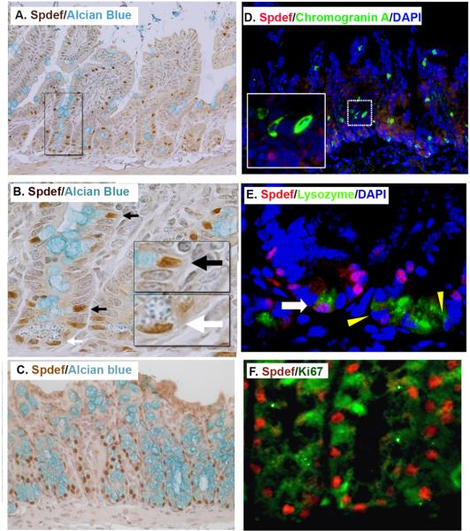
Background and aims: SPDEF (also termed PDEF or PSE) is an ETS family transcription factor that regulates gene expression in the prostate and goblet cell hyperplasia in the lung. Spdef has been reported to be expressed in the intestine. In this paper, we identify an important role for Spdef in regulating intestinal epithelial cell homeostasis and differentiation.

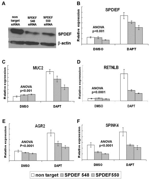
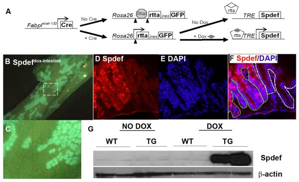
Methods: SPDEF expression was inhibited in colon cancer cells to determine its ability to control goblet cell gene activation. The effects of transgenic expression of Spdef on intestinal differentiation and homeostasis were determined.

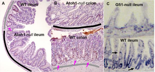
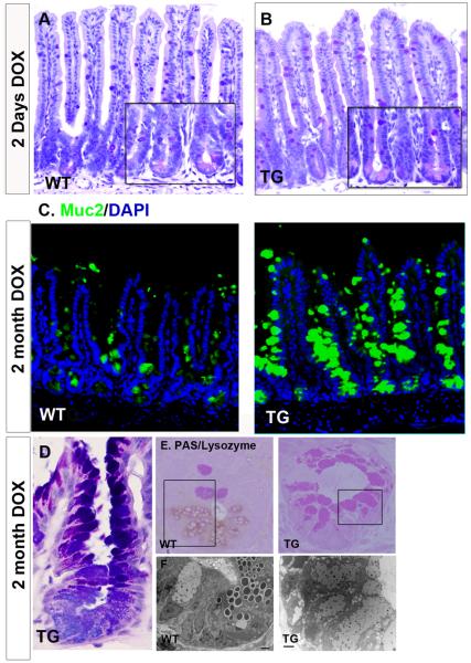
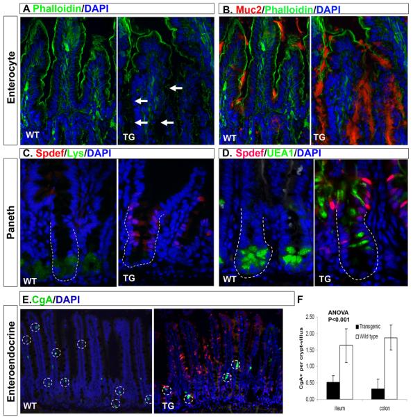
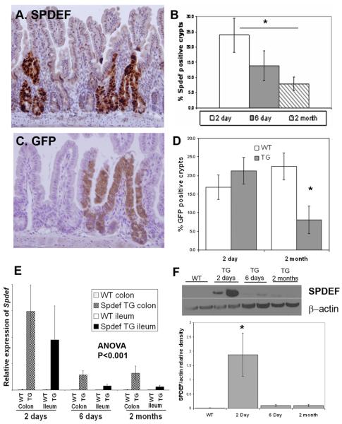
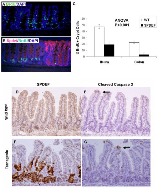
Results: In LS174T colon cancer cells treated with Notch/gamma-secretase inhibitor to activate goblet cell gene expression, shRNAs that inhibited SPDEF also repressed expression of goblet cell genes AGR2, MUC2, RETLNB, and SPINK4. Transgenic expression of Spdef caused the expansion of intestinal goblet cells and corresponding reduction in Paneth, enteroendocrine, and absorptive enterocytes. Spdef inhibited proliferation of intestinal crypt cells without induction of apoptosis. Prolonged expression of the Spdef transgene caused a progressive reduction in the number of crypts that expressed Spdef, consistent with its inhibitory effects on cell proliferation.

Conclusions: Spdef was sufficient to inhibit proliferation of intestinal progenitors and induce differentiation into goblet cells; SPDEF was required for activation of goblet cell associated genes in vitro. These data support a model in which Spdef promotes terminal differentiation into goblet cells of a common goblet/Paneth progenitor.


Figures

Figure 1
Figure 1. Spdef is expressed in intestinal goblet cells and in a subset Paneth cells
(A-C) Immunohistochemistry stained for Spdef in brown, counterstained with Alcian blue for goblet cells in (A-B) and (C) colon. (B) Higher magnification of the area boxed in (A). Black arrows show goblet cells nuclei stained with Spdef and white arrow shows Paneth cell nuclei stained with Spdef. Insets show higher magnification of boxed areas. (D) Immunofluorescence staining for Spdef (red), the enteroendocrine cell marker chromogranin A (green), and DNA stained with DAPI (blue). The inset is a higher magnification of the dotted area. No colocalization of Spdef with chromogranin A is seen. (E) Immunofluorescence staining for Spdef (red), lysozyme (green), and DAPI (blue). The white arrow shows a rare Paneth cell with Spdef staining; yellow triangles show Spdef-negative Paneth cells. (F) Immunofluorescence staining for Spdef (red) and Ki67 (green) in colon. Most Spdef positive cells do not stain with the proliferation marker Ki67.
Figure 2
Figure 2. Protein and mRNA localization of Spdef shows loss in Atoh1- and Gfi1-null intestines
Immunohistochemical labeling of Spdef (brown) in ileum (A) and colon (B) from Atoh1Δintestine mice [32]. These mosaic animals contain both wild type (underlined in gray) and Atoh-null (underlined in black) crypts. Spdef is only seen in wild type crypts (pink arrows show nuclei stained for Spdef). (C) In situ hybridization for Spdef shows localization of the mRNA in crypt progenitors and goblet cells in wild type ileum (black arrows), which is largely absent from Gfi1-null intestine.
Figure 3
Figure 3. Inhibition of SPDEF expression in LS174T blocks activation of goblet cell associated genes
(A) SPDEF and β-actin immunoblot on protein lysate from LS174T cells transduced with lentivirus encoding SPDEF-targeted shRNAs or non-target control shRNA. (B-F) RT-qPCR analysis of LS174T cells transduced with lentivirus expressing non-targeting shRNA (white bars) or SPDEF-targeting shRNA-548 and -550 (shaded bars), then treated with DAPT or DMSO. SPDEF shRNA-548 and 550 modestly but significantly reduced SPDEF induction by DAPT, and significantly reduced MUC2, SPINK4, AGR2, and RETNLB mRNA following DAPT treatment. Y-axis shows relative expression values normalized to GAPDH. P values are for 2-way ANOVA for shRNA (non target vs siRNA 548 or 550) and treatment (DAPT vs. DMSO). Error bars show SEM.
Figure 4
Figure 4. Doxycycline-inducible Spdef expression in Spdefdox-intestine colon
(A) Schematic of how the three alleles in Spdefdox-intestine mice cause intestine-specific, DOX-inducible expression of Spdef. (B-C) GFP fluorescence of whole colon from a Spdefdox-intestine mouse following 2 days of DOX, showing a mosaic pattern of GFP expression. (C) Close-up view of the area outlined in (B) shows the patchy crypt-by-crypt mosaic of cre-mediated recombination. (D-F) Colon section from mouse in (B), stained for Spdef (red in (D and F)) and DAPI (blue in (E and F) shows Spdef-expressing crypts (F: outlined in green) adjacent to unrecombined crypts (F: outlined in white). (G) Immunoblot of colonic tissue shows robust induction of Spdef following 2 days of DOX administration to Spdefdox-intestine (TG) but not SpdefWT (WT) or untreated mice. Immunoblot for β-actin was used to show equivalent total protein in each lane.
Figure 5
Figure 5. Spdef increases goblet cell production
Histologic analysis of DOX-treated SpdefWT (WT) and Spdefdox-intestine (TG) mice. (A-B) Hematoxylin and periodic acid Schiff/ Alcian blue (PAS/AB) stained ileum shows no overt differences between (A) WT and (B) TG after 2 days of DOX. Insets show high power image of crypts. (C) Immunofluorescence images of ileum from WT and TG after 2 months of DOX. Muc2 staining (green) for goblet cells is increased in TG ileum. (D) High power image of a crypt stained with PAS/AB shows only goblet cells in a TG mouse after 2 months of DOX. (E) Plastic embedded ileum stained for lysozyme and PAS. Lack of lysozyme staining combined with an increased PAS staining identified Spdef-expressing crypts (see Figure 6). (F) Transmission electron microscopy analysis of the serial sections of the area enclosed with black rectangles in (E) (scale bar: 2 micron). Loss of lysozyme staining along with the loss of electron dense secretory granules indicates a loss of Paneth cells in TG crypts. TG crypts are instead occupied by goblet cells with electron lucent mucin granules.
Figure 6
Figure 6. Spdef expression decreases absorptive, Paneth, and enteroendocrine cell numbers
Immunofluorescence analysis of ileum from SpdefWT (WT) and Spdefdox-intestine mice (TG) treated for 2 months with DOX. DAPI staining is shown in blue and Spdef staining is shown in red except for (B) where Muc2 staining is shown in red. Green staining shows phalloidin (A, B), lysozyme (C), UEA1 (D), or chromogranin A (E). White arrows in (A) show gaps in phalloidin staining where Muc2 staining is increased (B). Dotted lines in (C,D) outline crypts. (E) Chromogranin A positive cells are circled with dotted lines. (F) Quantitation of chromogranin A positive cells in SpdefWT and Spdefdox-intestine ileum. Error bars show SEM. Intestinal epithelia that express Spdef show decreased staining with phalloidin, chromogranin A, lysozyme, and Paneth cell-specific UEA1, and increased staining with Muc2, suggesting re-specification of cell lineages due to Spdef expression.
Figure 7
Figure 7. Progressive loss of Spdef expressing crypts during continuous transgene expression
Immunohistochemistry (in brown) for Spdef (A) and GFP (C) in mice treated with DOX for 2 days. (B&D) Quantitation of Spdef-expressing and GFP-expressing crypts at time points indicated. Both Spdef-expressing and GFP-expressing crypts decreased significantly after 2 months of DOX compared to 2 days of DOX. (E) RT-qPCR of Spdef expression normalized to GAPDH from SpdefWT (WT) and Spdefdox-intestine (TG) mice treated with DOX for up to 2 months. Duration of DOX treatment and tissue analysed are indicated on the graph. (F) Representative immunoblot for Spdef and Actin on cecal lysates from WT and TG mice treated with DOX as shown. Densitometry of band intensity for Spdef normalized to Actin is shown in the graph below. Spdef mRNA (E) and protein (F) levels were decreased upon continuous DOX administration to Spdefdox-intestine mice. * P<0.05.
Figure 8
Figure 8. Spdef inhibits crypt cell proliferation
(A-B) Immunofluorescence staining for BrdU (green), Spdef (red), and DAPI (blue), is shown for Spdefdox-intestine (B) and SpdefWT (A) mice treated with DOX for 2 days and sacrificed following a 2hr BrdU pulse. Crypts that express Spdef are outlined in (B) by the red line. (C) Quantitation of BrdU-labeled crypt cells from SpdefWT mice (white bars) and from Spdef-expressing crypts in Spdefdox-intestine mice (black bars), showed less proliferation of Spdef-expressing crypts in both colon and ileum. (D-G) Immunohistochemistry for Spdef (D,F) and the apoptotic marker cleaved-caspase-3 (E,G) is shown in brown, counterstained with hematoxylin. Serial sections from Spdefdox-intestine (labeled as transgenic, F&G) and SpdefWT (labeled as wild type, D&E) were used. Black arrows point to cleaved caspase 3 positive cells at the villus tip. No difference in apoptosis was observed.

References

    1. Fre S, Huyghe M, Mourikis P, Robine S, Louvard D, Artavanis-Tsakonas S. Notch signals control the fate of immature progenitor cells in the intestine. Nature. 2005;435:964–968. - PubMed
    1. Stanger BZ, Datar R, Murtaugh LC, Melton DA. Direct regulation of intestinal fate by Notch. Proc Natl Acad Sci U S A. 2005;102:12443–12448. - PMC - PubMed
    1. van Es JH, van Gijn ME, Riccio O, van den Born M, Vooijs M, Begthel H, Cozijnsen M, Robine S, Winton DJ, Radtke F, Clevers H. Notch/gamma-secretase inhibition turns proliferative cells in intestinal crypts and adenomas into goblet cells. Nature. 2005;435:959–963. - PubMed
    1. Yang Q, Bermingham NA, Finegold MJ, Zoghbi HY. Requirement of Math1 for secretory cell lineage commitment in the mouse intestine. Science. 2001;294:2155–2158. - PubMed
    1. Lee CS, Perreault N, Brestelli JE, Kaestner KH. Neurogenin 3 is essential for the proper specification of gastric enteroendocrine cells and the maintenance of gastric epithelial cell identity. Genes Dev. 2002;16:1488–1497. - PMC - PubMed

Publication types

MeSH terms