. 2010 Jan;138(1):185-96.
doi: 10.1053/j.gastro.2009.09.045. Epub 2009 Sep 26.

Toll-like receptor-4 inhibits enterocyte proliferation via impaired beta-catenin signaling in necrotizing enterocolitis

Affiliations

Toll-like receptor-4 inhibits enterocyte proliferation via impaired beta-catenin signaling in necrotizing enterocolitis

Chhinder P Sodhi et al. Gastroenterology. 2010 Jan.

Abstract

Background & aims: Necrotizing enterocolitis (NEC), the leading cause of gastrointestinal death from gastrointestinal disease in preterm infants, is characterized by exaggerated TLR4 signaling and decreased enterocyte proliferation through unknown mechanisms. Given the importance of beta-catenin in regulating proliferation of many cell types, we hypothesize that TLR4 impairs enterocyte proliferation in NEC via impaired beta-catenin signaling.

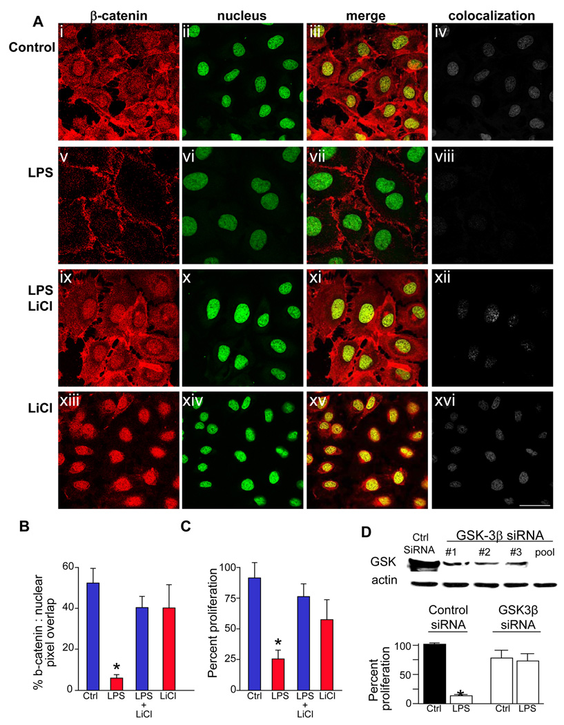
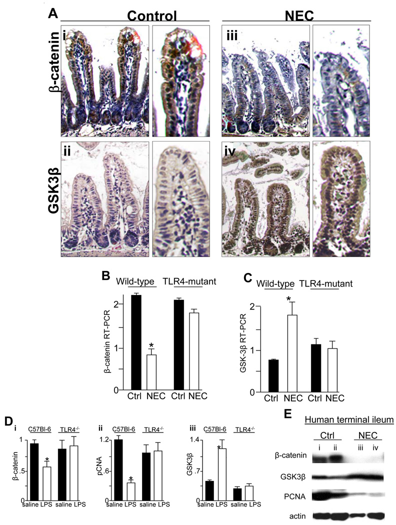
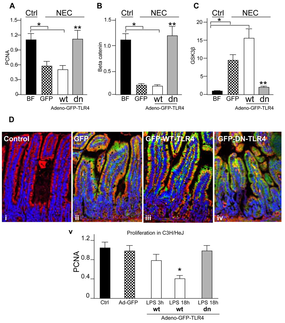
Methods: Enterocyte proliferation was detected in IEC-6 cells or in ileum or colon from wild-type, TLR4-mutant, or TLR4(-/-) mice after induction of NEC or endotoxemia. beta-Catenin signaling was assessed by cell fractionation or immunoconfocal microscopy to detect its nuclear translocation. Activation and inhibition of beta-catenin were achieved via cDNA or small interfering RNA, respectively. TLR4 in the intestinal mucosa was inhibited with adenoviruses expressing dominant-negative TLR4.

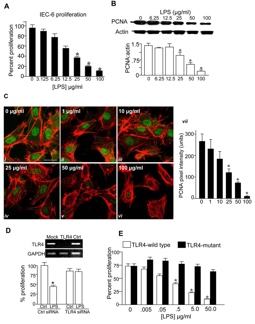
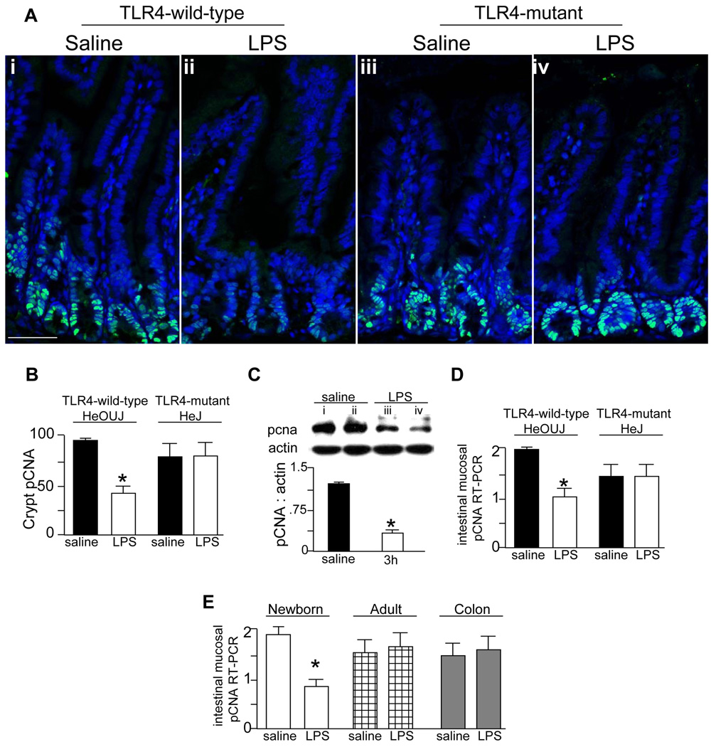
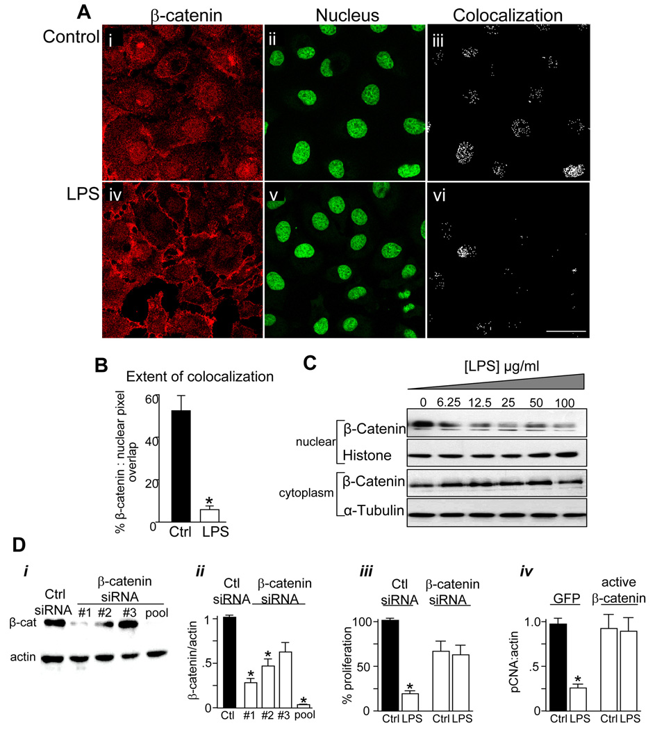
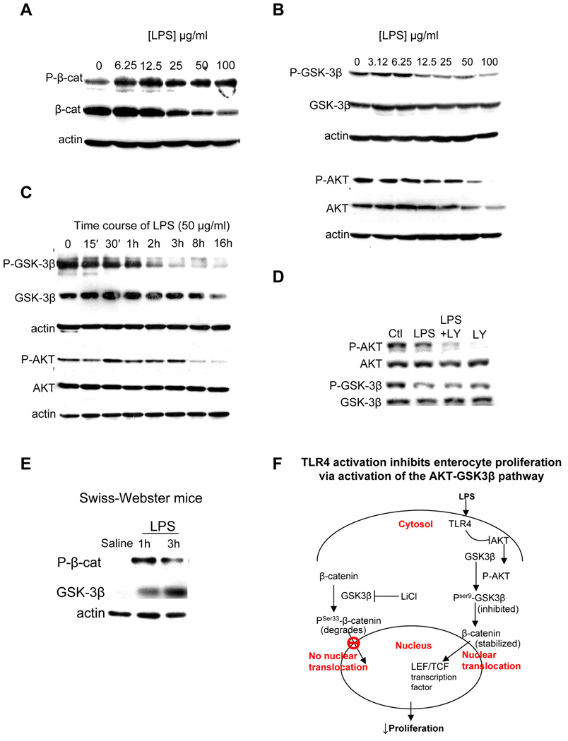
Results: TLR4 activation significantly impaired enterocyte proliferation in the ileum but not colon in newborn but not adult mice and in IEC-6 enterocytes. beta-Catenin activation reversed these effects in vitro. To determine the mechanisms involved, TLR4 activation phosphorylated the upstream inhibitory kinase GSK3beta, causing beta-catenin degradation. NEC in both mouse and humans was associated with decreased beta-catenin and increased mucosal GSK3beta expression. Strikingly, the inhibition of enterocyte beta-catenin signaling in NEC could be reversed, and enterocyte proliferation restored, through adenoviral-mediated inhibition of TLR4 signaling in the small intestinal mucosa.

Conclusion: We now report a novel pathway linking TLR4 with inhibition of beta-catenin signaling via GSK3beta activation, leading to reduced enterocyte proliferation in vitro and in vivo. These data provide additional insights into the pathogenesis of diseases of intestinal inflammation such as NEC.


Conflict of interest statement

Conflicts of interest:

Xia-hua Shi – no conflicts exist; Chhinder P. Sodhi - – no conflicts exist ; Ward M. Richardson - – no conflicts exist; Steven C. Gribar - – no conflicts exist; Thomas Prindle Jr - – no conflicts exist; Maria Branca – no conflicts exist; Anthony Russo - – no conflicts exist; Congrong Ma - – no conflicts exist ; Richard Shapiro – no conflicts exist; Zachary Grant – no conflicts exist; David J. Hackam – no conflicts exist.

Transcript profiling – Not applicable

Writing assistance – Not applicable.

Figures

Figure 1
Figure 1. TLR4 activation inhibits enterocyte proliferation in vitro
A: Percent proliferation in serum-starved IEC-6 cells treated with LPS (3h) at the indicated dosage as determined by XTT assay. B: SDS-PAGE showing pCNA and actin expression and their quantified expression ratio, in IEC-6 cells treated with LPS. *p<0.01 by ANOVA. C: Confocal micrographs showing expression of pCNA (green) and actin (red) treated with LPS for 3h at the indicated concentration; quantification of pCNA pixel density is shown, size bar=10µm. p<0.05 vs. untreated cells, ANOVA. D: RT-PCR showing expression of TLR4 and GAPDH in IEC-6 cells that were either untreated (“mock”), or treated with siRNA to TLR4 (“TLR4”) or to no known targets (“ctrl”), and percent proliferation in IEC-6 cells treated as indicated. E: Effect of inhibition of TLR4 by siRNA on proliferation in embryonic fibroblasts from TLR4-wild-type (C3H/HeOUJ) or TLR4-mutant (C3H/HeJ) mice treated with LPS for 16h as indicated. p<0.05 vs. untreated cells, ANOVA
Figure 2
Figure 2. TLR4 activation leads to an inhibition of enterocyte proliferation in vivo
A: Confocal micrographs showing sections of terminal ileum from TLR4-wild-type (i–ii) and TLR4-mutant mice (iii–iv) that were injected with LPS or saline and immunostained with pCNA. Size bar = 250 µm. B: Quantification of pCNA staining in crypts in A, *p<0.01 vs. saline C3H/HeOUJ by ANOVA. Representative of 3 separate experiments with at least 5 mice per group. C: SDS-PAGE and quantification showing the expression of pCNA and actin in terminal ileal mucosal scrapings from TLR4-wild-type mice injected with either saline (lanes i–ii) or LPS (lanes iii–iv) for 3h,*p<0.05. Representative of 3 separate experiments with at least 5 mice per group. D: Quantitative RT-PCR showing pCNA expression in terminal ileal mucosal scrapings from TLR4-wild-type and TLR4-mutant mice injected with saline (black bars) or LPS (white bars) for 3h,*p<0.005 vs. saline in C3H/HeOUJ by ANOVA. Representative of 3 separate experiments with at least 5 mice per group. E: RT-PCR expression of pCNA in the terminal ileal mucosa of newborn (white bars) and adult mice (checkered bars), and the colon of newborn mice (grey bars) after injection with LPS. *p<0.005 by ANOVA. Representative of 3 separate experiments with at least 5 mice per group.
Figure 3
Figure 3. TLR4 activation inhibits enterocyte proliferation via inhibition of β-catenin signaling
A: Confocal images of IEC-6 cells showing β-catenin (red, i, iv) and the nuclear marker Draq-5 (green, ii, v). Size bar=10 µm. B: Extent of colocalization (in iii and vi) was calculated using ImageJ,*p<0.05, representative of 5 separate experiments. C: SDS-PAGE of β-catenin in nuclear and cytoplasmic fractions obtained from IEC-6 cells treated with LPS as indicated, along with the nuclear marker histone and the cytoplasmic marker α-tubulin. Representative of 3 separate experiments. D: SDS-PAGE (i) and quantification of β-catenin:actin expression (ii) in IEC-6 cells transfected with siRNA to no known target (“Ctrl”) or to β-catenin. Three siRNA's (#1, #2, #3) were used either individually or in combination (“pooled”), as indicated. iii: Proliferation in IEC-6 cells treated with the indicated siRNA (XTT assay). iv: RT-PCR showing fold change in pCNA:actin in IEC-6 cells transfected with either GFP or constitutively active β-catenin and treated with LPS. *p<0.05 vs. control, representative of 3 separate experiments.
Figure 4
Figure 4. LPS activates the AKT-GSK3β signaling pathway in enterocytes
A–E: SDS-PAGE showing expression of the indicated protein in IEC-6 cells treated with LPS at the concentrations and durations indicated (A–D) or mucosal scrapings from the terminal ileum of mice injected with saline or LPS for the durations indicated (E). All blots are representative of 3 separate experiments; over 5 mice per group in E. F: Schematic summarizing panels A–D showing mechanism by which TLR4 inhibits enterocyte proliferation.
Figure 5
Figure 5. TLR4 activation inhibits enterocyte proliferation via GSK3β
A: Confocal images showing immunolocalization of β-catenin (red, i, v, ix, xiii), the nuclear marker draq-5 (ii, vi, x, xiv), merged images (iii, vii, xi, xv) and colocalization of nuclear and β-catenin (iv, viii, xii, xvi) using ImageJ in IEC-6 cells treated with media (control), or the indicated treatment (LPS and/or the GSK3β inhibitor LiCl). Size bar=10µm. B: Quantification of colocalization between the nucleus and β-catenin using ImageJ.*p<0.05 vs. control cells, ANOVA. C: Proliferation of IEC-6 cells treated as indicated (XTT assay) *p<0.05 vs. control cells, ANOVA. D: upper: SDS-PAGE showing GSK3β in IEC-6 cells transfected with indicated siRNA's to GSK3β, either alone (#1,#2,#3) or in combination (“pool”). lower: proliferation of IEC-6 cells (XTT) after treatment with indicated siRNA and LPS or media (“ctrl”)*p<0.01 vs. control cells, ANOVA. Representative of 3 separate experiments.
Figure 6
Figure 6. Necrotizing enterocolitis is associated with decreased β-catenin and increased GSK3β expression in enterocytes in mice and humans
A: Immunostaining of β-catenin (brown staining, i, iii) and GSK3β (brown staining, ii, iv) in either breast fed or mice with NEC. Representative of 4 separate experiments with at least 5 mice per group. B–C: Quantitative RT-PCR showing β-catenin (B) and GSK3β (C) in wild-type and TLR4-mutant mice with (white bars) and without (black bars) NEC. Representative of 3 separate experiments with at least 5 mice per group. D: RT-PCR showing the intestinal mucosal expression of pCNA, GSK3β and β-catenin in wild-type (C57Bl-6) and TLR4−/− mice after injection of saline (black bars) or LPS (white bars). *p<0.05, ANOVA. Representative of 3 separate experiments with at least 5 mice per group. E: SDS-PAGE showing β-catenin in mucosal scrapings obtained from two infants without (lanes i, ii) and two infants with (lanes iii, iv) NEC. Blots were stripped and re-probed for GSK3β, pCNA and actin.
Figure 7
Figure 7. Inhibition of enterocyte TLR4 signaling in mice with NEC restores enterocyte proliferation and reverses the changes in β-catenin and GSK3β expression
A–C: Newborn mice were either breast fed (black bars) or were induced to develop NEC and administered adenoviruses expressing GFP (checkered bars), GFP-wild-type TLR4 (white bars), or GFP—dominant-negative TLR4 by enteral gavage (grey bars). The expression by quantitative RT-PCR of pCNA (A), β-catenin (B) and GSK3β (C) in mucosal scrapings in each group is shown. *p<0.005 vs. breast fed, **p<0.001 vs. both NEC mice administered adeno-GFP and adeno-GFP-wild-type-TLR4, ANOVA. Representative of 5 separate experiments with over 5 mice per group D: i–v: Confocal micrographs showing GFP (green), actin (red) and nuclei (blue) in untreated (“control”) mice, or mice treated by gavage with the indicated adenovirus. v: RT-PCR of pCNA in TLR4 mutant mice that were administered GFP (checkered bars), GFP-wild-type-TLR4 (white bars), GFP-dominant-negative-TLR4 (grey bars) or control (black bars) and LPS as indicated. Representative of 3 separate experiments with over 10 mice per group.

Comment in

References

    1. Gribar SCAR, Sodhi CP, Hackam DJ. The role of epithelial Toll-like receptor signaling in the pathogenesis of intestinal inflammation. J Leukoc Biol. 2008;83:493–498. - PubMed
    1. Anand RJ, Leaphart CL, Mollen KP, Hackam DJ. The role of the intestinal barrier in the pathogenesis of necrotizing enterocolitis. Shock. 2007;27:124–133. - PubMed
    1. Grave GD, Nelson SA, Walker WA, Moss RL, Dvorak B, Hamilton FA, Higgins R, Raju TN. New therapies and preventive approaches for necrotizing enterocolitis: report of a research planning workshop. Pediatr Res. 2007;62:510–514. - PubMed
    1. Gribar SC, Sodhi CP, Richardson WM, Anand RJ, Gittes GK, Branca MF, Jakub A, Shi XH, Shah S, Ozolek JA, Hackam DJ. Reciprocal expression and signaling of TLR4 and TLR9 in the pathogenesis and treatment of necrotizing enterocolitis. J Immunol. 2009;182:636–646. - PMC - PubMed
    1. Cetin S, Ford HR, Sysko LR, Agarwal C, Wang J, Neal MD, Baty C, Apodaca G, Hackam DJ. Endotoxin inhibits intestinal epithelial restitution through activation of Rho-GTPase and increased focal adhesions. J Biol Chem. 2004;279:24592–24600. - PubMed

Publication types

MeSH terms