Review
. 2009 Nov;34(11):579-88.
doi: 10.1016/j.tibs.2009.06.009. Epub 2009 Sep 26.

Mammalian two-hybrids come of age

Affiliations
Review

Mammalian two-hybrids come of age

Sam Lievens et al. Trends Biochem Sci. 2009 Nov.

Abstract

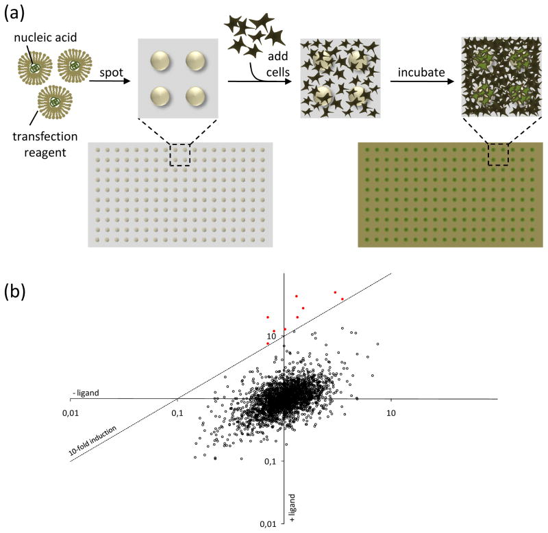
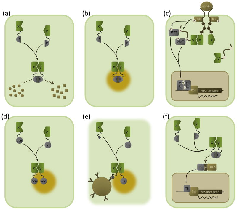
A diverse series of mammalian two-hybrid technologies for the detection of protein-protein interactions have emerged in the past few years, complementing the established yeast two-hybrid approach. Given the mammalian background in which they operate, these assays open new avenues to study the dynamics of mammalian protein interaction networks, i.e. the temporal, spatial and functional modulation of protein-protein associations. In addition, novel assay formats are available that enable high-throughput mammalian two-hybrid applications, facilitating their use in large-scale interactome mapping projects. Finally, as they can be applied in drug discovery and development programs, these techniques also offer exciting new opportunities for biomedical research.


Figures

Figure I
Figure I. Outline of the Y2H concept
(a) Modularity of eukaryotic transcription factors. DB and AD are coupled by a flexible linker, thereby allowing simultaneous binding of the DB to its cognate promoter region and the AD with RNA polymerase II (Pol). Both events are required to activate gene transcription, (b) The Y2H assay. Interaction of proteins X and Y reconstitutes transcription factor activity by complementing its DB and AD, respectively, and activating reporter gene transcription.
Figure I
Figure I. Reverse transfected cell arrays and their application in MAPPIT
(a) Generation of a cell array. Reverse transfection mixtures containing nucleic acids complexed with a transfection reagent are spotted onto a carrier surface. After drying, these arrays are overlaid with cells and incubated. Cells growing on top of the spots are (reverse) transfected with the nucleic acids present in the dried reverse transfection complexes, resulting in an array of transfected cells. (b) Result of an array MAPPIT screen. MAPPIT is an inducible assay that requires stimulation with an appropriate cytokine ligand. To exploit this additional level of control, luciferase signals obtained from samples treated with ligand are corrected for the background signal from samples that were not ligand-stimulated. The resulting induction factor is used to score interactions. In the conceptual example depicted here, each dot represents a different prey being combined with the bait that is being screened. For each bait–prey combination the normalized luciferase activity obtained with and without addition of the ligand are plotted against each other. Bait–prey combinations yielding a signal above a user-defined cut-off (dashed line; here corresponding to 10-fold induction of ligand treated versus untreated) score positive (red dots).
Figure 1
Figure 1. Overview of M2H assay principles
Schematic outline of the M2H assays discussed in the text. The interacting proteins X and Y are depicted in green; complemented protein fragments or proteins are grey. Please note that the M2H (sensu stricto) assay is not depicted here as it is conceptually identical to the Y2H system (Box 1). (a) Enzyme complementation systems. The interaction between two proteins of interest (X and Y) brings together two fragments of a reporter enzyme (F and F′); this reconstitutes the enzyme’s catalytic activity, converting a substrate (circles) into a detectable product (squares). The enzyme fragments can be pre-folded subunits (e.g. the split β-galactosidase assay) or fragments that fold only upon complementation (e.g. the β-lactamase and luciferase protein fragment complementation assays). (b) BiFC. Association of proteins X and Y reconstitutes a fluorescent protein from two fragments (F and F′), resulting in fluorescent emission upon excitation at a suitable wavelength (yellow halo). (c) MAPPIT. Bait X is coupled to a signalling-deficient cytokine receptor (via substituting tyrosine residues in the receptor tail which are critical for recruiting signal transducer and activator of transcription (STAT) proteins (brown dots)). The prey Y is tethered to a portion of another receptor containing intact recruitment sites. Upon bait–prey interaction, a functional receptor is reconstituted which can be activated by the appropriate cytokine ligand. Upon ligand binding, pre-associated Janus kinases (Jak) are activated by cross-phosphorylation (P). Activated Jaks phosphorylate (P) tyrosine residues in the receptor fragment coupled to the prey (white dots), which then act as docking sites for STATs. Recruited STATs are in turn phosphorylated by the Jaks, leading to their activation and subsequent dissociation and translocation to the nucleus. In the nucleus, STAT dimers induce STAT-dependent reporter gene transcription. In the MASPIT setup, dihydrofolate reductase (DHFR) is coupled to the signalling deficient receptor chain and a chemical compound is fused to methotrexate, a molecule that binds to DHFR with very high affinity. Addition of such a methotrexate fusion compound to cells expressing the DHFR-coupled receptor results in the chemical compound being displayed as a bait. (d) FRET and BRET. Association of proteins X and Y brings into proximity an energy donor (don) and acceptor protein (acc). In FRET, (intact) fluorescent proteins are used as donor and acceptor. Excitation of the donor fluorophore causes nonradiative energy transfer to occur between the two, resulting in fluorescent emission of the acceptor fluorophore (yellow halo). Instead of a fluorescent protein, a luciferase enzyme is used as a donor in BRET. Enzymatic oxidation of a luciferase substrate generates bioluminescence, and energy transfer to the acceptor protein produces a fluorescent signal. (e) LUMIER. Interaction between a Flag-tagged (triangle) bait protein X and a prey protein Y fused to Renilla luciferase is detected by bioluminescence measurements (yellow halo) of immunoprecipitates obtained from cell lysates using anti-Flag antibodies coupled to sepharose beads (brown sphere). (f) Split TEV assay. Association between proteins X and Y complements a functional TEV protease from fragments F and F′. At the site of a TEV recognition sequence, the protease cleaves a chimeric protein consisting of a transcription factor (TF) coupled to an ERT2 domain, thus retaining the transcription factor in the cytosol. After cleavage, the released transcription factor migrates into the nucleus where it induces reporter gene transcription. Note that the split TEV assay is a flexible system and that the setup shown here is only one of the possible choices.

References

    1. Goehler H, et al. A protein interaction network links GIT1, an enhancer of huntingtin aggregation, to Huntington’s disease. Mol Cell. 2004;15:853–865. - PubMed
    1. Lim J, et al. A protein-protein interaction network for human inherited ataxias and disorders of Purkinje cell degeneration. Cell. 2006;125:801–814. - PubMed
    1. Taylor IW, et al. Dynamic modularity in protein interaction networks predicts breast cancer outcome. Nat Biotechnol. 2009;27:199–204. - PubMed
    1. Kocher T, Superti-Furga G. Mass spectrometry-based functional proteomics: from molecular machines to protein networks. Nat Methods. 2007;4:807–815. - PubMed
    1. Suter B, et al. Two-hybrid technologies in proteomics research. Curr Opin Biotechnol. 2008;19:316–323. - PubMed

Publication types