. 2009 Oct 15;183(8):5079-84.
doi: 10.4049/jimmunol.0901514. Epub 2009 Sep 28.

The survival of memory CD4+ T cells within the gut lamina propria requires OX40 and CD30 signals

Affiliations

The survival of memory CD4+ T cells within the gut lamina propria requires OX40 and CD30 signals

David R Withers et al. J Immunol. .

Abstract

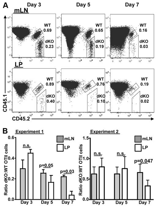
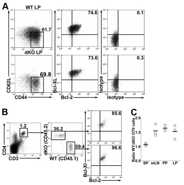
Although CD4(+) memory T cells reside within secondary lymphoid tissue, the major reservoir of these cells is in the lamina propria of the intestine. In this study, we demonstrate that, in the absence of signals through both OX40 and CD30, CD4(+) T cells are comprehensively depleted from the lamina propria. Deficiency in either CD30 or OX40 alone reduced CD4(+) T cell numbers, however, in mice deficient in both OX40 and CD30, CD4(+) T cell loss was greatly exacerbated. This loss of CD4(+) T cells was not due to a homing defect because CD30 x OX40-deficient OTII cells were not impaired in their ability to express CCR9 and alpha(4)beta(7) or traffic to the small intestine. There was also no difference in the priming of wild-type (WT) and CD30 x OX40-deficient OTII cells in the mesenteric lymph node after oral immunization. However, following oral immunization, CD30 x OX40-deficient OTII cells trafficked to the lamina propria but failed to persist compared with WT OTII cells. This was not due to reduced levels of Bcl-2 or Bcl-XL, because expression of these was comparable between WT and double knockout OTII cells. Collectively, these data demonstrate that signals through CD30 and OX40 are required for the survival of CD4(+) T cells within the small intestine lamina propria.


Figures

FIGURE 1
FIGURE 1
CD4+ T cell numbers in small intestine villi of WT, CD30KO, OX40KO, and dKO mice. Sections of small intestine from WT, CD30KO, OX40KO, and dKO mice were stained for expression of CD3 and CD4 and numbers of CD4+ and CD4 T cells enumerated. A, Expression of CD3 (green) and CD4 (red), counterstained with DAPI (gray). Scale bar, 50 μm. B, Numbers of CD4+ T cells per mm2 small intestine LP. C, Numbers of CD4 T cells per mm2 small intestine LP. D, Numbers of IELs per mm2 small intestine epithelium. Each data point represents an individual micrograph from which cells were counted. Sections were cut from tissues from at least four mice of each type. Statistical significances of differences shown were obtained using the Wilcoxon two sample test, *, p < 0.01; **, p < 0.00001. E, Expression of CD3 (green), CD4 (blue), and CD8β or γδ TCR (red) in sections of WT and dKO small intestine. CD8β+ and γδ TCR+ T cells circled, outline of villus shown in white. Scale bar, 50 μm.
FIGURE 2
FIGURE 2
Normal homing of dKO CD4+ T cells to small intestine. To investigate whether dKO OTII T cells proliferated normally and expressed gut-homing molecules, CFSE-labeled WT (CD45.1+) and dKO (CD45.2+) OTII T cells were transferred (1:1 ratio) to CD45.1+CD45.2+ mice that were orally immunized with OVA. A, Ratio of dKO:WT OTII cells 3 days post immunization in the mLN and spleen. Data represent mean (with SD) of two experiments, each with three mice. B, Expression of CCR9 and α4β7 vs CFSE in WT and dKO OTII T cells. Data are representative of three mice. C, Expression of CCR9 and α4β7 by WT and dKO OTII T cells cultured in vitro with splenic DCs, OVA, and retinoic acid for 6 days. Data are representative of two experiments. D, Ratio of dKO:WT in vitro-cultured OTII cells in mLN, small intestine, LP, and spleen, 24 h after transfer into allotype-marked mice. Data represent mean (with SD) of two experiments, each with three mice.
FIGURE 3
FIGURE 3
In vivo activated WT and dKO cells acquire similar expression of CCR9 and α4β7 in the mLN. Recipient mice (CD45.1+) were adoptively transferred with dKO (CD45.2+) and WT (CD45.1+CD45.2+) OTII cells, input ratio 0.8. The OTII cells were then activated in vivo by oral administration of OVA and the mLN was analyzed at day 3. A, Expansion of both dKO OTII (CD45.2) and WT (CD45.1+CD45.2+) cells after OVA administration. B, Ratio of dKO:WT OTII cells with and without OVA administration. C, CFSE profile of dKO and WT OTII cells. D, Representative expression of CCR9 and α4β7 of dKO and WT cells. E, Expression CCR9 and α4β7 of dKO and WT cells, data represent mean (with SD) of three mice per group.
FIGURE 4
FIGURE 4
dKO OTII cells fail to persist in the LP after in vivo activation. Recipient mice (CD45.1) were adoptively transferred with dKO (CD45.2+) and WT (CD45.1+CD45.2+) cells, input ratio 0.8. The OTII cells were then activated in vivo by oral administration of OVA. The ratio of dKO OTII cells to WT OTII cells in the mLN and LP was then assayed at days 3, 5 and 7 post immunization. A, Detection of WT and dKO OTII cells in the mLN and LP, numbers show percentage of CD4+ T cells. B, Ratio of dKO:WT OTII cells in mLN and LP from two independent experiments; data is mean (with SD) of two to three mice per group.
FIGURE 5
FIGURE 5
Normal Bcl-2 and Bcl-xL expression in dKO LP CD4+ T cells. To investigate whether the failure of dKO CD4+ T cells to survive in the LP was due to impaired expression of Bcl-2 and Bcl-xL, intracellular staining was done. A, Expression of Bcl-2 and Bcl-xL by CD62LCD44high CD4+ T cells of LP isolated from WT and dKO mice. B, WT (CD45.1) and dKO (CD45.2) OTII cells were transferred into CD3εTg26 mice which were then orally immunized with OVA and analyzed 4 days later. Expression of Bcl-2 and Bcl-xL by WT and dKO OTII cells of LP. C, Ratio of WT:dKO OTII cells in spleen, mLN, PP, and LP at 4 days after oral immunization. Numbers show percentage of cells in gate, data from three mice.

Similar articles

Cited by

References

    1. Mowat AM, Viney JL. The anatomical basis of intestinal immunity. Immunol. Rev. 1997;156:145–166. - PubMed
    1. Reinhardt RL, Khoruts A, Merica R, Zell T, Jenkins MK. Visualizing the generation of memory CD4 T cells in the whole body. Nature. 2001;410:101–105. - PubMed
    1. Iijima H, Takahashi I, Kiyono H. Mucosal immune network in the gut for the control of infectious diseases. Rev. Med. Virol. 2001;11:117–133. - PubMed
    1. Strobel S, Mowat AM. Oral tolerance and allergic responses to food proteins. Curr. Opin. Allergy Clin. Immunol. 2006;6:207–213. - PubMed
    1. Dubois B, Goubier A, Joubert G, Kaiserlian D. Oral tolerance and regulation of mucosal immunity. Cell Mol. Life Sci. 2005;62:1322–1332. - PMC - PubMed

Publication types

MeSH terms