. 2009 Oct 15;183(8):5379-87.
doi: 10.4049/jimmunol.0900228. Epub 2009 Sep 28.

Mitochondrial dysfunction increases allergic airway inflammation

Affiliations

Mitochondrial dysfunction increases allergic airway inflammation

Leopoldo Aguilera-Aguirre et al. J Immunol. .

Abstract

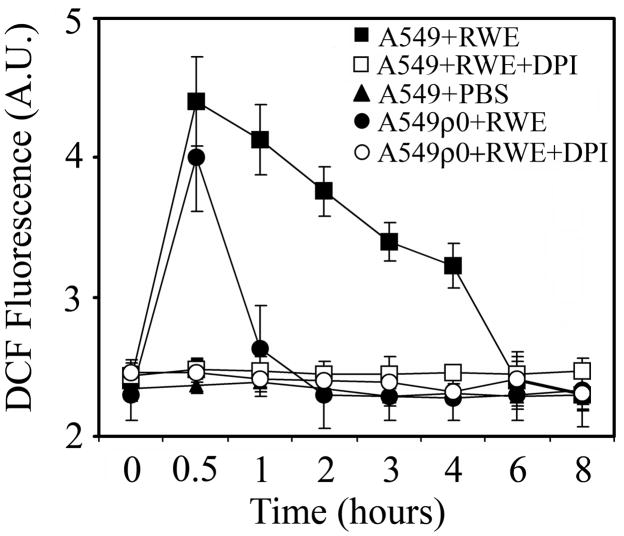
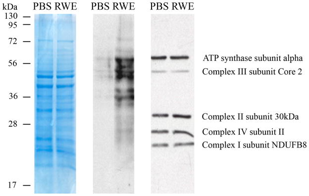
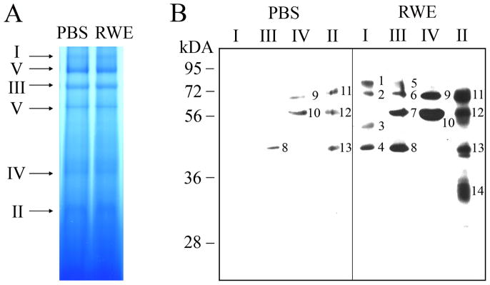
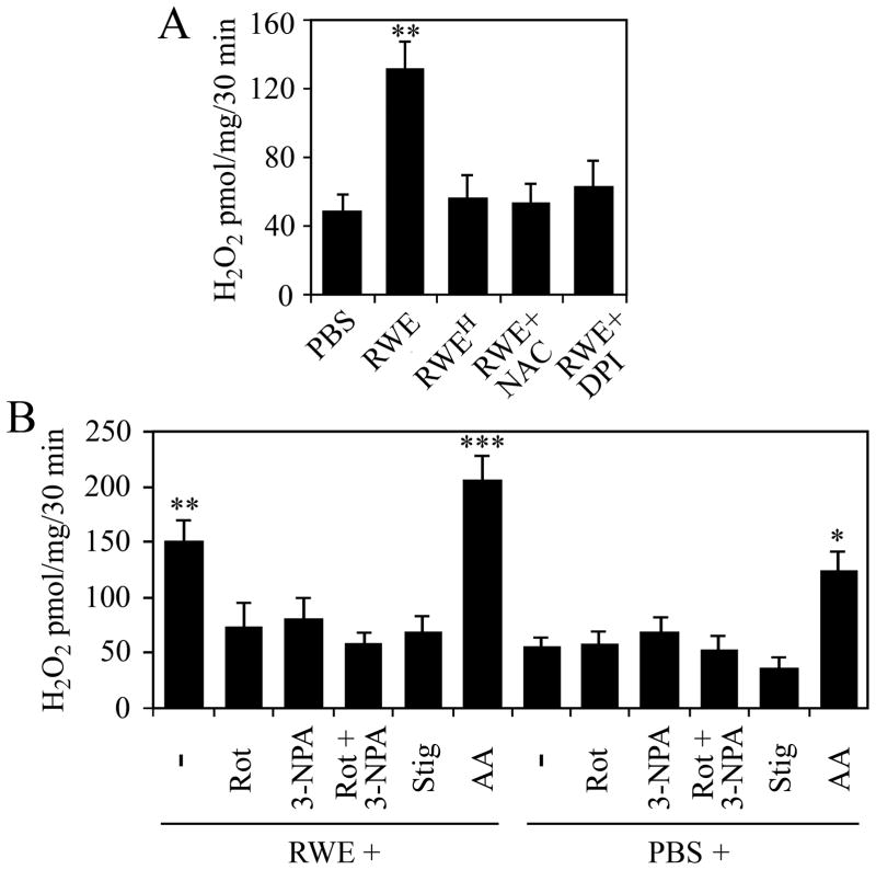
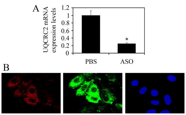
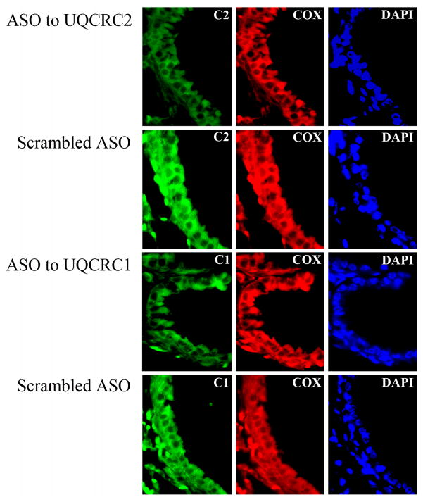
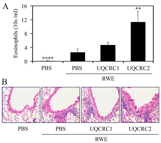
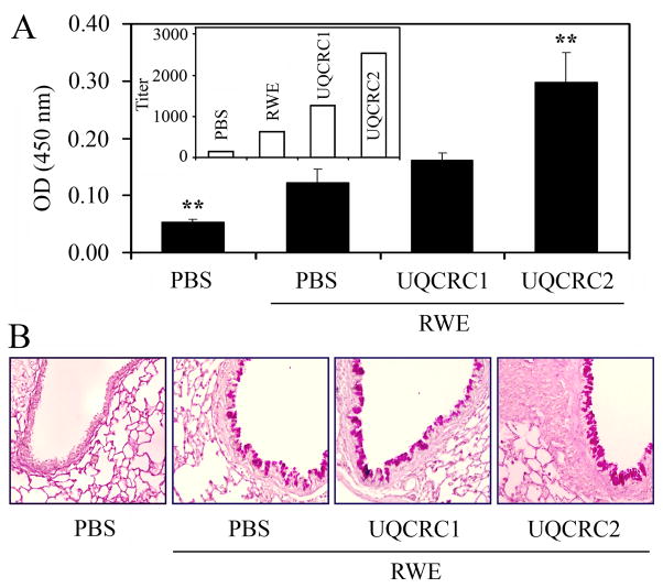
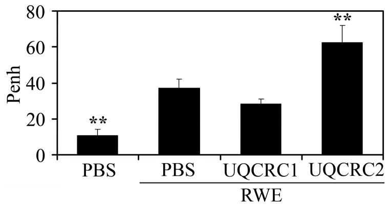
The prevalence of allergies and asthma among the world's population has been steadily increasing due to environmental factors. It has been described that exposure to ozone, diesel exhaust particles, or tobacco smoke exacerbates allergic inflammation in the lungs. These environmental oxidants increase the levels of cellular reactive oxygen species (ROS) and induce mitochondrial dysfunction in the airway epithelium. In this study, we investigated the involvement of preexisting mitochondrial dysfunction in the exacerbation of allergic airway inflammation. After cellular oxidative insult induced by ragweed pollen extract (RWE) exposure, we have identified nine oxidatively damaged mitochondrial respiratory chain-complex and associated proteins. Out of these, the ubiquinol-cytochrome c reductase core II protein (UQCRC2) was found to be implicated in mitochondrial ROS generation from respiratory complex III. Mitochondrial dysfunction induced by deficiency of UQCRC2 in airway epithelium of sensitized BALB/c mice prior the RWE challenge increased the Ag-induced accumulation of eosinophils, mucin levels in the airways, and bronchial hyperresponsiveness. Deficiency of UQCRC1, another oxidative damage-sensitive complex III protein, did not significantly alter cellular ROS levels or the intensity of RWE-induced airway inflammation. These observations suggest that preexisting mitochondrial dysfunction induced by oxidant environmental pollutants is responsible for the severe symptoms in allergic airway inflammation. These data also imply that mitochondrial defects could be risk factors and may be responsible for severe allergic disorders in atopic individuals.


Figures

FIG. 1
FIG. 1
Exposure of cells with functional mitochondria results in sustained increase in the intracellular ROS levels. Airway epithelial cells (A549) cells and mitochondrial DNA depleted A549ρ0 cells were loaded with H2DCF-DA and treated with 100 μg/ml RWE in the presence or absence of DPI, a NAD(P)H oxidase inhibitor. Changes in intracellular DCF fluorescence were determined by flow cytometry. The data points represent mean values of three independent experiments.
FIG. 2
FIG. 2
Treatment of epithelial cells with RWE increases the levels of carbonylated proteins in mitochondria. Mitochondria isolated from mock-treated and RWE-exposed (100 μg/ml) cells were purified (Materials and Methods). Mitochondrial lysates were DNPH derivatized and subjected to SDS-PAGE (left panel). After blotting onto membrane, oxidatively-damaged proteins were detected using DNP-specific antibody (middle panel). In parallel experiments the relative levels of respiratory complex proteins in mitochondrial preparations were analyzed using OXPHOS monoclonal antibody cocktail (right panel). Results are from a representative experiment from three repeats.
FIG. 3
FIG. 3
Detection and identification of carbonylated mitochondrial respiratory complex proteins. A, Mitochondria isolated from mock-treated and RWE-exposed (100 μg/ml) cells were purified. Equal amounts of mitochondrial lysates were loaded and MRCs were isolated on blue native PAGE. B, Complexes (I–IV) were excised from blue native gels and DNPH derivatized prior to separation on 10% SDS-PAGE. Carbonylated proteins were visualized using antibody to DNP. Oxidatively damaged proteins identified by MALDI-TOF/MS analysis are shown in Table I.
FIG. 4
FIG. 4
Mitochondria isolated from RWE-treated cells released higher amounts of H2O2 than mitochondria isolated from mock-treated cells. A, Pretreatment of cells with antioxidant (NAC) as well as physical (heat-treatment, RWEH) or chemical (DPI) inactivation of pollen NAD(P)H oxidases abolishes the ability of RWE to increase mitochondrial ROS generation. B, Identification of respiratory complex III, as a major site of ROS production in mitochondria from RWE-treated cells. Administration of rotenone (Rot), 3-NPA, as well as stigmatellin (Stig) to mitochondria decreases, while addition of antimycin A (AA) enhances RWE-induced mitochondrial ROS production. Data are presented as means ± SEM of 3 independent experiments. *P< 0.05, **P< 0.01, ***P< 0.001 vs. H2O2 release from mitochondria of mock- treated cells.
FIG. 5
FIG. 5
Down-regulation of UQCRC2 increases cellular ROS levels. A, ASO to UQCRC2 down-regulated its expression in LA-4 cells by more than 80% (Materials and Methods). B, Increased ROS levels in cells efficiently transfected with ASO to UQCRC2. Cells were transfected with Texas Red-labeled ASO and various times thereafter cellular ROS levels were assessed by H2DCF-DA using microscopic analysis. Only cells with red fluorescence (Texas Red) show increased DCF signal (green fluorescence) in transfected cell cultures. *P< 0.05 vs. mock-treated cells.
FIG. 6
FIG. 6
Inhibition of the expression of mitochondrial respiratory complex III core proteins in the lungs by local ASO treatment. ASO were administered intranasally to RWE-sensitized mice and expression of UQCRC1 and UQCRC2 was analyzed in lung sections by fluorescent microscopy. In the bronchial epithelium focal down-regulation of both UQCRC1 and UQCRC2 was observed (left panel); however, the expression of mitochondrially synthesized cytochrome c oxidase subunit IIb was not affected (middle panel). Scrambled ASO treatment did not modified the expression of either core proteins or cytochrome c oxidase subunit IIb (left and middle panels). C1, UQCRC1; C2, UQCRC2; Cox, cytochrome c oxidase subunit IIb; DAPI, 4,6'-diamidino-2-phenylindole.
FIG. 7
FIG. 7
Preexisting mitochondrial dysfunction induced by UQCRC2 downregulation increases RWE-induced accumulation of inflammatory cells in the airways. Antisense oligonucleotide treatment, specific for UQCRC2 but not for UQCRC1, increases the number of eosinophils in the bronchoalveolar lavage fluids (A) and enhances accumulation of inflammatory cells in the peribronchial area (B) in RWE-challenged mice. **P< 0.01, ****P< 0.0001 vs. mock-treated, RWE-challenged mice.
FIG. 8
FIG. 8
Mitochondrial dysfunction induced by UQCRC2 downregulation enhances RWE-induced mucin production in the airways. Down-regulation of UQCRC2, but not UQCRC1, increases the levels of MUC5AC in the bronchoalveolar lavage fluids (A) and increases the metaplasia of mucuos cells in airway epithelium (B) of RWE-challenged mice. Inset: endpoint titers of MUC5AC in the samples. **P< 0.01 vs. mock-treated, RWE-challenged mice.
FIG. 9
FIG. 9
Preexisting mitochondrial dysfunction mediated by ASO to UQCRC2 increases RWE-induced airway hyperresponsiveness. Changes in pause of breathing (Penh) as an index of airway obstruction were measured by barometric whole-body plethysmography. **P< 0.01 vs. mock-treated, RWE-challenged mice.
FIG. 10
FIG. 10

References

    1. Bacsi A, Dharajiya N, Choudhury BK, Sur S, Boldogh I. Effect of pollen-mediated oxidative stress on immediate hypersensitivity reactions and late-phase inflammation in allergic conjunctivitis. J Allergy Clin Immunol. 2005;116:836–843. - PMC - PubMed
    1. Bacsi A, Choudhury BK, Dharajiya N, Sur S, Boldogh I. Subpollen particles: carriers of allergenic proteins and oxidases. J Allergy Clin Immunol. 2006;118:844–850. - PMC - PubMed
    1. Boldogh I, Bacsi A, Choudhury BK, Dharajiya N, Alam R, Hazra TK, Mitra S, Goldblum RM, Sur S. ROS generated by pollen NADPH oxidase provide a signal that augments antigen-induced allergic airway inflammation. J Clin Invest. 2005;115:2169–2179. - PMC - PubMed
    1. Dharajiya N, Choudhury BK, Bacsi A, Boldogh I, Alam R, Sur S. Inhibiting pollen reduced nicotinamide adenine dinucleotide phosphate oxidase-induced signal by intrapulmonary administration of antioxidants blocks allergic airway inflammation. J Allergy Clin Immunol. 2007;119:646–653. - PMC - PubMed
    1. Bowler RP. Oxidative stress in the pathogenesis of asthma. Curr Allergy Asthma Rep. 2004;4:116–122. - PubMed

Publication types

MeSH terms