. 2009 Oct 5;187(1):71-9.
doi: 10.1083/jcb.200907109. Epub 2009 Sep 28.

Autophagy promotes synapse development in Drosophila

Affiliations

Autophagy promotes synapse development in Drosophila

Wei Shen et al. J Cell Biol. .

Abstract

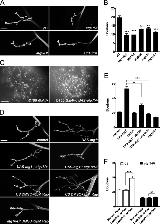
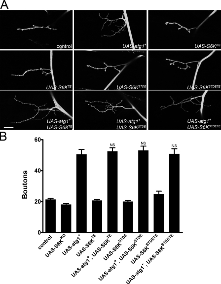
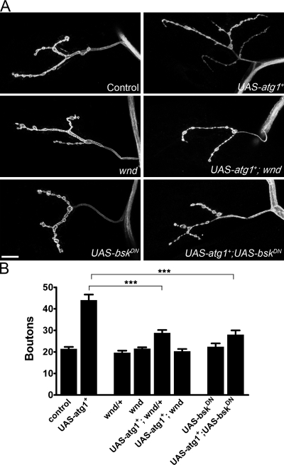
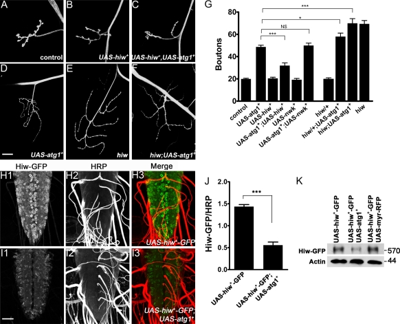
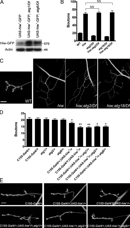
Autophagy, a lysosome-dependent degradation mechanism, mediates many biological processes, including cellular stress responses and neuroprotection. In this study, we demonstrate that autophagy positively regulates development of the Drosophila melanogaster larval neuromuscular junction (NMJ). Autophagy induces an NMJ overgrowth phenotype closely resembling that of highwire (hiw), an E3 ubiquitin ligase mutant. Moreover, like hiw, autophagy-induced NMJ overgrowth is suppressed by wallenda (wnd) and by a dominant-negative c-Jun NH(2)-terminal kinase (bsk(DN)). We show that autophagy promotes NMJ growth by reducing Hiw levels. Thus, autophagy and the ubiquitin-proteasome system converge in regulating synaptic development. Because autophagy is triggered in response to many environmental cues, our findings suggest that it is perfectly positioned to link environmental conditions with synaptic growth and plasticity.


Figures

Figure 1.
Figure 1.
Autophagy promotes NMJ growth in Drosophila. (A) Confocal images of NMJ 4 labeled with FITC-HRP. Compared with control (w1118), atg1/Df, atg2/Df, and atg18/Df mutants exhibit smaller NMJs. (B) Quantification of bouton numbers at NMJ 4 in larvae of the genotypes shown. (C) Fluorescent images of larval brain hemisphere of control (C155-Gal4/+) and atg1+-overexpressing larvae (C155-Gal4/UAS-atg1+) stained with LysoTracker. Substantially increased staining is observed in the latter, indicating elevated levels of autophagy. (D) Confocal images of NMJ 4 labeled with FITC-HRP. Compared with control (elav-Gal4/+), neuronal overexpression of atg1+ (UAS-atg1+) results in strong NMJ overgrowth. Overgrowth is reduced by loss of one copy of atg18+ (UAS-atg1+; atg18/+) and almost completely suppressed by removing both copies of atg18+ (UAS-atg1+; atg18/Df). Larvae fed on 2 µM rapamycin (Rap) to induce autophagy exhibit NMJ overgrowth, which is completely suppressed in an atg18/Df background. (E and F) Quantification of bouton numbers at NMJ 4 in the indicated larvae. (B, E, and F) Error bars denote SEM. **, P < 0.01; ***, P < 0.001. CS, Canton-S; WT, wild type. Bars: (A and D) 15 µm; (C) 50 µm.
Figure 2.
Figure 2.
Atg1-dependent changes in NMJ growth are independent of effects on translation. (A) Confocal images of NMJ 4 labeled with FITC-HRP. Blocking or activating translation by overexpressing a dominant-negative S6K (UAS-S6KKQ) or a constitutively activated S6K transgene (UAS-S6KTE, UAS-S6KSTDE, or UAS-S6KSTDETE) has little affect on NMJ growth. None of three constitutively activated S6K transgenes suppresses NMJ overgrowth when coexpressed with atg1+. (B) Quantification of bouton numbers at NMJ 4 in larvae of genotypes shown. Comparisons are made with elav-Gal4/UAS-atg1+. Error bars denote SEM. Bar, 15 µm.
Figure 3.
Figure 3.
Autophagy-dependent NMJ overgrowth is suppressed by wnd and bskDN. (A) Confocal images of NMJ 4 labeled with FITC-HRP. Compared with control (C155-Gal4/+), neuronal overexpression of atg1+ (C155-Gal4/UAS-atg1+) results in NMJ overgrowth. Both wnd and bskDN suppress NMJ overgrowth caused by atg1+ overexpression. (B) Quantification of bouton numbers at NMJ 4 for larvae of genotypes shown. Error bars denote SEM. ***, P < 0.001. Bar, 15 µm.
Figure 4.
Figure 4.
Autophagy promotes synaptic growth via down-regulation of Hiw. (A–F) Confocal images of NMJ 4 labeled with FITC-HRP. (A) elav-Gal4/+. (B–D) Neuronal overexpression of hiw+ alone (B) via elav-Gal4 driver has no affect on NMJ growth but is sufficient to suppress NMJ overgrowth caused by overexpression of atg1+ (C and D). (E and F) Neuronal overexpression of atg1+ in a hiw mutant background (F) does not further enhance NMJ growth compared with hiw mutants alone (E). (G) Quantification of bouton numbers at NMJ 4 in larvae of the genotypes shown. Comparisons are made with elav-Gal4/UAS-atg1+. (H and I) Confocal images of larval ventral ganglia expressing a Hiw-GFP fusion protein labeled with anti-GFP (H1 and I1) and anti-HRP (H2 and I2). Hiw-GFP fusion protein is present at high levels in C155-Gal4/UAS-hiw+-GFP larvae (H1–H3). However, Hiw levels are reduced by coexpression of atg1+ in C155-Gal4/UAS-hiw+-GFP; UAS-atg1+ larvae (I1–I3), whereas HRP levels remain unchanged. (J) Quantification of Hiw-GFP and HRP fluorescence intensities in C155-Gal4/UAS-hiw+-GFP larvae (1.44 ± 0.05; n = 4) and in C155-Gal4/UAS-hiw+-GFP; UAS-atg1+ larvae (0.55 ± 0.07; n = 5). (K) Western blots of extracts from larval brains probed for Hiw-GFP and actin confirm the reduction of Hiw-GFP levels caused by overexpression of atg1+. Molecular mass is indicated in kilodaltons. (G and J) Error bars denote SEM. *, P < 0.05; ***, P < 0.001. Bars: (A–F) 25 µm; (H and I) 50 µm.
Figure 5.
Figure 5.
Accumulation of Hiw contributes to NMJ undergrowth when autophagy is impaired. (A) Western blots of larval brain extracts from the indicated genotypes probed for Hiw-GFP and actin show significant increases of Hiw-GFP levels in atg1/Df and atg6/Df mutants. Molecular mass is indicated in kilodaltons. (B) Quantification of bouton numbers at NMJ 4 in larvae of the genotypes shown. (C) Confocal images of NMJ 4 labeled with FITC-HRP. hiw; atg2/Df and hiw; atg18/Df NMJ overgrowth in double mutants is the same as hiw alone, which is consistent with the idea that Hiw is down-regulated by autophagy. (D) Quantification of bouton numbers at NMJ 4 in larvae of the genotypes shown. (E) Confocal images of NMJ 4 labeled with FITC-HRP. Moderate overexpression of Hiw in C155-Gal4/+ females results in mild NMJ undergrowth. Stronger overexpression of Hiw in C155-Gal4/Y males leads to more marked NMJ undergrowth. When Hiw is overexpressed, loss of one copy of atg1+, -2+, or -6+ further exacerbates NMJ undergrowth. (B and D) Error bars denote SEM. *, P < 0.05; **, P < 0.01. Bars: (C) 25 µm; (E) 15 µm.

Similar articles

Cited by

References

    1. Aberle H., Haghighi A.P., Fetter R.D., McCabe B.D., Magalhães T.R., Goodman C.S. 2002. wishful thinking encodes a BMP type II receptor that regulates synaptic growth in Drosophila.Neuron. 33:545–558 doi:10.1016/S0896-6273(02)00589-5 - DOI - PubMed
    1. Barcelo H., Stewart M.J. 2002. Altering Drosophila S6 kinase activity is consistent with a role for S6 kinase in growth.Genesis. 34:83–85 doi:10.1002/gene.10132 - DOI - PubMed
    1. Berry D.L., Baehrecke E.H. 2007. Growth arrest and autophagy are required for salivary gland cell degradation in Drosophila.Cell. 131:1137–1148 doi:10.1016/j.cell.2007.10.048 - DOI - PMC - PubMed
    1. Collins C.A., DiAntonio A. 2007. Synaptic development: insights from Drosophila.Curr. Opin. Neurobiol. 17:35–42 doi:10.1016/j.conb.2007.01.001 - DOI - PubMed
    1. Collins C.A., Wairkar Y.P., Johnson S.L., DiAntonio A. 2006. Highwire restrains synaptic growth by attenuating a MAP kinase signal.Neuron. 51:57–69 doi:10.1016/j.neuron.2006.05.026 - DOI - PubMed

Publication types

MeSH terms