

Three-dimensional MR volumetric analysis of the posterior fossa CSF space in hemifacial spasm
- PMID: 19786697
- PMCID: PMC2754328
- DOI: 10.1212/WNL.0b013e3181b9c8ce
Three-dimensional MR volumetric analysis of the posterior fossa CSF space in hemifacial spasm
Abstract
Background: We hypothesize that a smaller posterior fossa (PF) CSF space may be a risk factor for hemifacial spasm (HFS).
Objective: We conducted a case-control 3-dimensional magnetic resonance (MR) volumetric study in patients with HFS and determined the clinical predictive factors of PF CSF volume.
Methods: Patients with clinically diagnosed HFS and controls matched for age, sex, race, and hypertension underwent MRI/magnetic resonance angiography examination. The PF CSF space was segmented and quantified on a heavily T2-weighted high-resolution 3-dimensional MR volume slab, centered over the porus acusticus.
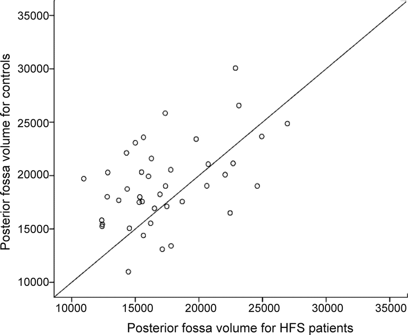
Results: Eighty-two study subjects (41 patients and 41 controls) were included. The mean PF CSF volume in patients with HFS and controls was 17,303.0 +/- 3,900.0 vs 19,216.0 +/- 3,912.0 mm(3). The mean volume in patients with HFS was 11.4% smaller than in controls (p = 0.015). Analysis of differences between individually matched pairs and controls also revealed that PF CSF for controls was larger than that for patients with HFS (p = 0.007). A multivariate linear regression analysis revealed that a small PF CSF volume was associated with HFS (p = 0.01). Decreasing age (p = 0.001) and female gender (p < 0.0005), but not hypertension (p = 0.892), were also found to be predictors of a low PF CSF volume.
Conclusions: Our results showed that the posterior fossa (PF) CSF volume was lower in patients with HFS compared with matched controls. HFS, female gender, and younger age were associated with smaller PF CSF volume. These observations could explain the strong female preponderance in both clinic- and population-based epidemiologic studies.
Figures


References
-
- Wang A, Jankovic J. Hemifacial spasm: clinical findings and treatment. Muscle Nerve 1998;21:1740–1747. - PubMed
-
- Stamey W, Jankovic J. The other Babinski sign in hemifacial spasm. Neurology 2007;69:402–404. - PubMed
-
- Chan LL, Lo YL, Lee E, Fook-Chong S, Tan EK. Ventrolateral medullary compression in hypertensive patients with hemifacial spasm. Neurology 2005;65:1467–1470. - PubMed
-
- Felber S, Birbamer G, Aichner F, Poewe W, Kampfl A. Magnetic resonance imaging and angiography in hemifacial spasm. Neuroradiology 1992;34:413–416. - PubMed
Publication types
MeSH terms
LinkOut - more resources
Full Text Sources
Medical