. 2009 Sep 29;4(9):e7217.
doi: 10.1371/journal.pone.0007217.

The trypanosome Rab-related proteins RabX1 and RabX2 play no role in intracellular trafficking but may be involved in fly infectivity

Affiliations

The trypanosome Rab-related proteins RabX1 and RabX2 play no role in intracellular trafficking but may be involved in fly infectivity

Senthil Kumar A Natesan et al. PLoS One. .

Abstract

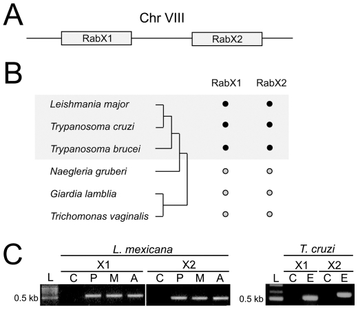
Background: Rab GTPases constitute the largest subgroup of the Ras superfamily and are primarily involved in vesicle targeting. The full extent of Rab family function is unexplored. Several divergent Rab-like proteins are known but few have been characterized. In Trypanosoma brucei there are sixteen Rab genes, but RabX1, RabX2 and RabX3 are divergent within canonical sequence regions. Where known, trypanosome Rab functions are broadly conserved when orthologous relationships may be robustly established, but specific functions for RabX1, X2 and X3 have yet to be determined. RabX1 and RabX2 originated via tandem duplication and subcellular localization places RabX1 at the endoplasmic reticulum, while RabX2 is at the Golgi complex, suggesting distinct functions. We wished to determine whether RabX1 and RabX2 are involved in vesicle transport or other cellular processes.

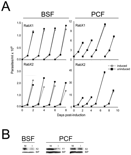
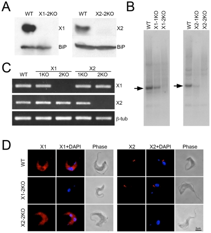
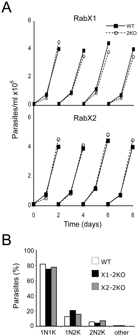
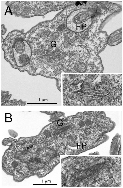
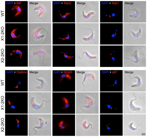
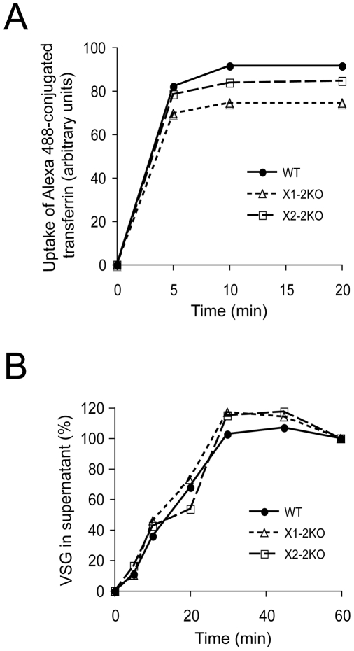
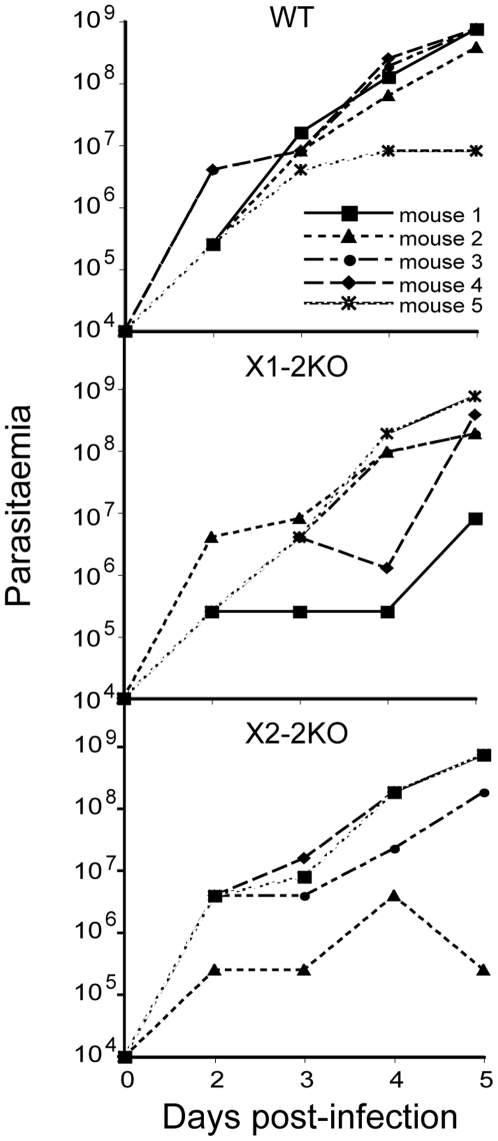
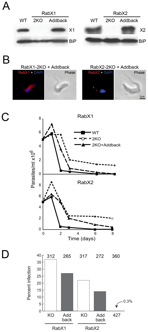
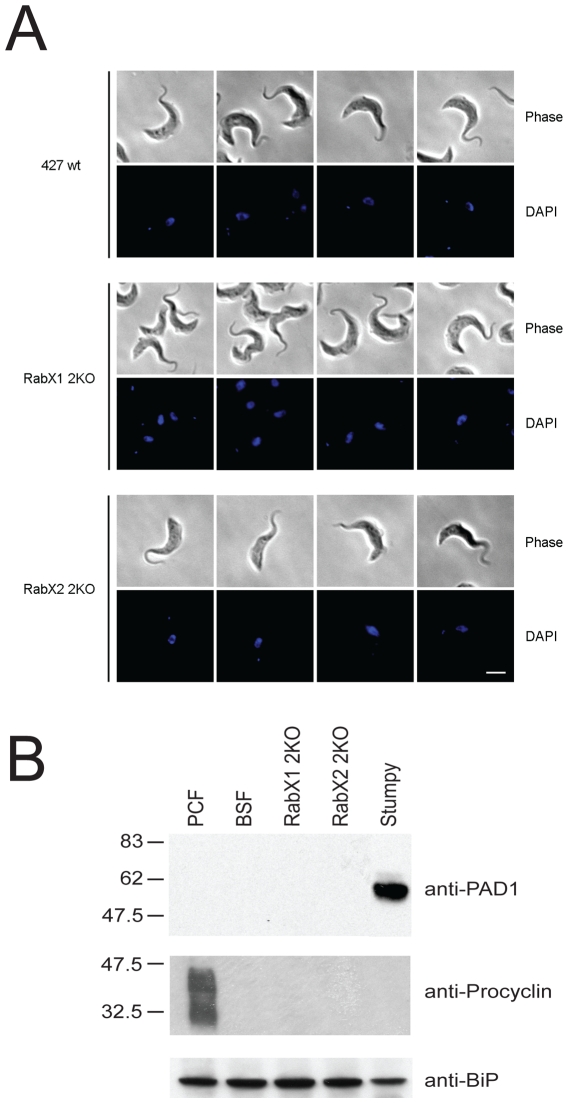
Methodology/principal findings: Using comparative genomics we find that RabX1 and RabX2 are restricted to trypanosomatids. Gene knockout indicates that RabX1 and RabX2 are non-essential. Simultaneous RNAi knockdown of both RabX1 and RabX2, while partial, was also non-lethal and may suggest non-redundant function, consistent with the distinct locations of the proteins. Analysis of the knockout cell lines unexpectedly failed to uncover a defect in exocytosis, endocytosis or in the morphology or location of multiple markers for the endomembrane system, suggesting that neither RabX1 nor RabX2 has a major role in intracellular transport. However, it was apparent that RabX1 and RabX2 knockout cells displayed somewhat enhanced survival within flies.

Conclusions/significance: RabX1 and RabX2, two members of the trypanosome Rab subfamily, were shown to have no major detectable role in intracellular transport, despite the localization of each gene product to highly specific endomembrane compartments. These data extend the functional scope of Rab proteins in trypanosomes to include non-canonical roles in differentiation-associated processes in protozoa.


Conflict of interest statement

Competing Interests: The authors have declared that no competing interests exist.

Figures

Figure 1
Figure 1. RabX1 and RabX2 are specific to trypanosomatids.
(A) Schematic representation of the RabX1 and RabX2 locus arranged on Trypanosoma brucei chromosome VIII. (B) Presence of RabX1 and RabX2 encoding sequences from trypanosomes in relation to additional fully sequenced Excavata lineages. RabX1 and RabX2 are found only in trypanosomatids, but not Naegleria gruberi, Giardia lamblia and Trichomonas vaginalis. Closed dots indicate gene is found, open dots not found. Schematic phylogenetic tree to indicate relationships between the lineages is on the left. (C) RT-PCR showing the presence of RabX1 and RabX2 in the major life stages of L. mexicana and T. cruzi. L, DNA ladder; C, no reverse transcriptase negative control; P, promastigote; M, metacyclic; A, amastigote; E, epimastigote.
Figure 2
Figure 2. RNAi indicates that RabX1 and RabX2 are non-essential for normal growth of T. brucei.
Growth curves of BSF SMB and PCF PTT parasites after tetracycline-induced RNAi for RabX1 and RabX2. Top left panel, Growth curve for BSF SMB parasites transfected with p2T7TAblue-RabX1 RNAi construct. Bottom left panel, growth curve for BSF SMB parasites transfected with p2T7TAblue-RabX2. Top right panel, growth curve for PCF PTT parasites transfected with p2T7-177-RabX1. Bottom right panel, growth curve for PCF PTT parasites transfected with p2T7-177-RabX2. RNAi was induced in BSF SMB parasites by the addition of 1 µg/ml and in PCF PTT lines by the addition of 10 µg/ml of tetracycline. BSF or PCF cells were cultured in the absence (open symbol) or presence (closed symbol) of tetracycline. Insets in the respective growth curves show Western blots demonstrating the RNAi-mediated down regulation of RabX1 or RabX2 along with BiP, an ER marker, as a loading control. Western blot for RabX1 and RabX2 was performed with 1×10⁷ cells after two days of incubation with 1 µg/ml tetracycline in BSF and four days of incubation with 10 µg/ml tetracycline in PCF. NI, non-induced; I, induced. The experiments have been repeated at least twice.
Figure 3
Figure 3. Generation of RabX1-2KO and RabX2-2KO lines.
(A) Western blots using 1×10⁷ cells demonstrating the absence of RabX1 and RabX2 protein in their respective 2KO mutants but the ER marker BiP is equally present in wild type and the 2KO mutants. (B) Southern blots using 5 µg of genomic DNA shows the absence of RabX1 and RabX2 genes in their respective 2KO mutants, while the 1KO mutants show reduced amounts of RabX1 and RabX2 hybridization compared to wild type DNA. (C) RT-PCR using RNA obtained from wild type, 1KO and 2KO mutants indicates the absence of RabX1 RNA in X1-2KO and RabX2 RNA in X2-2KO, while the levels of beta-tubulin are unaltered between the wild type, 1KOs and 2KOs. (D) Immunofluoresence indicating absence of RabX1 in X1-2KO and RabX2 in X2-2KO. Parasites were stained with affinity-purified antibody to RabX1 or RabX2 (red) and cells were counterstained with DAPI (blue) for DNA. Phase-contrast images are shown next to the respective fluorescence images. Scale bar 2 µm.
Figure 4
Figure 4. Knockout of neither RabX1 nor RabX2 has an effect on proliferation.
(A) Growth curve for RabX1- and RabX2-2KO BSF mutants. RabX1- and RabX2-2KO have a similar growth rate to the wild type. Closed symbols, wild type; open symbols, knockout mutant. The analysis has been repeated twice (B). RabX1- and RabX2-2KO mutants have no defect in their cell cycle. RabX1- and RabX2-2KO mutant cells were cultured to 1×10⁶ cells/ml, fixed with 4% paraformaldehyde, adhered to poly-lysine slides and stained with DAPI. Their position in the cell cycle was determined by counting the number of nuclei and kinetoplasts in at least 200 individual cells.
Figure 5
Figure 5. RabX1 and RabX2 are non-essential for normal endomembrane morphology.
Ultrastructural analysis of RabX1- and RabX2-2KO mutants was performed using electron microscopy. (A) Electron micrograph of X1-2KO mutant showing no changes to the endomembrane compartments and the presence of unaltered ERGIC (inset). (B) Electron micrograph of X2-2KO mutant showing the presence of an unaltered Golgi apparatus (inset) with no changes to other endomembrane compartments. FP, flagellar pocket; G, Golgi apparatus.
Figure 6
Figure 6. Knockout of RabX1 and RabX2 does not affect the location of endomembrane compartment markers.
Immunofluorescence demonstrating the locations of BiP, clathrin, ISG65, p67, Rab1 and Rab2 (red) in both RabX1- and RabX2-2KO parasites. Parasites were counterstained with DAPI (blue) for DNA. Scale bar 2 µm. Note that the location of BiP and Rab2 with the ER, Rab1 with the Golgi complex, clathrin and ISG65 with endosomes and p67 with lysosomes is unchanged in both RabX1- and RabX2-2KO in comparison to wild type parasites. Multiple cells were analyzed and representative examples shown.
Figure 7
Figure 7. Endocytosis and exocytosis is not significantly altered in RabX1- and RabX2-2KO mutants.
(A) Uptake of Alexa 488-conjugated transferrin in wild type, RabX1- and Rab X2-2KO mutants. Parasites grown to log phase were washed and incubated with Alexa 488-conjugated transferrin in their growth medium. Aliquots were taken at 0, 5, 10 and 20 minutes of incubation, cells washed to remove unbound transferrin and levels of intracellular transferrin determined by FACS. Uptake of Alexa 488-conjugated transferrin reached a maximum by 10 minutes in wild type, RabX1-2KO and RabX2-2KO parasites. Further, there was no significant difference in uptake between the wild type and the knockout lines. (B) Export of newly synthesized VSG in wild type, RabX1- and RabX2-2KO mutants. Parasites grown to log phase were pulse labeled with 35S-methionine. Samples were taken at 0, 20, 40 and 60 minutes of incubation and soluble VSG was hydrolyzed by GPI-PLC after hypotonic lysis of the parasites. Soluble VSG was quantified by analyzing VSG intensity using NIH ImageJ. Results are shown as percentage of total recovered soluble VSG after background subtraction. Note that there is no significant difference in the export of VSG by RabX1- and RabX2-2KO mutants compared to wild type parasites. Each analysis was performed twice with highly similar results.
Figure 8
Figure 8. RabX1 and RabX2 are not required for mouse infectivity.
1×10⁴ wild type, RabX1- and RabX2-2KO parasites were used to infect five different mice. Parasites per ml of blood were recorded by tail bleeding from two to five days post-infection and parasitaemia levels from individual mice are shown. There was no significant difference in the ability of RabX1- and RabX2-2KO mutants to infect mice compared to the wild type parasites. Note that for RabX1- and RabX2-2KO infections similar levels of parasitaemia in individual mice have led to overlapping of the lines in the representative graph.
Figure 9
Figure 9. RabX1 and RabX2 play a role in differentiation of BSF to PCF.
(A) Western blot analysis indicating over-expression of RabX1 and RabX2 in the add-back version of the respective knockout mutants. RabX1 and RabX2 are not detected in the respective 2KO mutants. BiP, an ER-marker was used as a loading control. (B) Immunofluorescence analysis showing the location of RabX1 to the ER and RabX2 to the Golgi apparatus in the add-back RabX1-2KO and RabX2-2KO cell lines respectively. Parasites were counterstained with DAPI (blue) for DNA. Phase-contrast images are shown adjacent to the respective fluorescence images. Scale bar 2 µm. (C) Proliferation of wild type, knockout mutants and the add-back lines of RabX1 and RabX2 during in vitro differentiation from BSF to PCF. BSF parasites grown to logarithmic phase were washed and incubated with SDM-79 medium at 5×10⁶ cells/ml and incubated at 27°C to induce differentiation to PCF. The number of parasites was counted up to eight days post-initiation of differentiation. Note that the knockout mutants were able to proliferate and maintain cell density more efficiently when compared to the wild type and the add-back lines. The experiment has been performed twice, with similar results. (D) Infection rates of wild type, knockout and add-back lines of RabX1 and RabX2 in tsetse flies dissected 4 to 5 days post-infection. Values at top indicate the numbers of flies dissected for the respective cell lines. The knockout mutants were able to infect tsetse midguts at significantly higher rates than wild type parasites (P<0.0001, Chi-squared). The add-back lines had lower infection rates that were significantly different from the knockout mutants (P = 0.02, Chi-squared).
Figure 10
Figure 10. Stumpy and procyclic markers are not expressed in RabX1 or RabX2 gene knockouts.
(A) Cell morphology comparison of knockout lines against wild type background cells. Cells were stained with DAPI and morphology was observed by phase contrast. Scale bar 2 µm. (B) Lysates from 427 PCFs, 427 BSFs, RabX1-2KO BSFs, RabX2-2KO BSFs and stumpy cells were probed with either anti-PAD1 or anti-procyclin antibodies. Molecular weight marker is shown on the left. BiP was used as loading control.

References

    1. Barrett MP, Burchmore RJ, Stich A, Lazzari JO, Frasch AC, et al. The trypanosomiases. Lancet. 2003;362:1469–1480. - PubMed
    1. Natesan SK, Peacock L, Matthews K, Gibson W, Field MC. Activation of endocytosis as an adaptation to the mammalian host by trypanosomes. Eukaryot Cell. 2007;6:2029–2037. - PMC - PubMed
    1. Reuner B, Vassella E, Yutzy B, Boshart M. Cell density triggers slender to stumpy differentiation of Trypanosoma brucei bloodstream forms in culture. Mol Biochem Parasitol. 1997;90:269–280. - PubMed
    1. Vassella E, Reuner B, Yutzy B, Boshart M. Differentiation of African trypanosomes is controlled by a density sensing mechanism which signals cell cycle arrest via the cAMP pathway. J Cell Sci. 1997;110:2661–2671. - PubMed
    1. Matthews KR. The developmental cell biology of Trypanosoma brucei. J Cell Sci. 2005;118:283–290. - PMC - PubMed

Publication types

Substances