. 2009 Nov;24(5):633-43.
doi: 10.3892/ijmm_00000274.

Novel synthetic inhibitors of 3-hydroxy-3-methylglutaryl-coenzyme A (HMG-CoA) reductase activity that inhibit tumor cell proliferation and are structurally unrelated to existing statins

Affiliations

Novel synthetic inhibitors of 3-hydroxy-3-methylglutaryl-coenzyme A (HMG-CoA) reductase activity that inhibit tumor cell proliferation and are structurally unrelated to existing statins

Jean-Pierre H Perchellet et al. Int J Mol Med. 2009 Nov.

Abstract

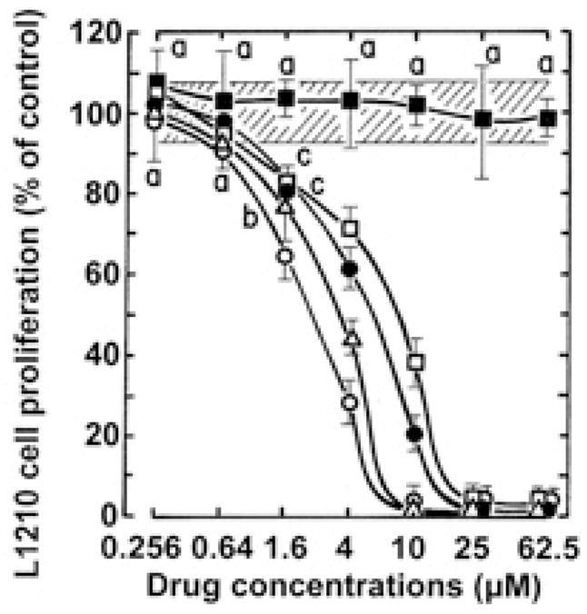
Pilot-scale libraries of eight-membered medium ring lactams (MRLs) and related tricyclic compounds (either seven-membered lactams, thiolactams or amines) were screened for their ability to inhibit the catalytic activity of human recombinant 3-hydroxy-3-methylglutaryl-coenzyme A (HMG-CoA) reductase in vitro. A dozen of the synthetic compounds mimic the inhibition of purified HMG-CoA reductase activity caused by pravastatin, fluvastatin and sodium salts of lovastatin, mevastatin and simvastatin in this cell-free assay, suggesting direct interaction with the rate-limiting enzyme of cholesterol biosynthesis. Moreover, several MRLs inhibit the metabolic activity of L1210 tumor cells in vitro to a greater degree than fluvastatin, lovastatin, mevastatin and simvastatin, whereas pravastatin is inactive. Although the correlation between the concentration-dependent inhibitions of HMG-CoA reductase activity over 10 min in the cell-free assay and L1210 tumor cell proliferation over 4 days in culture is unclear, some bioactive MRLs elicit interesting combinations of statin-like (IC50: 7.4-8.0 microM) and anti-tumor (IC50: 1.4-2.3 microM) activities. The HMG-CoA reductase-inhibiting activities of pravastatin and an MRL persist in the presence of increasing concentrations of NADPH. But increasing concentrations of HMG-CoA block the HMG-CoA reductase-inhibiting activity of pravastatin without altering that of an MRL, suggesting that MRLs and existing statins may have different mechanisms of enzyme interaction and inhibition. When tested together, suboptimal concentrations of synthetic MRLs and existing statins have additive inhibitory effects on HMG-CoA reductase activity. Preliminary molecular docking studies with MRL-based inhibitors indicate that these ligands fit sterically well into the HMG-CoA reductase statin-binding receptor model and, in contrast to mevastatin, may occupy a narrow channel housing the pyridinium moiety on NADP+.


Figures

Figure 1
Figure 1
Spectrophotometric time-scans demonstrating the ability of DL-II-D7 (A) to mimic the HMG-CoA reductase-inhibiting activity of pravastatin (B), using the HMG-CoA reductase screening assay kit from Sigma-Aldrich in vitro. Reactions, containing 4 μl of NADPH and 12 μl of HMG-CoA substrate in a final volume of 0.2 ml of assay buffer, were initiated (time 0) by the addition of 2 μl of the catalytic domain of human recombinant HMG-CoA reductase and incubated in quartz microcells at 37°C in the presence or absence (control) of 1-μl aliquots of drugs dissolved in DMSO. The rates of NADPH consumed were monitored every 20 sec for up to 500–600 sec by scanning the decreases in absorbance at 340 nm. (A) The concentration of the enzyme stock solution was 0.85 mg protein/ml and the specific activity of the control reaction calculated between 20–300 sec was 0.555268 μmol of NADPH oxidized/min/mg protein. (B) The concentration of the enzyme stock solution was 0.6 mg protein/ml and the specific activity of the control reaction calculated between 0–340 sec was 0.583976 μmol of NADPH oxidized/min/mg protein.
Figure 2
Figure 2
Comparison of the abilities of serial concentrations (plotted on a logarithmic scale) of GG-II-Ala29 (○), GG-II-G3 (□), GG-II-G7 (△), DL-II-D4 (■) and DL-II-D7 (▲) to directly inhibit the catalytic activity of human HMG-CoA reductase in a cell-free assay in vitro. The concentration of the HMG-CoA reductase stock solution was 0.6 mg protein/ml. Reactions were incubated at 37°C in quartz microcells and the rates of NADPH consumed were calculated from the decreases in spectrophotometric absorbance at 340 nm between 20 and 300 sec after addition of the enzyme. Results are expressed as % of the control specific activity of the enzyme in the absence of drugs (0.564405±0.044024 μmole of NADPH oxidized/min/mg protein, 100±7.8%, striped area). Bars, means ± SD (n=3). aNot different from control; bP<0.05, cP<0.025 and dP<0.005, smaller than control.
Figure 3
Figure 3
Comparison of the abilities of serial concentrations (logarithmic scale) of pravastatin (◇), fluvastatin (▽), lovastatin (○), mevastatin (□), simvastatin (△) and sodium salts of lovastatin (●), mevastatin (■) and simvastatin (▲) to directly inhibit the catalytic activity of human HMG-CoA reductase in a cell-free assay in vitro. The concentration of the HMG-CoA reductase stock solution was 0.85 mg protein/ml. Reactions were incubated at 37°C in quartz microcells and the rates of NADPH consumed were calculated from the decreases in spectrophotometric absorbance at 340 nm between 20 and 340 sec after addition of the enzyme. Results are expressed as % of the control specific activity of the enzyme in the absence of drugs (0.529439±0.043084 μmole of NADPH oxidized/min/mg protein, 100±8.1%, striped area). Bars, means ± SD (n=3). aNot different from control; bP<0.05, smaller than control.
Figure 4
Figure 4
Comparison of the abilities of 25 μM DL-II-D7 (striped columns) and 256 nM pravastatin (open columns) to directly inhibit the catalytic activity of human HMG-CoA reductase in the presence of increasing concentrations of HMG-CoA substrate in a cell-free assay in vitro. Reactions, containing 4 μl of NADPH (to obtain a fixed final concentration of 400 μM) and 12 μl of HMG-CoA substrate (to obtain variable final concentrations of 400–6,400 μM) in a final volume of 0.2 ml of assay buffer, were initiated (time 0) by the addition of 2 μl of the catalytic domain of human recombinant HMG-CoA reductase and incubated in quartz micro-cells at 37°C in the presence or absence (control) of 1-μl aliquots of drugs dissolved in DMSO. The concentration of the HMG-CoA reductase stock solution was 0.52 mg protein/ml. The rates of NADPH consumed were calculated from the decreases in spectrophotometric absorbance at 340 nm between 20 and 620 sec after addition of the enzyme. Results are expressed as % of the respective control specific activities of the enzyme in the absence of drugs (100±8.2%, striped area). The control specific activity of HMG-CoA reductase in the presence of 400 μM HMG-CoA substrate was 0.487231±0.040148 μmole of NADPH oxidized/min/mg protein. Bars, means ± SD (n=3). aNot different from control; bP<0.05, smaller than control.
Figure 5
Figure 5
Comparison of the abilities of single or combined treatments with 64 nM pravastatin or simvastatin (sodium salt) and 10 μM DL-II-D7 or GG-II-G3 to directly inhibit the catalytic activity of human HMG-CoA reductase in a cell-free assay in vitro. Reactions, containing 4 μl of NADPH (to obtain a final concentration of 400 μM) and 12 μl of HMG-CoA substrate (to obtain a final concentration of 400 μM) in a final volume of 0.2 ml of assay buffer, were initiated (time 0) by the addition of 2 μl of the catalytic domain of human recombinant HMG-CoA reductase and incubated in quartz microcells at 37°C in the presence or absence (control) of 1-μl aliquots of drugs dissolved in DMSO. The concentration of the HMG-CoA reductase stock solution was 0.52 mg protein/ml. The rates of NADPH consumed were calculated from the decreases in spectrophotometric absorbance at 340 nm between 0 and 720 sec after addition of the enzyme. Results are expressed as % of the control specific activity of the enzyme in the absence of drugs (0.384534±0.023841 μmol of NADPH oxidized/min/mg protein, 100±6.2%, striped area). Bars, means ± SD (n=3).
Figure 6
Figure 6
Bound conformers of ligands interacting with the substrate binding sites of HMG-CoA reductase, including (A) the docked conformer of DL-II-D4, and (B) crystallographically resolved positions of mevastatin (lower left) and NADP+ (upper right) as derived by superimposing relevant crystal structures 1HW8 (9) and 1DQ9 (10) onto our receptor model. All ligands are rendered as CPK-colored sticks. The receptor surface is colored as follows: red = O, blue = polar N, cyan = polar H, white = polar alkyls, and yellow = non-polar alkyls.
Figure 7
Figure 7
Chemical structure of 6 novel HMG-CoA reductase inhibitors that inhibit tumor cell proliferation. The structure of 2 clinically useful statins is shown for the sake of comparison.
Figure 8
Figure 8
Comparison of the abilities of serial concentrations (logarithmic scale) of GG-II-Ala29 (○), GG-II-G3 (□), GG-II-G7 (△), DL-II-D4 (■) and DL-II-D7 (▲) to inhibit the metabolic activity of L1210 tumor cells at day 4 in vitro. Cell proliferation results are expressed as % of the net absorbance of MTS/formazan after bioreduction by vehicle-treated control cells after 4 days in culture (A490 nm=1.373±0.077, 100±5.6%, striped area). The blank value (A490 nm=0.351 at day 4) for cell-free culture medium supplemented with MTS:PMS reagent was substracted from the results. Bars, means ± SD (n=3). aNot different from control; bP<0.005, smaller than control.
Figure 9
Figure 9
Comparison of the abilities of serial concentrations (logarithmic scale) of fluvastatin (○), lovastatin (●), mevastatin (□), pravastatin (■) and simvastatin (△) to inhibit the metabolic activity of L1210 tumor cells at day 4 in vitro. Cell proliferation results are expressed as % of the net absorbance of MTS/formazan after bioreduction by vehicle-treated control cells after 4 days in culture (A490 nm=1.138±0.083, 100±7.3%, striped area). The blank value (A490 nm=0.391) for cell-free culture medium supplemented with MTS:PMS reagent was substracted from the results. Bars, means ± SD (n=3). aNot different from control; bP<0.05 and cP<0.025, smaller than control.
Figure 10
Figure 10
Chemical structures that inhibit tumor cell proliferation but not HMG-CoA reductase activity.
Scheme 1
Scheme 1
Scheme 2
Scheme 2

References

    1. Tapiolas DM, Roman M, Fenical W, Stout TJ, Clardy J. Octalactins A and B: cytotoxic eight-membered-ring lactones from a marine bacterium, Streptomyces sp. J Am Chem Soc. 1991;113:4682–4683.
    1. Perchellet JP, Perchellet EM, Newell SW, Freeman JA, Ladesich JB, Jeong Y, Sato N, Buszek KR. Antitumor activity of novel octalactin A analogs in murine leukemia cells in vitro. Anticancer Res. 1998;18:97–106. - PubMed
    1. Brown N, Xie B, Markina N, VenderVelde D, Perchellet JP, Perchellet EM, Crow KR, Buszek KR. Synthesis of a natural product-inspired eight-membered ring lactam library via ring-closing metathesis. Bioorg Med Chem Lett. 2008;18:4876–4679. - PMC - PubMed
    1. Brown N, Gao G, Minatoya M, Xie B, VanderVelde D, Lushington GH, Perchellet JP, Perchellet EM, Crow KR, Buszek KR. Design and synthesis of medium-ring lactam libraries inspired by octalactin A. A convergent-divergent approach. J Comb Chem. 2008;10:628–631. - PMC - PubMed
    1. Buszek KR. First intramolecular benzyne Diels-Alder reaction with an acyclic diene. Unusual effect of diene geometry on the course of the reaction. Tetrahedron Lett. 1995;36:9125–9128.

Publication types

MeSH terms

Substances