. 2009 Dec;15(12):3899-910.
doi: 10.1089/ten.tea.2008.0677.

Magnetic resonance imaging of chondrocytes labeled with superparamagnetic iron oxide nanoparticles in tissue-engineered cartilage

Affiliations

Magnetic resonance imaging of chondrocytes labeled with superparamagnetic iron oxide nanoparticles in tissue-engineered cartilage

Sharan Ramaswamy et al. Tissue Eng Part A. 2009 Dec.

Abstract

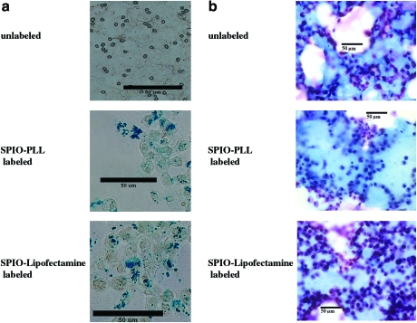
The distribution of cells within tissue-engineered constructs is difficult to study through nondestructive means, such as would be required after implantation. However, cell labeling with iron-containing particles may prove to be a useful approach to this problem, because regions containing such labeled cells have been shown to be readily detectable using magnetic resonance imaging (MRI). In this study, we used the Food and Drug Administration-approved superparamagnetic iron oxide (SPIO) contrast agent Feridex in combination with transfection agents to label chondrocytes and visualize them with MRI in two different tissue-engineered cartilage constructs. Correspondence between labeled cell spatial location as determined using MRI and histology was established. The SPIO-labeling process was found not to affect the phenotype or viability of the chondrocytes or the production of major cartilage matrix constituents. We believe that this method of visualizing and tracking chondrocytes may be useful in the further development of tissue engineered cartilage therapeutics.


Figures

FIG. 1.
FIG. 1.
(a) Prussian blue stained unlabeled (control) chondrocytes (top), superparamagnetic iron oxide (SPIO)- poly-L-lysine (PLL)–labeled chondrocytes (middle), and SPIO-lipofectamine labeled chondrocytes (bottom) after 6 days of 2D culture. Blue stain indicates presence of iron (Fe). (b) Masson's Trichrome stained slides of unlabeled (control) chondrocytes (top), SPIO-PLL-labeled chondrocytes (middle), and SPIO-lipofectamine–labeled chondrocytes (bottom) after 6 days of 2D culture. Blue stain indicates presence of collagen, and cell nuclei have been counter-stained with hematoxylin (purple color). Color images available online at www.liebertonline.com/ten.
FIG. 2.
FIG. 2.
(a) TUNEL assay of SPIO-lipofectamine labeled chondrocytes (top) and unlabeled (control) chondrocytes (middle). There is an absence of positive brownish-pink staining in both these groups. Positive control (pink stain) of skin cells showing apoptotic behavior (bottom). (b) Live-Dead assay of SPIO-labeled chondrocytes within a hydrogel construct, with green circles representing live cells and red circles representing dead cells. Color images available online at www.liebertonline.com/ten.
FIG. 3.
FIG. 3.
Histology of a 4-week-old hollow-fiber bioreactor (HFBR) inoculated with SPIO-labeled chondrocytes showing positive staining for (a) glycosaminoglycan (GAG; Alcian blue) and (b) collagen (Masson's Trichrome). A blue stain represents positive staining for GAG or collagen. Color images available online at www.liebertonline.com/ten.
FIG. 4.
FIG. 4.
Immunohistochemistry confirming phenotypic stability of SPIO-labeled chondrocytes in the HFBR system: (a) aggrecan, (b) type II collagen, and (c) type I collagen. No Cy3 fluorescence was evident in the negative controls. Color images available online at www.liebertonline.com/ten.
FIG. 5.
FIG. 5.
Histology of cellular hydrogels showing positive staining for (a) GAG (Safranin O) and (b) collagen (Masson's Trichrome) at approximately 5 weeks of growth. Hydrogels seeded with SPIO-labeled chondrocytes (top) and hydrogels seeded with unlabeled chondrocytes (bottom). Safranin O stains GAGs red and Masson's Trichrome stains collagen blue. Color images available online at www.liebertonline.com/ten.
FIG. 6.
FIG. 6.
(a) Magnetic resonance (MR) image of a HFBR inoculated with SPIO-labeled chondrocytes demonstrating SPIO-labeled and unlabeled regions of the tissue at 2 days. (b) Corresponding Prussian blue stained histological slice confirming the presence of SPIO-labeled cells within the outer ring of the tissue and lack of SPIO-labeled cells within the inner ring. (c) Left: Axial MR image of a HFBR inoculated with SPIO-labeled chondrocytes acquired after 35 days of tissue culture. The entire outer ring showed the presence of SPIO nanoparticles, whereas these were absent in the entire inner ring. Right: Axial MR image of a HFBR inoculated with unlabeled chondrocytes showing the absence of a dark outer ring after 31 days of tissue culture. Color images available online at www.liebertonline.com/ten.
FIG. 7.
FIG. 7.
(a) MR image of a hydrogel seeded with unlabeled chondrocytes after 30 days of growth, showing the absence of signal voids within the construct. (b) MR images (axial slice) of representative hydrogel seeded with SPIO-labeled chondrocytes at 0 days (top) and at 30 days after inoculation (bottom). (c) MR image (left) and corresponding histological section (right) of a hydrogel seeded with SPIO-labeled chondrocytes after 30 days of growth. Note the close correspondence between signal voids in the MR image and cells in the histological image.

References

    1. Kuo C.K. Li W.J. Mauck R.L. Tuan R.S. Cartilage tissue engineering: its potential and uses. Curr Opin Rheumatol. 2006;18:64. - PubMed
    1. Elisseeff J. Injectable cartilage tissue engineering. Expert Opin Biol Ther. 2004;4:1849. - PubMed
    1. Neves A.A. Medcalf N. Brindle K. Functional assessment of tissue-engineered meniscal cartilage by magnetic resonance imaging and spectroscopy. Tissue Eng. 2003;9:51. - PubMed
    1. Novotny J.E. Turka C.M. Jeong C. Wheaton A.J. Li C. Presedo A. Richardson D.W. Reddy R. Dodge G.R. Biomechanical and magnetic resonance characteristics of a cartilage-like equivalent generated in a suspension culture. Tissue Eng. 2006;12:2755. - PubMed
    1. Trattnig S. Pinker K. Krestan C. Plank C. Millington S. Marlovits S. Matrix-based autologous chondrocyte implantation for cartilage repair with HyalograftC: two-year follow-up by magnetic resonance imaging. Eur J Radiol. 2006;57:9. - PubMed

Publication types

MeSH terms