. 2009 Dec;9(12):2679-96.
doi: 10.1111/j.1600-6143.2009.02831.x. Epub 2009 Sep 25.

TNF receptors differentially signal and are differentially expressed and regulated in the human heart

Affiliations

TNF receptors differentially signal and are differentially expressed and regulated in the human heart

R S Al-Lamki et al. Am J Transplant. 2009 Dec.

Abstract

Tumor necrosis factor (TNF) utilizes two receptors, TNFR1 and 2, to initiate target cell responses. We assessed expression of TNF, TNFRs and downstream kinases in cardiac allografts, and compared TNF responses in heart organ cultures from wild-type ((WT)C57BL/6), TNFR1-knockout ((KO)), TNFR2(KO), TNFR1/2(KO) mice. In nonrejecting human heart TNFR1 was strongly expressed coincidentally with inactive apoptosis signal-regulating kinase-1 (ASK1) in cardiomyocytes (CM) and vascular endothelial cells (VEC). TNFR2 was expressed only in VEC. Low levels of TNF localized to microvessels. Rejecting cardiac allografts showed increased TNF in microvessels, diminished TNFR1, activation of ASK1, upregulated TNFR2 co-expressed with activated endothelial/epithelial tyrosine kinase (Etk), increased apoptosis and cell cycle entry in CM. Neither TNFR was expressed significantly by cardiac fibroblasts. In (WT)C57BL/6 myocardium, TNF activated both ASK1 and Etk, and increased both apoptosis and cell cycle entry. TNF-treated TNFR1(KO) myocardium showed little ASK1 activation and apoptosis but increased Etk activation and cell cycle entry, while TNFR2(KO) myocardium showed little Etk activation and cell cycle entry but increased ASK1 activation and apoptosis. These observations demonstrate independent regulation and differential functions of TNFRs in myocardium, consistent with TNFR1-mediated cell death and TNFR2-mediated repair.


Conflict of interest statement

Disclosure

There are no conflicts of interests

Figures
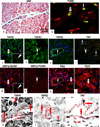
Figure 1
Figure 1
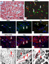
Normal human myocardium; H&E section shows normal histology (a) with negligible TUNEL staining in a few CM (open arrow) and in CD31-positive VEC (arrow) (b). A marked co-expression for TNFR1 and ASK1Ser967 is seen in (open arrows), in resident MNC and in VEC (inset; arrowheads) (c,d). Co-expression of TNFR1 and ASK1pThr845 is seen in resident MNC (arrowheads) (e,f). TNFR2 and activation of Etkp is demonstrated in VEC (arrows) and Etkp is also detected in a few CM (open arrow) (g,h). TNF is confined to microvessels (arrows) and in MNC (arrowhead) and TACE is present in CM (open arrow) (I,j). TNFR1 mRNA is present in CM (open arrow) and in VEC (arrows) (k) while mRNA for TNFR2 and TNF is confined to VEC (arrows) and in MNC (arrowheads) (l,m). Dotted lines mark out the vessels. Images are representative of 10 different samples, each of which gave similar results. Cardiomyocytes (CM); Vascular endothelial cells (VEC); Mononuclear cells (MNC). (Original Mags: a, ×112; b; ×63; c–d; ×40; e–f; ×63; g–I; ×40; j; ×63; k–m, ×235).
Figure 2
Figure 2
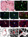
Mild rejection (grade 1A); H&E show focal perivascular or interstitial infiltrates with mild intensity and lack of CM damage (a) and an increased TUNEL-reactive CM (open arrows) and in CD31-positive VEC (arrows) (b). Co-expression of TNFR1 and ASK1p-Thr845 is seen in VEC (arrows), with ASK1p-Thr845 also detected in some CM, negative for TNFR1 (open arrow) (c,f). TNFR2 and activation of Etkp is strongly demonstrated in CM with some staining present in regions of the intercalated discs (open arrows) (d,g). TNF is confined to microvessels (arrow) and MNC (arrowhead) and TACE is present in CM (open arrow) and in VEC (arrow) (e,h). TNFR1 mRNA is seen in VEC (arrow) (i) while TNFR2 and TNF mRNA is present in CM (open arrows) and in VEC (arrows) (j,k). Dotted lines mark out the vessels. Images are representative of 6 experiments different samples with similar results (Original Mags; a; ×112; c–h; ×63; I–j; ×112; k; ×235).
Figure 3
Figure 3
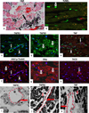
Focal moderate rejection (grade 2); H&E stained section show aggressive infiltration and/or focal CM damage (a) with increased TUNEL-reactive CM (open arrows) and VEC (arrows) (b). TNFR1 and ASK1p-Thr845 co-expression in VEC (arrows) with ASK1p-Thr845 also detected in some CM (open arrow) (c,f). TNFR2 and Etkp activation is evident in CM with some staining in regions of the intercalated discs (open arrows) (d,g). TNF is confined to microvessels (arrows) and MNC (arrowhead) (e) and TACE is demonstrated in CM (open arrows) (h). TNFR1 mRNA is confined to VEC (arrows) and TNFR2 and TNF mRNA are strongly expressed in CM (open arrows) and in VEC (arrow). Dotted lines mark out the vessels. Images are representative of 6 independent experiments with similar results. (Original Mags: a, ×112; b–h, ×63; i–k, ×235).
Figure 4
Figure 4
Low moderate rejection (grade 3A); H&E stained section show multifocal aggressive infiltrates and/or CM damage with a few TUNEL-positive CM (b). Co-expression of TNFR1 and ASK1p-Thr845 is evident in VEC (arrows) and in MNC (arrowheads) with ASK1p-Thr845 also detected in some CM (open arrows) (c,f). TNFR2 and Etkp activation is evident in CM (open arrows) (d,g) while TNF is confined to microvessels (arrow) and in MNC (arrowhead) (e). TACE is noticeably diminished in CM with a strongly signal detected in VEC (arrow) (h). TNFR1 mRNA is confined to VEC (arrow) (i) and TNFR2 and TNF mRNA is seen in CM (open arrows) and in VEC (arrows) (j,k). Dotted lines mark out the vessels. Images are representative of 6 independent experiments with similar results (Original Mags: a,i. ×235; b–h, ×63; j,k, ×112). Apoptotic index (l) presented as means ± SEM show statistical significant differences between control (normal human myocardium) and the rejection grades; with the highest level of apoptotic cell death observed in grades 1A and 2 as compared to control. Controls Vs each rejection grade is indicated in the graph and ranges from p<0.001 to p<0.05.
Figure 4
Figure 4
Low moderate rejection (grade 3A); H&E stained section show multifocal aggressive infiltrates and/or CM damage with a few TUNEL-positive CM (b). Co-expression of TNFR1 and ASK1p-Thr845 is evident in VEC (arrows) and in MNC (arrowheads) with ASK1p-Thr845 also detected in some CM (open arrows) (c,f). TNFR2 and Etkp activation is evident in CM (open arrows) (d,g) while TNF is confined to microvessels (arrow) and in MNC (arrowhead) (e). TACE is noticeably diminished in CM with a strongly signal detected in VEC (arrow) (h). TNFR1 mRNA is confined to VEC (arrow) (i) and TNFR2 and TNF mRNA is seen in CM (open arrows) and in VEC (arrows) (j,k). Dotted lines mark out the vessels. Images are representative of 6 independent experiments with similar results (Original Mags: a,i. ×235; b–h, ×63; j,k, ×112). Apoptotic index (l) presented as means ± SEM show statistical significant differences between control (normal human myocardium) and the rejection grades; with the highest level of apoptotic cell death observed in grades 1A and 2 as compared to control. Controls Vs each rejection grade is indicated in the graph and ranges from p<0.001 to p<0.05.
Figure 5
Figure 5
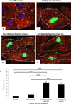
Confocal images of human cardiac biopsies. Nuclear staining for pH3 is illustrated in combination with α-sarcomeric-actin staining of the CM cytoplasm (green and red, respectively). (a). pH3 is absent in normal human myocardium. In comparison, a strong signal is apparent in CM in sections with mild grade 1A (b), focal moderate grade 2 (c), and low moderate grade 3A rejection (d). Proliferative indices (e); results are presented as means ± SEM. A significant difference is seen between normal myocardium and all three rejection episodes (***p<0.001) but not between grade 2 and 3A rejection. (Original Mags; ×63; Controls: n = 5; Allograft rejections, n= 5).
Figure 6
Figure 6
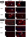
(A). Untreated WTC57BL/6 cultures show positive staining for ASK1p-Ser967 and ASK1p-Thr845 in CM (open arrows) and in VEC (arrows) (a,b). Untreated TNFR1KO cultures are positive for ASK1p-Ser967 (arrowhead) but not for ASK1p-Thr845 (c,d). In contrast, TNF-treated WTC57BL/6 cultures show a diminished ASK1p-Ser967 (g) but a strong signal for ASK1p-Thr845 in CM (open arrow) and in VEC (arrowhead) (h) while TNF-treated TNFR1KO are negative (i,j). In contrast, CM (open arrow) and VEC (arrow) are strongly positive for ASK1p-Thr845 but not for ASK1p-Ser967 in TNF-treated TNFR2KO cultures (k–l). (B). A strong signal for total Etk and phosphorylated Etk (Etkp) is seen in CM (open arrow) and in VEC (arrow) in untreated WTC57BL/6 (a,b) and in TNFR1KO cultures (c,d), while untreated TNFR2KO cultures are negative (e,f). TNF-treated WTC57BL/6 and TNFR1KO cultures show Etk and Etkp in VEC (arrows) and in CM (open arrows) (g–j). In contrast, TNF-treated TNFR2KO cultures are negative (k,l). Images are representative of 3 independent experiments with similar results (Original Mags, ×63).
Figure 6
Figure 6
(A). Untreated WTC57BL/6 cultures show positive staining for ASK1p-Ser967 and ASK1p-Thr845 in CM (open arrows) and in VEC (arrows) (a,b). Untreated TNFR1KO cultures are positive for ASK1p-Ser967 (arrowhead) but not for ASK1p-Thr845 (c,d). In contrast, TNF-treated WTC57BL/6 cultures show a diminished ASK1p-Ser967 (g) but a strong signal for ASK1p-Thr845 in CM (open arrow) and in VEC (arrowhead) (h) while TNF-treated TNFR1KO are negative (i,j). In contrast, CM (open arrow) and VEC (arrow) are strongly positive for ASK1p-Thr845 but not for ASK1p-Ser967 in TNF-treated TNFR2KO cultures (k–l). (B). A strong signal for total Etk and phosphorylated Etk (Etkp) is seen in CM (open arrow) and in VEC (arrow) in untreated WTC57BL/6 (a,b) and in TNFR1KO cultures (c,d), while untreated TNFR2KO cultures are negative (e,f). TNF-treated WTC57BL/6 and TNFR1KO cultures show Etk and Etkp in VEC (arrows) and in CM (open arrows) (g–j). In contrast, TNF-treated TNFR2KO cultures are negative (k,l). Images are representative of 3 independent experiments with similar results (Original Mags, ×63).
Figure 7
Figure 7
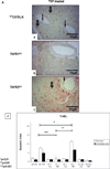
(A). TNF-treated WTC57BL/6 cultures (a) but not TNF-treated TNFR1KO cultures (b) show increased TUNEL-positive CM (open arrows) and VEC (arrows). In comparison, an increased TUNEL staining is seen in CM (arrowhead) and in VEC (arrow) in TNF-treated TNFR2KO cultures (c). Apoptotic index (d) show statistically significant difference between cultures with the highest level of apoptotic cell death observed in TNF-treated TNFR2KO cultures as compared to TNF-treated WTC57BL/6 (*p<0.01) or -TNFR1KO (**p<0.01) or TNFR1/TNFR2KO cultures. (B). A high level of pH3 expression is seen in α-sarcomeric actin-positive CM (open arrows) and in CD31-positive VEC (arrow) in TNF-treated WTC57BL/6 cultures (a,b) with a similar pattern but higher level of expression is evident in TNFR1KO cultures (c,d). In contrast, TNF-treated TNFR2KO cultures show only occasional pH3 expression (e,f). Nuclei stained with To-Pro-3-iodide. (Original Mags; A- a–c; ×112; B- a–f; ×63).
Figure 7
Figure 7
(A). TNF-treated WTC57BL/6 cultures (a) but not TNF-treated TNFR1KO cultures (b) show increased TUNEL-positive CM (open arrows) and VEC (arrows). In comparison, an increased TUNEL staining is seen in CM (arrowhead) and in VEC (arrow) in TNF-treated TNFR2KO cultures (c). Apoptotic index (d) show statistically significant difference between cultures with the highest level of apoptotic cell death observed in TNF-treated TNFR2KO cultures as compared to TNF-treated WTC57BL/6 (*p<0.01) or -TNFR1KO (**p<0.01) or TNFR1/TNFR2KO cultures. (B). A high level of pH3 expression is seen in α-sarcomeric actin-positive CM (open arrows) and in CD31-positive VEC (arrow) in TNF-treated WTC57BL/6 cultures (a,b) with a similar pattern but higher level of expression is evident in TNFR1KO cultures (c,d). In contrast, TNF-treated TNFR2KO cultures show only occasional pH3 expression (e,f). Nuclei stained with To-Pro-3-iodide. (Original Mags; A- a–c; ×112; B- a–f; ×63).

Similar articles

Cited by

References

    1. Tartaglia LA, Weber RF, Figari IS, Reynolds C, Palladino MA, Jr, Goeddel DV. The two different receptors for tumor necrosis factor mediate distinct cellular responses. Proc Natl Acad Sci U S A. 1991;88(20):9292–9296. - PMC - PubMed
    1. Sandborn WJ, Hanauer SB. Antitumor necrosis factor therapy for inflammatory bowel disease: a review of agents, pharmacology, clinical results, and safety. Inflamm Bowel Dis. 1999;5(2):119–133. - PubMed
    1. Nikolaus S, Schreiber S. Anti-TNF biologics in the treatment of chronic inflammatory bowel disease. Internist (Berl) 2008;49(8):947–943. - PubMed
    1. Meldrum DR. Tumor necrosis factor in the heart. Am J Physiol. 1998;274(3 Pt 2):R577–R595. - PubMed
    1. Muller-Ehmsen J, Schwinger RH. TNF and congestive heart failure: therapeutic possibilities. Expert Opin Ther Targets. 2004;8(3):203–209. - PubMed

Publication types

MeSH terms