. 2009 Sep 29;10(1):88.
doi: 10.1186/1465-9921-10-88.

PKA and Epac cooperate to augment bradykinin-induced interleukin-8 release from human airway smooth muscle cells

Affiliations

PKA and Epac cooperate to augment bradykinin-induced interleukin-8 release from human airway smooth muscle cells

Sara S Roscioni et al. Respir Res. .

Abstract

Background: Airway smooth muscle contributes to the pathogenesis of pulmonary diseases by secreting inflammatory mediators such as interleukin-8 (IL-8). IL-8 production is in part regulated via activation of Gq-and Gs-coupled receptors. Here we study the role of the cyclic AMP (cAMP) effectors protein kinase A (PKA) and exchange proteins directly activated by cAMP (Epac1 and Epac2) in the bradykinin-induced IL-8 release from a human airway smooth muscle cell line and the underlying molecular mechanisms of this response.

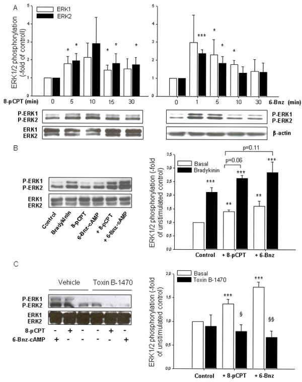
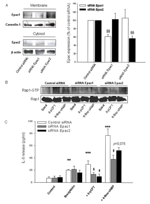
Methods: IL-8 release was assessed via ELISA under basal condition and after stimulation with bradykinin alone or in combination with fenoterol, the Epac activators 8-pCPT-2'-O-Me-cAMP and Sp-8-pCPT-2'-O-Me-cAMPS, the PKA activator 6-Bnz-cAMP and the cGMP analog 8-pCPT-2'-O-Me-cGMP. Where indicated, cells were pre-incubated with the pharmacological inhibitors Clostridium difficile toxin B-1470 (GTPases), U0126 (extracellular signal-regulated kinases ERK1/2) and Rp-8-CPT-cAMPS (PKA). The specificity of the cyclic nucleotide analogs was confirmed by measuring phosphorylation of the PKA substrate vasodilator-stimulated phosphoprotein. GTP-loading of Rap1 and Rap2 was evaluated via pull-down technique. Expression of Rap1, Rap2, Epac1 and Epac2 was assessed via western blot. Downregulation of Epac protein expression was achieved by siRNA. Unpaired or paired two-tailed Student's t test was used.

Results: The beta2-agonist fenoterol augmented release of IL-8 by bradykinin. The PKA activator 6-Bnz-cAMP and the Epac activator 8-pCPT-2'-O-Me-cAMP significantly increased bradykinin-induced IL-8 release. The hydrolysis-resistant Epac activator Sp-8-pCPT-2'-O-Me-cAMPS mimicked the effects of 8-pCPT-2'-O-Me-cAMP, whereas the negative control 8-pCPT-2'-O-Me-cGMP did not. Fenoterol, forskolin and 6-Bnz-cAMP induced VASP phosphorylation, which was diminished by the PKA inhibitor Rp-8-CPT-cAMPS. 6-Bnz-cAMP and 8-pCPT-2'-O-Me-cAMP induced GTP-loading of Rap1, but not of Rap2. Treatment of the cells with toxin B-1470 and U0126 significantly reduced bradykinin-induced IL-8 release alone or in combination with the activators of PKA and Epac. Interestingly, inhibition of PKA by Rp-8-CPT-cAMPS and silencing of Epac1 and Epac2 expression by specific siRNAs largely decreased activation of Rap1 and the augmentation of bradykinin-induced IL-8 release by both PKA and Epac.

Conclusion: Collectively, our data suggest that PKA, Epac1 and Epac2 act in concert to modulate inflammatory properties of airway smooth muscle via signaling to the Ras-like GTPase Rap1 and to ERK1/2.


Figures

Figure 1
Figure 1
cAMP-elevating agent fenoterol augments bradykinin-induced release of IL-8. hTERT-airway smooth muscle cells were stimulated for 18 hrs with the indicated concentrations of bradykinin (A). Cells were incubated for 30 min without (Basal) or with 1 μM fenoterol. Then, cells were stimulated with 10 μM bradykinin for 18 hrs. IL-8 release was assessed by ELISA as described in Materials and Methods. Results are expressed as mean ± SEM of separate experiments (n = 3-10). *P < 0.05, ***P < 0.001 compared to unstimulated control; #P < 0.05 compared to bradykinin-stimulated control.
Figure 2
Figure 2
Bradykinin-induced IL-8 release is increased by the PKA activator 6-Bnz-cAMP. hTERT-airway smooth muscle cells were stimulated with the indicated concentrations of 6-Bnz-cAMP in the absence (Basal) or presence of 10 μM bradykinin for 18 hrs. IL-8 release was assessed by ELISA. Results are expressed as mean ± SEM of separate experiments (n = 3-10). ***P < 0.001 compared to unstimulated control; #P < 0.05, ##P < 0.01 compared to bradykinin-stimulated control.
Figure 3
Figure 3
Bradykinin-induced IL-8 release is increased by the Epac activators 8-pCPT-2'-O-Me-cAMP and Sp-8-pCPT-2'-O-Me-cAMPS. hTERT-airway smooth muscle cells were stimulated with the indicated concentrations of 8-pCPT-2'-O-Me-cAMP (A) or with 100 μM of 8-pCPT-2'-O-Me-cAMP, Sp-8-pCPT-2'-O-Me-cAMPS and 8-pCPT-2'-O-Me-cGMP (B) in the absence (Basal) or presence of 10 μM bradykinin for 18 hrs. IL-8 release was measured by ELISA. Results are expressed as mean ± SEM of separate experiments (n = 3-9). **P < 0.01, ***P < 0.001 compared to unstimulated control; #P < 0.05, ##P < 0.01 compared to bradykinin-stimulated control.
Figure 4
Figure 4
Effects of cyclic nucleotide analogs and cAMP-elevating agents on VASP phosphorylation. Phosphorylation of the PKA effector VASP in the absence (Control) and presence of 8-pCPT-2'-O-Me-cAMP, Sp-8-pCPT-2'-O-Me-cAMPS, 8-pCPT-2'-O-Me-cGMP (each 100 μM), 500 μM 6-Bnz-cAMP for 15 min was evaluated by using a VASP-specific antibody. Equal loading was verified by analysis of β-actin. Representative blots are shown (A). hTERT-airway smooth muscle cells were stimulated for 15 minutes without (Control) or with forskolin, 8-pCPT-2'-O-Me-cAMP (each 100 μM), 500 μM 6-Bnz-cAMP, 1 μM fenoterol and 10 μM bradykinin in the absence or presence of 100 μM Rp-8-CPT-cAMPS (B). Representative blots are shown above. Equal loading was verified by analysis of β-actin. Below are the densitometric quantifications of n = 3-6 independent experiments. Data are expressed as percentage of phospho-VASP over total VASP. **P < 0.01, ***P < 0.001 compared to unstimulated control; §P < 0.05 compared to basal condition.
Figure 5
Figure 5
Role of Epac and PKA in GTP-loading of Rap1 and Rap2. hTERT-airway smooth muscle cells were fractioned as described in Material and Methods. Expression of membrane-associated or cytosolic Rap1 (A) and Rap2 (B) was evaluated and normalized to the content of the cell fraction-specific marker proteins caveolin-1 and β-actin, respectively. hTERT-airway smooth muscle cells were stimulated for 10 min without (Control) and with 100 μM 8-pCPT-2'-O-Me-cAMP or 500 μM 6-Bnz-cAMP. Thereafter, GTP-loaded and total Rap1 (A) or Rap2 (B) were determined as described in Material and Methods. Representative immunoblots are shown above with the respective densitometric quantifications underneath them (n = 3-5). *P < 0.05, **P < 0.01 compared to unstimulated control.
Figure 6
Figure 6
Impact of Ras-like GTPases on bradykinin-induced IL-8 release. hTERT cells were treated for 24 hrs without (Control) and with 100 pg/ml of Clostridium difficile toxin B-1470 (Toxin B-1470). Then, cell morphology was assessed by phase-contrast microscopy (A). Cell number was measured on the same cells by Alamar blue as described in Material and Methods. Data represent percentage of unstimulated control (B; upper panel). In addition, IL-8 release was measured on supernatant of cells treated with 10 μM bradykinin alone or in combination with 100 μM 8-pCPT-2'-O-Me-cAMP or 500 μM 6-Bnz-cAMP in the absence (Basal) or presence of 100 pg/ml Toxin B-1470 by using ELISA (B; lower panel). Results are expressed as mean ± SEM of separate experiments (n = 4). *P < 0.05, **P < 0.01 compared to unstimulated control; #P < 0.05 compared to bradykinin-stimulated condition; §P < 0.05 compared to basal condition.
Figure 7
Figure 7
Role of Epac and PKA in basal and bradykinin-induced ERK1/2 phosphorylation. Impact of Ras-like GTPases. hTERT-airway smooth muscle cells were stimulated for the indicated period of time (A) or for 5 min without and with 100 μM 8-pCPT-2-O-Me-cAMP (8-pCPT) or 500 μM 6-Bnz-cAMP in the absence or presence of 10 μM bradykinin (10 min) (B) or 100 pg/ml Clostridium difficile toxin B-1470 or its vehicle (24 hrs) (C). Phosphorylated ERK1/2 (P-ERK1/2), total ERK1/2 or β-actin were detected by specific antibodies. Representative immunoblots are shown with the respective densitometric quantifications. Data are expressed as fold of ERK1/2 phosphorylation over unstimulated control and represent mean ± SEM of separate experiments (n = 5-7). *P < 0.05, **P < 0.01, ***P < 0.001 compared to unstimulated control; §P < 0.05, §§P < 0.01 compared to basal condition.
Figure 8
Figure 8
Impact of ERK1/2 on bradykinin-induced IL-8 release and its augmentation by cAMP analogs. Cells were pretreated for 30 min with 3 μM U0126 or vehicle before the addition of 10 μM bradykinin (15 min), 100 μM 8-pCPT-2-O-Me-cAMP or 500 μM 6-Bnz-cAMP (each 5 min) (A). Phosphorylated ERK1/2 (P-ERK1/2) and total ERK1/2 were detected by specific antibodies. Representative immunoblots are shown on the left with the respective densitometric quantifications on the right (n = 5). Alternatively, cells were treated with bradykinin alone or in combination with 100 μM 8-pCPT-2'-O-Me-cAMP or 500 μM 6-Bnz-cAMP for 18 hrs. Thereafter, IL-8 release was measured by ELISA (B). Results represent mean ± SEM of separate experiments (n = 3-9). *P < 0.05, **P < 0.01, ***P < 0.001 compared to unstimulated control; §P < 0.05, §§P < 0.01, §§§P < 0.001 compared to basal condition.
Figure 9
Figure 9
Cooperativity of 8-pCPT-2'-O-Me-cAMP and 6-Bnz-cAMP on bradykinin-induced IL-8 release. hTERT-airway smooth muscle cells were incubated with 50 μM 6-Bnz-cAMP alone or in combination with the indicated concentrations of 8-pCPT-2'-O-Me-cAMP (A). Alternatively, cells were stimulated with 10 μM 8-pCPT-2'-O-Me-cAMP alone or in combination with the indicated concentrations of 6-Bnz-cAMP (B). After that, 10 μM bradykinin was added for 18 hrs and IL-8 levels were measured by ELISA. Results represent mean ± SEM of separate experiments (n = 3). *P < 0.05, **P < 0.01 compared to unstimulated control.
Figure 10
Figure 10
Impact of PKA inhibition on Rap1 activation and bradykinin-induced IL-8 release. Cells were treated for 30 min without (Basal) or with 100 μM Rp-8-CPT-cAMPS. In A, cells were first incubated with 100 μM 8-pCPT-2'-O-Me-cAMP or 500 μM 6-Bnz-cAMP for 5 min followed by measurement of GTP-loading of Rap1 as described in Material and Methods. Shown is a representative immunoblot. Alternatively, cells were stimulated with 10 μM bradykinin alone or in combination with 100 μM 8-pCPT-2'-O-Me-cAMP or 500 μM 6-Bnz-cAMP for 18 hrs (B). IL-8 release was then assessed by ELISA. Results are expressed as mean ± SEM of separate experiments (n = 3-7). *P < 0.05, **P < 0.01, compared to unstimulated control, §P < 0.05 compared to basal condition.
Figure 11
Figure 11
Impact of Epac silencing on Rap1 activation and bradykinin-induced IL-8 release. hTERT-airway smooth muscle cells were transfected for 72 hrs with control siRNA, Epac1 or Epac2 specific siRNAs (each 200 pmol). Thereafter, expression of membrane-associated Epac1 or cytosolic Epac2 was evaluated and normalized to the content of the cell fraction-specific marker proteins caveolin-1 and β-actin, respectively. Representative immunoblots are shown on the left with the respective densitometric quantifications on the right. Results are expressed as mean ± SEM of separate experiments (n = 5-7). Transfected cells were treated with 100 μM 8-pCPT-2'-O-Me-cAMP (8-pCPT) or 500 μM 6-Bnz-cAMP for 5 min and the amount of GTP-Rap1 was determined as described in Material and Methods. Shown is a representative immunoblot. In C, transfected cells were incubated with 10 μM bradykinin alone or in combination with 100 μM 8-pCPT-2'-O-Me-cAMP (8-pCPT) or 500 μM 6-Bnz-cAMP for 18 hrs. IL-8 release was then assessed by ELISA. Results are expressed as mean ± SEM of separate experiments (n = 3-7). **P < 0.01, ***P < 0.001 compared to unstimulated control; §P < 0.05, §§P < 0.01 compared to control siRNA.
Figure 12
Figure 12
Augmentation of bradykinin-induced IL-8 release in hTERT-airway smooth muscle cells by Epac and PKA. Activation of ERK1/2 is mediated via different GPCRs. The β2-agonist fenoterol acts on Gs-coupled receptors inducing cAMP elevation via activation of adenylyl cyclase (AC) while forskolin directly activates AC. cAMP activates two distinct cellular effectors: PKA and Epac, followed by activation of Ras-like GTPases, such as Rap1, and ERK1/2, and subsequently induction of specific transcription factors resulting in the production of IL-8. Bradykinin also elicits ERK1/2 phosphorylation most likely via activation of Gq-coupled receptors. The dotted line represents a potential pathway which has not been fully addressed in our study. ⊥ indicates inactivation and → indicates activation, see text for further details.

References

    1. Bousquet J, Jeffery PK, Busse WW, Johnson M, Vignola AM. Asthma. From bronchoconstriction to airways inflammation and remodeling. Am J Respir Crit Care Med. 2000;161:1720–1745. - PubMed
    1. Jeffery PK. Remodeling in asthma and chronic obstructive lung disease. Am J Respir Crit Care Med. 2001;164:S28–S38. - PubMed
    1. Barnes PJ. The cytokine network in asthma and chronic obstructive pulmonary disease. J Clin Invest. 2008;118:3546–3556. doi: 10.1172/JCI36130. - DOI - PMC - PubMed
    1. Halayko AJ, Solway J. Molecular mechanisms of phenotypic plasticity in smooth muscle cells. J Appl Physiol. 2001;90:358–368. - PubMed
    1. Panettieri RA Jr. Airway smooth muscle: immunomodulatory cells that modulate airway remodeling? Respir Physiol Neurobiol. 2003;137:277–293. doi: 10.1016/S1569-9048(03)00153-8. - DOI - PubMed

Publication types

MeSH terms

LinkOut - more resources