

Liver regeneration: alternative epithelial pathways
- PMID: 19788929
- PMCID: PMC2888836
- DOI: 10.1016/j.biocel.2009.09.014
Liver regeneration: alternative epithelial pathways
Abstract
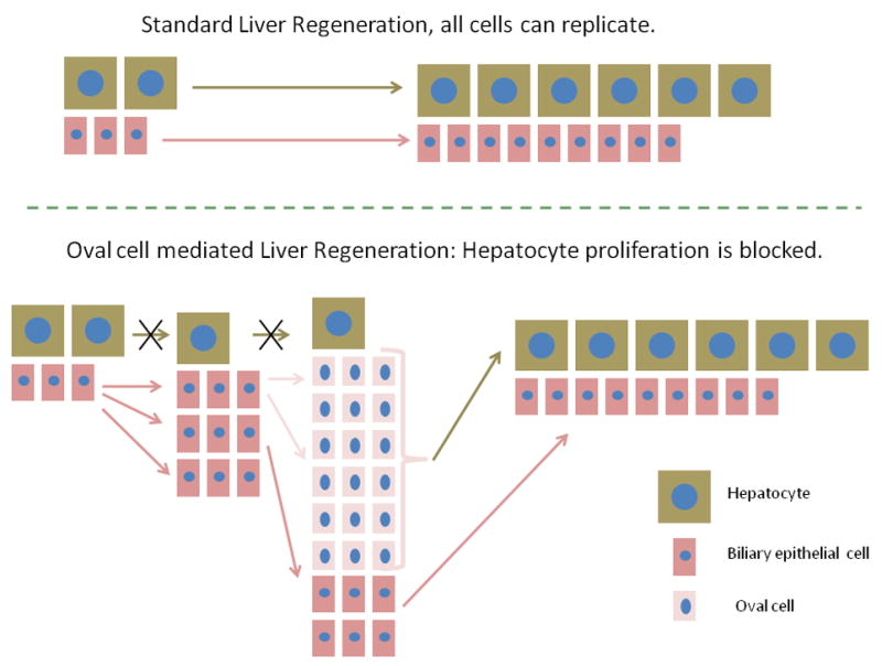
Loss of hepatic tissue triggers a regenerative response in the whole organ. Under typical normal conditions, all hepatic cells (epithelial: hepatocytes and biliary epithelial cells; non-epithelial: stellate cells, macrophages and endothelial cells) undergo one to three rounds of replication to establish the original number of cells and restore organ size. The review summarizes the literature of regenerative patterns in situations in which proliferation of either hepatocytes or biliary epithelial cells is inhibited. The evidence strongly suggests that under these circumstances, hepatocytes or biliary epithelial cells can function as facultative stem cells for each other and replenish the inhibited cellular compartment by a process of transdifferentiation, involving complex signaling pathways. These pathways are activated under experimental conditions in rodents and in fulminant hepatitis associated with liver failure in humans. Mechanistic analysis of these pathways has implications for liver biology and for potential therapeutic modalities in human liver disease.
Copyright © 2009 Elsevier Ltd. All rights reserved.
Figures
References
-
- Akhurst B, Matthews V, Husk K, Smyth MJ, Abraham LJ, Yeoh GC. Differential lymphotoxin-beta and interferon gamma signaling during mouse liver regeneration induced by chronic and acute injury. Hepatology. 2005;41:327–335. - PubMed
-
- Alison M, Golding M, Lalani EN, Nagy P, Thorgeirsson S, Sarraf C. Wholesale hepatocytic differentiation in the rat from ductular oval cells, the progeny of biliary stem cells. J Hepatol. 1997;26:343–352. - PubMed
-
- Alison MR, Poulsom R, Jeffery R, Anilkumar TV, Jagoe R, Sarraf CE. Expression of hepatocyte growth factor mRNA during oval cell activation in the rat liver. J Pathol. 1993;171:291–299. - PubMed
-
- Bisgaard HC, Nagy P, Santoni-Rugiu E, Thorgeirsson SS. Proliferation, apoptosis, and induction of hepatic transcription factors are characteristics of the early response of biliary epithelial (oval) cells to chemical carcinogens. Hepatology. 1996;23:62–70. - PubMed
Publication types
MeSH terms
Substances
Grants and funding
LinkOut - more resources
Full Text Sources
