. 2009 Sep;21(9):2655-71.
doi: 10.1105/tpc.109.069740. Epub 2009 Sep 29.

Cellular pathways regulating responses to compatible and self-incompatible pollen in Brassica and Arabidopsis stigmas intersect at Exo70A1, a putative component of the exocyst complex

Affiliations

Cellular pathways regulating responses to compatible and self-incompatible pollen in Brassica and Arabidopsis stigmas intersect at Exo70A1, a putative component of the exocyst complex

Marcus A Samuel et al. Plant Cell. 2009 Sep.

Abstract

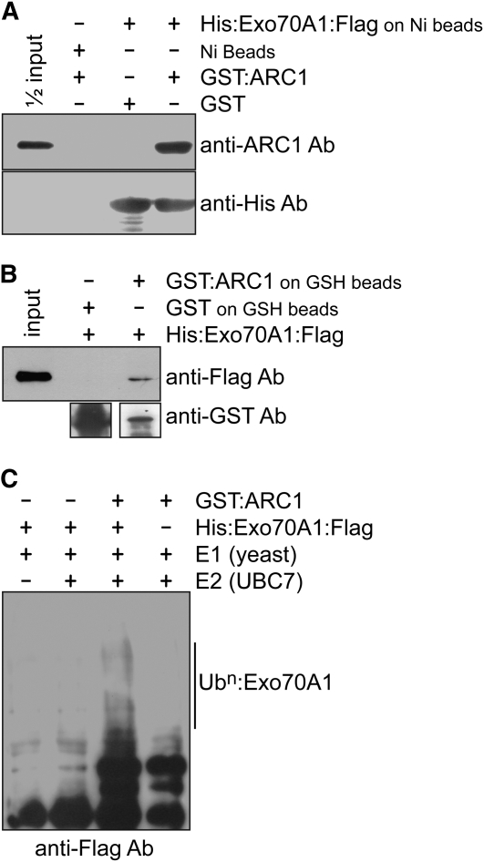
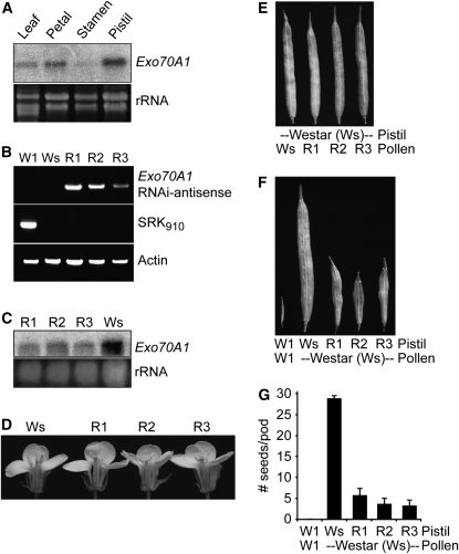
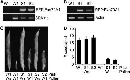
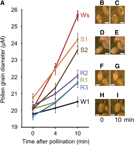
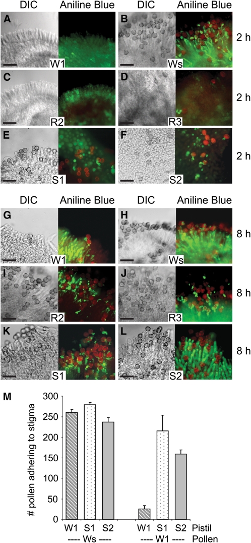
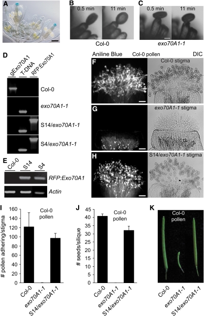
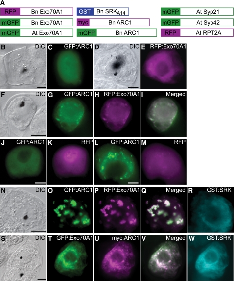
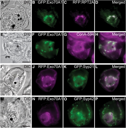
In the Brassicaceae, compatible pollen-pistil interactions result in pollen adhesion to the stigma, while pollen grains from unrelated plant species are largely ignored. There can also be an additional layer of recognition to prevent self-fertilization, the self-incompatibility response, whereby self pollen grains are distinguished from nonself pollen grains and rejected. This pathway is activated in the stigma and involves the ARM repeat-containing 1 (ARC1) protein, an E3 ubiquitin ligase. In a screen for ARC1-interacting proteins, we have identified Brassica napus Exo70A1, a putative component of the exocyst complex that is known to regulate polarized secretion. We show through transgenic studies that loss of Exo70A1 in Brassica and Arabidopsis thaliana stigmas leads to the rejection of compatible pollen at the same stage as the self-incompatibility response. A red fluorescent protein:Exo70A1 fusion rescues this stigmatic defect in Arabidopsis and is found to be mobilized to the plasma membrane concomitant with flowers opening. By contrast, increased expression of Exo70A1 in self-incompatible Brassica partially overcomes the self pollen rejection response. Thus, our data show that the Exo70A1 protein functions at the intersection of two cellular pathways, where it is required in the stigma for the acceptance of compatible pollen in both Brassica and Arabidopsis and is negatively regulated by Brassica self-incompatibility.


Figures

Figure 1.
Figure 1.
In Vitro Binding and Ubiquitination Assays with Brassica ARC1 and Exo70A1. (A) In vitro binding assay with purified GST or GST:ARC1 added to Ni-NTA–bound His6:Exo70A1:FLAG or Ni-NTA alone. ARC1 binds specifically to Exo70A1. (B) In vitro binding assay with purified His6:Exo70A1:FLAG protein added to GSH beads bound to GST or GST:ARC1. ARC1 binds specifically to Exo70A1. (C) In vitro ubiquitination assay of Exo70A1 by the ARC1 E3 ubiquitin ligase. His6:Exo70A1:FLAG was detected using an anti-FLAG antibody. Multi-ubiquitinated Exo70A1 is only detected when all components of the ubiquitination assay are present (lane 3). Due to the long exposure time, some cross-reacting bands from the ARC1 sample are seen at the bottom of the blot in lanes 3 and 4.
Figure 2.
Figure 2.
Reduced Expression Levels of Exo70A1 in the Stigmas of Compatible B. napus Westar Causes Decreased Seed Production. (A) Exo70A1 RNA expression in different B. napus tissues. Exo70A1 RNA is detected by RNA gel blot analysis in several tissues, with highest levels of expression in the pistil and lowest levels in the stamens. RNA was extracted from the W1 self-incompatible cultivar. (B) DNA genotyping of the Exo70A1 RNAi Westar lines. Three independent transgenic B. napus Westar lines, R1 to R3, carrying the SLR1proExo70A1 RNAi construct were generated. The presence of the Exo70A1 RNAi construct in the R1-R3 lines was detected by PCR using sense and antisense primer combinations. Actin served as a positive control for the PCR reaction. The self-incompatible B. napus W1 line is identified by the presence of the S910 allele through the PCR detection of the SRK910 gene (Silva et al., 2001). The compatible B. napus Westar (Ws) line does not carry this self-incompatibility allele. (C) Exo70A1 expression in the stigmas of the Exo70A1 RNAi Westar lines. All three independent transgenic Exo70A1 RNAi Westar lines, R1 to R3, have reduced Exo70A1 mRNA levels in the stigmas as shown by RNA gel blot analysis. (D) The Exo70A1 RNAi Westar lines produce flowers of wild-type appearance. (E) The Exo70A1 RNAi Westar lines produce wild-type pollen, which in turn produces full seedpods when used in pollinations with wild-type Westar pistils. (F) and (G) The Exo70A1 RNAi Westar stigmas, with reduced Exo70A1 expression, have significantly reduced seed production in comparison to Westar stigmas when pollinated with compatible Westar pollen (t test, P < 0.05). Seedpods (F) and average number of seeds per pod (G) from the Exo70A1 RNAi Westar (R1 to R3) plants. n = 5; error bars indicate ±se.
Figure 3.
Figure 3.
Expression of RFP:Exo70A1 in the Stigmas of Self-Incompatible B. napus W1 Disrupts the Self-Incompatibility Response and Leads to Increased Seed Production. (A) DNA genotyping of the RFP:Exo70A1 W1 lines. Two independent transgenic B. napus W1 lines, S1 and S2, carrying the SLR1proRFP:Exo70A1 (B. napus Exo70A1) construct were generated. The presence of RFP:Exo70A1 in the S1 and S2 lines was detected using PCR with primer combinations specific to the SLR1proRFP:Exo70A1 construct. The self-incompatible B. napus W1 line was identified by the presence of the S910 allele through the PCR detection of the SRK910 gene (Silva et al., 2001). The compatible B. napus Westar (Ws) line does not carry this self-incompatibility allele. (B) RFP:Exo70A1 expression in the stigmas of the RFP:Exo70A1 W1 lines. The two independent transgenic W1 lines, S1 and S2, express RFP:Exo70A1 in the stigmas, as shown by RT-PCR analysis (32 cycles; two biological replicates and three technical replicates). (C) and (D) The RFP:Exo70A1-expressing W1 stigmas have significantly increased seed production in comparison to W1 stigmas when pollinated with self-incompatible W1 pollen (t test, P < 0.05). The RFP:Exo70A1-expressing W1 stigmas also produce full seedpods when pollinated with compatible Westar pollen, as expected. Seedpods (C) and average number of seeds per pod (D) from the RFP:Exo70A1 W1 (S1 and S2) plants. n = 5; error bars indicate ±se.
Figure 4.
Figure 4.
Altered Rates of Pollen Hydration on the Exo70A1 RNAi Westar Stigmas and the RFP:Exo70A1-Expressing W1 Stigmas. (A) Pollen hydration was detected by measuring the increasing pollen grain diameters (μM) measured at 0, 4, and 10 min after pollination. The Exo70A1 RNAi Westar (R1 to R3) and control Westar (Ws) stigmas were pollinated with compatible Westar pollen. The RFP:Exo70A1-expressing W1 (S1 and S2) and control W1 stigmas were pollinated with self-incompatible W1 pollen. n > 20; error bars are ±se. (B) to (I) Representative images of hydrating pollen grains are shown at 0 and 10 min after pollination for a Westar stigma ([B] and [C]), an RFP:Exo70A1-expressing W1 stigma from the S2 line ([D] and [E]), an Exo70A1 RNAi Westar stigma from the R2 line ([F] and [G]), and a W1 stigma ([H] and [I]). Bars = 20 μm.
Figure 5.
Figure 5.
Altered Pollen Germination and Pollen Tube Growth on the Exo70A1 RNAi Westar Stigmas and the RFP:Exo70A1-Expressing W1 Stigmas. (A) to (L) Pollen attachment and pollen tube growth at 2 h ([A] to [F]) and 8 h ([G] to [L]) following pollination. The Exo70A1 RNAi Westar R2 and R3 stigmas ([C], [D], [I], and [J]) and control Westar (Ws) stigmas ([B] and [H]) were pollinated with compatible Westar pollen. The RFP:Exo70A1 W1 S1 and S2 stigmas ([E], [F], [K], and [L]) and control W1 stigmas ([A] and [G]) were pollinated with self-incompatible W1 pollen. Differential interference contrast (DIC) images and images of aniline blue–stained pistils are shown. The aniline blue fluorescence from the stigma and pollen tubes are shown in green and overlaid with the red autofluorescence from pollen grains. The Exo70A1 RNAi Westar stigmas support a reduced attachment of Westar pollen grains as well as reduced pollen tube growth ([C], [D], [I], and [J]). The RFP:Exo70A1-expressing W1 stigmas support increased attachment of self-incompatible W1 pollen as well as increased pollen tube growth ([E], [F], [K], and [L]). Bars = 100 μm. (M) Average number of self-incompatible W1 and compatible Westar pollen grains attached to the RFP:Exo70A1-expressing W1 stigmas. The number of pollen grains present on the RFP:Exo70A1-expressing W1 stigmas (S1 and S2) was counted 8 h following either a Westar or W1 pollination. The expression of RFP:Exo70A1 causes a significant increase in the number of self-incompatible W1 pollen grains attaching to the S1 and S2 stigmas in relation to W1 stigmas (t test, P < 0.05; n = 4).
Figure 6.
Figure 6.
The Arabidopsis Exo70A1 Gene Shares a Conserved Function with B. napus Exo70A1 in the Stigma during Pollen–Stigma Interactions. (A) Arabidopsis Exo70A1proGUS shows strong GUS expression in stigmatic tissues. Bar = 250 μm. (B) and (C) Pollen hydration on an Arabidopsis Col-0 or exo70A1 stigmatic papilla. Pollen hydration was observed following the placement of a single wild-type Col-0 pollen grain on top of either a wild-type Col-0 (B) or exo70A1-1 (C) stigmatic papilla using a micromanipulator. Images are shown at 0.5 and 11 min after pollination. Pollen grain hydration is impaired on the exo70A1-1 mutant stigma, while full hydration is observed for the wild-type compatible Col-0 stigma. Bars = 10 μm. (D) DNA genotyping of the RFP:Exo70A1-expressing Arabidopsis lines. Two independent transgenic Arabidopsis lines, S4 and S14, carrying the SLR1proRFP:Exo70A1 construct were generated. This construct was introduced into a heterozygous Exo70A1/exo70A1-1 Arabidopsis plant (see Supplemental Figures 3A to 3D online), and transformed heterozygous plants were recovered. The next generation was then screened for segregating wild-type Col-0 and homozygous exo70A1-1 plants carrying the RFP:Exo70A1 construct. The presence of RFP:Exo70A1 in the S4 and S14 lines was detected by PCR using sense and antisense primer combinations. The exo70A1-1 Arabidopsis plants were identified by the presence of T-DNA (see Supplemental Figures 3A and 3B online). The undisrupted Exo70A1 gene (gExo70A1) was detected using gene-specific primers. (E) RFP:Exo70A1 expression in the pistils of the RFP:Exo70A1 Arabidopsis lines. The two independent transgenic Arabidopsis lines, S4 and S14, express RFP:Exo70A1 in the pistils, as shown by RT-PCR analysis (28 cycles; two biological replicates and four technical replicates). (F) to (H) Pollen attachment and pollen tube growth in Arabidopsis pistils from a wild-type Col-0 (F), exo70A1-1 mutant (G), and the exo70A1-1 mutant expressing RFP:Exo70A1 (S14/exo70A1-1) (H). All pistils were pollinated with compatible Arabidopsis Col-0 pollen and then stained with aniline blue to visualize the pollen attachment and pollen tube growth. As expected, abundant pollen tube growth is observed on the compatible wild-type Col-0 stigma. The exo70A1-1 mutant stigma does not support any pollen attachment or pollen tube growth, and only background fluorescence is observed in the aniline blue–stained stigma. The expression of RFP:Exo70A1 in the exo70A1-1 stigma rescues this defect and restores pollen attachment and pollen tube growth. Bars = 50 μm. (I) Average number of attached compatible Col-0 pollen grains on stigmas from wild-type Col-0, the exo70A1-1 mutant, or the exo70A1-1 mutant expressing RFP:Exo70A1 (S14/exo70A1-1) (n = 5; error bars indicate ±se). The number of adhered pollen for the exo70A1-1 mutant is significantly lower than for either Col-0 or S14/exo70A1-1 (t test, P < 0.05), while the results for the rescued S14/exo70A1-1 stigmas are not significantly different from those for Col-0 (t test, P > 0.05). (J) and (K) Average number of seeds per silique ([J]; n = 7) and siliques (K) from Col-0, exo70A1-1, and the exo70A1-1 mutant expressing RFP:Exo70A1 (S14/exo70A1-1) pistils following a Col-0 pollination. Following a compatible Col-0 pollination, no seeds are produced from exo70A1-1 mutant pistils, while the expression of RFP:Exo70A1 in the exo70A1-1 mutant stigma restores full seed production. Seed set values for Col-0, the exo70A1-1 mutant, and S14/exo70A1-1 are all significantly different from each other (t test, P < 0.05).
Figure 7.
Figure 7.
Characterization of RFP:Exo70A1 Lines in Col-0 and the exo70A1-1 Mutant Background. RFP and RFP/DIC merged images of stigmas from different stages are marked. (A) and (B) In mature stigmas (stage 13) from the S14/Col-0 line, RFP:Exo70A1 is found at the plasma membrane. (C) In immature stigmas (stage 12) from the S14/Col-0 line, RFP:Exo70A1 is localized to internal structures that are reminiscent of Golgi. (D) In the S14/exo70A1-1 mature stigmas, RFP:Exo70A1 is localized to the plasma membrane, but some also remains inside the cells. (E) A stage 13 Col-0 stigmatic papilla expressing both RFP:Exo70A1 and a GFP:δ-TIP vacuolar marker. The GFP:δ-TIP marker outlines the membranes of the central vacuole as well as the vacuolar network in the apical region. The localization of the GFP:δ-TIP marker is distinct from that of RFP:Exo70A1, which confirms the plasma membrane localization of RFP:Exo70A1. (F) Stage 12 Col-0 stigmatic papillae expressing both RFP:Exo70A1 and a ST:GFP Golgi marker. The ST:GFP Golgi marker overlaps with RFP:Exo70A1, confirming the Golgi localization of RFP:Exo70A1 in immature stigmatic papillae. (G) and (H) Following a Col-0 pollination, RFP:Exo70A1 disappears from the plasma membrane in the S14/Col-0 lines. (G) A pollinated stigma at 8 min after pollination. (H) A different pollinated stigma at 10 min after pollination. The RFP image on the left and the corresponding merged image show an area with several attached pollen grains, while the right RFP image and the corresponding merged image show an area with a single attached pollen grain. Note the absence of RFP fluorescence at the plasma membrane in papillae that are in contact with a pollen grain. Bars = 50 μm in (A), 20 μm in (B) to (D), (G), and (H), and 10 μm in (E) and (F).
Figure 8.
Figure 8.
The Effect of SRK and ARC1 on the Subcellular Localization of Exo70A1 in BY-2 Cells. (A) Schematics of different constructs used to transiently transform tobacco BY-2 cells using particle bombardment. Full-length coding regions were fused to different tags, except for SRKA14, where the cytosolic kinase domain was fused to GST as described by Stone et al. (2003). Bn, Brassica napus; At, Arabidopsis thaliana. (B) to (E) Single transformations with either GFP:ARC1 ([B] and [C]) or RFP:Exo70A1 (B. napus Exo70A1) ([D] and [E]). Cytosolic localization is observed for both fusion proteins. (F) to (I) Double transformation and coexpression of GFP:ARC1 and RFP:Exo70A1 (B. napus Exo70A1). Cytosolic localization is observed for both proteins. (J) and (K) Control double transformation and coexpression of GFP:ARC1 and RFP alone. (L) and (M) Control triple transformation and coexpression of GST:SRK (kinase domain only), GFP:ARC1, and RFP alone. The coexpression of the active GST:SRK causes ARC1 to localize to punctate perinuclear structures as previously published (Stone et al., 2003; Samuel et al., 2008) but has no effect on RFP. GST:SRK remains cytosolic, as previously published (Stone et al., 2003; Samuel et al., 2008). (N) to (R) Triple transformation and coexpression of GST:SRK (kinase domain only), GFP:ARC1, and RFP:Exo70A1 (B. napus Exo70A1). Subcellular localization of Exo70A1 and ARC1 to the perinuclear region is observed. GST:SRK remains cytosolic. (S) to (W) Triple transformation and coexpression of GST:SRK (kinase domain only), GFP:ARC1, and RFP:Exo70A1 (Arabidopsis Exo70A1). Subcellular localization of Arabidopsis Exo70A1 and ARC1 to the perinuclear region is also observed. Black dots in the DIC images are the tungsten particles used for the bombardments. Areas of white in the merged images indicate overlapping localization patterns. Bars = 10 μm.
Figure 9.
Figure 9.
The Subcellular Localization of B. napus Exo70A1 in BY-2 Cells in Relation to Different Marker Proteins. (A) to (D) Quadruple transformation and coexpression of RFP:RPT2A with GFP:Exo70A1, myc:ARC1, and GST:SRK. Exo70A1 colocalizes with the proteasomal subunit, RPT2A. (E) to (H) Triple transformations and coexpression of GST:SRK, myc:ARC1, and GFP:Exo70A1, followed by ConA-594 staining. Concanavalin A stains the endoplasmic reticulum and overlaps with the Exo70A1/ARC1 subcellular structures. (I) to (P) Quadruple transformations and coexpression of GST:SRK, myc:ARC1, RFP:Exo70A1, and either GFP:Syp21 ([I] to [L]) or GFP:Syp42 ([M] to [P]). Syp21 is a marker for the late endosome/prevacuolar compartment, and Syp42 was shown to reside in the trans-Golgi network (Sanderfoot et al., 2001; Samaj et al., 2005). There is a bit of overlap of the Exo70A1/ARC1 subcellular structures with Syp21. GST:SRK remains cytosolic, and myc:ARC1 colocalizes with Exo70A1 to the punctate structures as shown in Figures 8N to 8R and previously published (Stone et al., 2003). Black dots in the DIC images are the tungsten particles used for the bombardments. Areas of white in the merged images indicate overlapping localization patterns. Bars = 10 μm.

References

    1. Barile, M., Pisitkun, T., Yu, M.J., Chou, C.L., Verbalis, M.J., Shen, R.F., and Knepper, M.A. (2005). Large scale protein identification in intracellular aquaporin-2 vesicles from renal inner medullary collecting duct. Mol. Cell. Proteomics 4 1095–1106. - PMC - PubMed
    1. Boevink, P., Oparka, K., Santa Cruz, S., Martin, B., Betteridge, A., and Hawes, C. (1998). Stacks on tracks: The plant Golgi apparatus traffics on an actin/ER network. Plant J. 15 441–447. - PubMed
    1. Boyd, C., Hughes, T., Pypaert, M., and Novick, P. (2004). Vesicles carry most exocyst subunits to exocytic sites marked by the remaining two subunits, Sec3p and Exo70p. J. Cell Biol. 167 889–901. - PMC - PubMed
    1. Chong, Y.T., Gidda, S.K., Sanford, C., Parkinson, J., Mullen, R.T., and Goring, D.R. (2009). Characterisation of the Arabidopsis exocyst complex gene families by phylogenetic, expression profiling, and subcellular localisation studies. New Phytol., in press. - PubMed
    1. Clough, S.J., and Bent, A.F. (1998). Floral dip: A simplified method for Agrobacterium-mediated transformation of Arabidopsis thaliana. Plant J. 16 735–743. - PubMed

Publication types

MeSH terms

Associated data

LinkOut - more resources