. 2009 Oct;2(10):895-902.
doi: 10.1158/1940-6207.CAPR-09-0125. Epub 2009 Sep 29.

UVR exposure sensitizes keratinocytes to DNA adduct formation

Affiliations

UVR exposure sensitizes keratinocytes to DNA adduct formation

Sudhir Nair et al. Cancer Prev Res (Phila). 2009 Oct.

Abstract

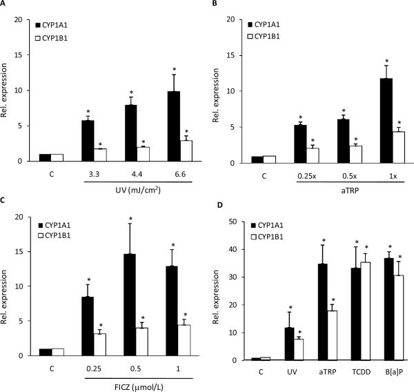
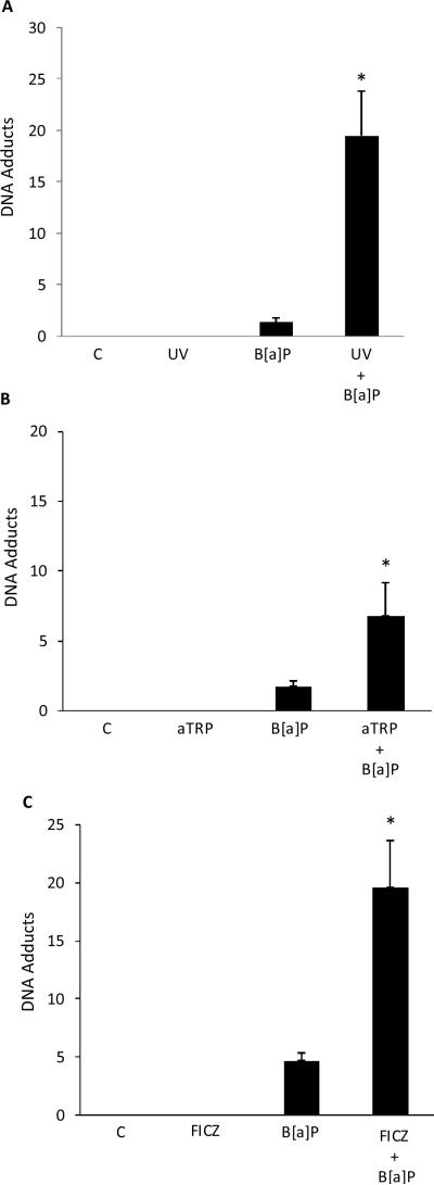
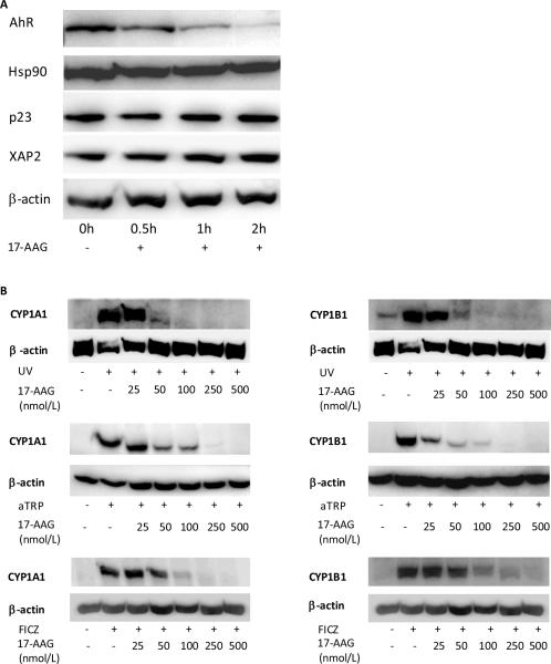
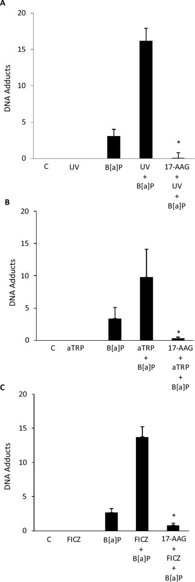
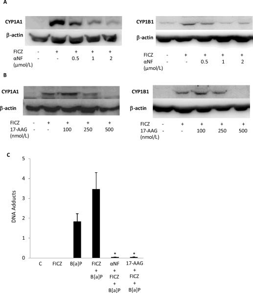
UV radiation (UVR) and exposure to tobacco smoke, a source of polycyclic aromatic hydrocarbons (PAH), have been linked to skin carcinogenesis. UVR-mediated activation of the aryl hydrocarbon receptor (AhR) stimulates the transcription of CYP1A1 and CYP1B1, which encode proteins that convert PAH to genotoxic metabolites. We determined whether UVR exposure sensitized human keratinocytes to PAH-induced DNA adduct formation. UVR exposure induced CYP1A1 and CYP1B1 in HaCaT cells, an effect that was mimicked by photooxidized tryptophan (aTRP) and FICZ, a component of aTRP. UVR exposure or pretreatment with aTRP or FICZ also sensitized cells to benzo(a)pyrene (B[a]P)-induced DNA adduct formation. alphaNF, an AhR antagonist, suppressed UVR-, aTRP-, and FICZ-mediated induction of CYP1A1 and CYP1B1 and inhibited B[a]P-induced DNA adduct formation. Treatment with 17-AAG, an Hsp90 inhibitor, caused a marked decrease in levels of AhR; inhibited UVR-, aTRP-, and FICZ-mediated induction of CYP1A1 and CYP1B1; and blocked the sensitization of HaCaT cells to B[a]P-induced DNA adduct formation. FICZ has been suggested to be a physiologic ligand of the AhR that may have systemic effects. Hence, studies of FICZ were also carried out in MSK-Leuk1 cells, a model of oral leukoplakia. Pretreatment with alpha-naphthoflavone or 17-AAG blocked FICZ-mediated induction of CYP1A1 and CYP1B1, and suppressed the increased B[a]P-induced DNA adduct formation. Collectively, these results suggest that sunlight may activate AhR signaling and thereby sensitize cells to PAH-mediated DNA adduct formation. Antagonists of AhR signaling may have a role in the chemoprevention of photocarcinogenesis.


Figures

Fig. 1
Fig. 1
UVR induces CYP1A1 and CYP1B1 mRNA levels in HaCaT cells. A, Cells were UV irradiated at the indicated doses and then cultured for 6 h prior to cell harvest. B and C, Cells were treated with the indicated concentrations of aTRP (B) or FICZ (C) for 6 h prior to cell harvest. D, Cells were UV irradiated (4.4 mJ/cm2) and then cultured for 6 h or treated with 1X aTRP, 1 nmol/L TCDD or 5 μmol/L B[a]P for 6 h prior to cell harvest. mRNA was isolated and then analyzed by qPCR. Values for CYP1A1 and CYP1B1 were normalized to the expression levels of β-actin. In A–D, control cells (C) were sham irradiated or treated with vehicle. A–D, means ± S.D. are shown, n=3. *, P<0.05.
Fig. 2
Fig. 2
UVR treatment sensitizes keratinocytes to B[a]P-mediated DNA adduct formation. HaCaT cells were UV (4 mJ/cm2) irradiated and then cultured for 12 h (A) or treated with 1X aTRP (B) or 1 μmol/L FICZ (C) for 12 h. Subsequently, 1 μmol/L B[a]P or vehicle was added and cells were incubated for another 6 h. DNA was then isolated for analysis of DNA adducts. In A–C, control cells (C) were sham irradiated or treated with vehicle. A–C, means ± S.D. are shown, n=3. *, P<0.05.
Fig. 3
Fig. 3
α-Naphthoflavone suppresses UVR-mediated induction of CYP1A1 and CYP1B1 and DNA adduct formation. HaCaT cells were pretreated with vehicle or the indicated concentration of αNF for 2 h. Subsequently, cells were UV-irradiated (4.4 mJ/cm2) and then cultured for 12 h (A) or treated with 1X aTRP (B) or 1 μmol/L FICZ (C) for 12 h prior to being harvested for Western blot analysis. Cellular lysate proteins (50 μg/lane) were separated by SDS-polyacrylamide gel electrophoresis and subjected to western blotting with antibodies specific for CYP1A1, CYP1B1 and β-actin. In D–F, HaCaT cells were pretreated with 2 μmol/L αNF or vehicle for 2 h. Cells were then UV-irradiated (4.4 mJ/cm2) and cultured for 12 h (D) or treated with 1X aTRP (E) or 1 μmol/L FICZ (F) for 12 h. Subsequently, 1 μmol/L B[a]P or vehicle was added and cells were incubated for another 6 h. DNA was then isolated for analysis of DNA adducts. In D–F, control cells (C) were sham irradiated or treated with vehicle. D–F, means ± S.D. are shown, n=3. *, P<0.05.
Fig. 4
Fig. 4
17-AAG, a Hsp90 inhibitor, suppresses AhR protein levels and blocks UVR-mediated induction of CYP1A1 and CYP1B1. A, HaCaT cells were treated with vehicle or 500 nmol/L 17-AAG for 0–2 h and were then harvested for Western blot analysis. Cellular lysate proteins (50 μg/lane) were separated by SDS-polyacrylamide gel electrophoresis and subjected to western blotting with antibodies specific for AhR, Hsp90, p23, XAP2 and β-actin. B, HaCaT cells were treated with vehicle or the indicated concentrations of 17-AAG for 2 h. Subsequently, cells were sham or UV-irradiated (4.4 mJ/cm2) and then cultured for 12 h or treated with 1X aTRP or 1 μmol/L FICZ for 12 h prior to being harvested for Western blot analysis. Cellular lysate proteins (50 μg/lane) were loaded onto a 10% SDS-polyacrylamide gel, electrophoresed, and subsequently transferred onto nitrocellulose. Immunoblots were probed with antibodies specific for CYP1A1, CYP1B1 and β-actin.
Fig. 5
Fig. 5
17-AAG suppresses UVR-mediated sensitization of keratinocytes to DNA adduct formation. In A–C, HaCaT cells were pretreated with 500 nmol/L 17-AAG or vehicle for 2 h. Cells were then UV-irradiated (4.4 mJ/cm2) and cultured for 12 h (A) or treated with 1X aTRP (B) or 1 μmol/L FICZ (C) for 12 h. Subsequently, 1 μmol/L B[a]P or vehicle was added and cells were incubated for another 6 h. DNA was then isolated for analysis of DNA adducts. In A–C, control cells (C) were sham irradiated or treated with vehicle. A–C, means ± S.D. are shown, n=3. *, P<0.05.
Fig. 6
Fig. 6
α-Naphthoflavone and 17-AAG suppress FICZ-mediated induction of CYP1A1 and CYP1B1 and DNA adduct formation in MSK-Leuk1 cells. Cells were pretreated with vehicle or the indicated concentration of αNF (A) or 17-AAG (B) for 2 h. Subsequently, cells were treated with 1 μmol/L FICZ for 12 h prior to being harvested for Western blot analysis. Cellular lysate proteins (100 μg/lane) were loaded onto a 10% SDS-polyacrylamide gel, electrophoresed, and subsequently transferred onto nitrocellulose. Immunoblots were probed with antibodies specific for CYP1A1, CYP1B1 and β-actin. C, Cells were pretreated with 2 μmol/L αNF, 500 nmol/L 17-AAG or vehicle for 2 h. Cells were then treated with 1 μmol/L FICZ or vehicle for 12 h. Subsequently, 1 μmol/L B[a]P or vehicle was added and cells were incubated for another 12 h. DNA was then isolated for analysis of DNA adducts. Means ± S.D. are shown, n=3. *, P<0.05.

Similar articles

Cited by

References

    1. Aubry F, MacGibbon B. Risk factors of squamous cell carcinoma of the skin. A case-control study in the Montreal region. Cancer. 1985;55:907–911. - PubMed
    1. Grodstein F, Speizer FE, Hunter DJ. A prospective study of incident squamous cell carcinoma of the skin in the nurses' health study. J Natl Cancer Inst. 1995;87:1061–1066. - PubMed
    1. De Hertog SA, Wensveen CA, Bastiaens MT, et al. Relation between smoking and skin cancer. J Clin Oncol. 2001;19:231–238. - PubMed
    1. Kanjilal S, Strom SS, Clayman GL, et al. p53 mutations in nonmelanoma skin cancer of the head and neck: molecular evidence for field cancerization. Cancer Res. 1995;55:3604–3609. - PubMed
    1. Perea-Milla López E, Miñarro-Del Moral RM, Martínez-García C, et al. Lifestyles, environmental and phenotypic factors associated with lip cancer: a case-control study in southern Spain. Br J Cancer. 2003;88:1702–7. - PMC - PubMed

Publication types

MeSH terms

Substances