. 2009 Oct 1;15(19):6148-57.
doi: 10.1158/1078-0432.CCR-09-1039. Epub 2009 Sep 29.

Vorinostat inhibits brain metastatic colonization in a model of triple-negative breast cancer and induces DNA double-strand breaks

Affiliations

Vorinostat inhibits brain metastatic colonization in a model of triple-negative breast cancer and induces DNA double-strand breaks

Diane Palmieri et al. Clin Cancer Res. .

Abstract

Purpose: As chemotherapy and molecular therapy improve the systemic survival of breast cancer patients, the incidence of brain metastases increases. Few therapeutic strategies exist for the treatment of brain metastases because the blood-brain barrier severely limits drug access. We report the pharmacokinetic, efficacy, and mechanism of action studies for the histone deactylase inhibitor vorinostat (suberoylanilide hydroxamic acid) in a preclinical model of brain metastasis of triple-negative breast cancer.

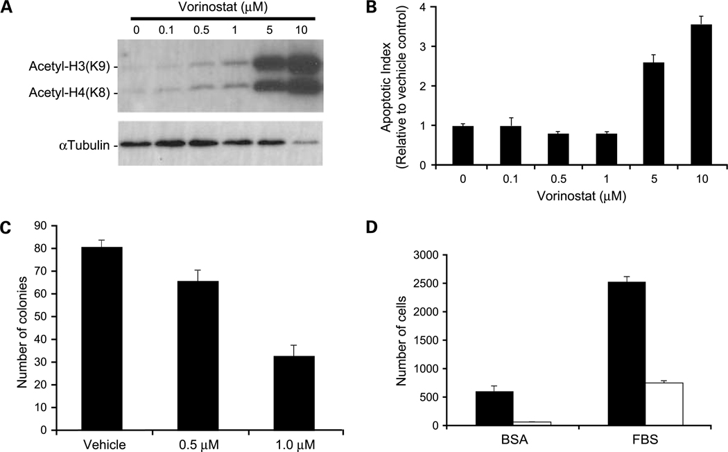
Experimental design: The 231-BR brain trophic subline of the MDA-MB-231 human breast cancer cell line was injected into immunocompromised mice for pharmacokinetic and metastasis studies. Pharmacodynamic studies compared histone acetylation, apoptosis, proliferation, and DNA damage in vitro and in vivo.

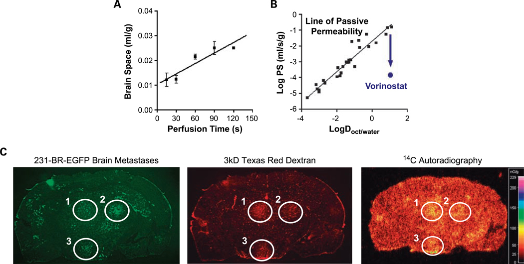
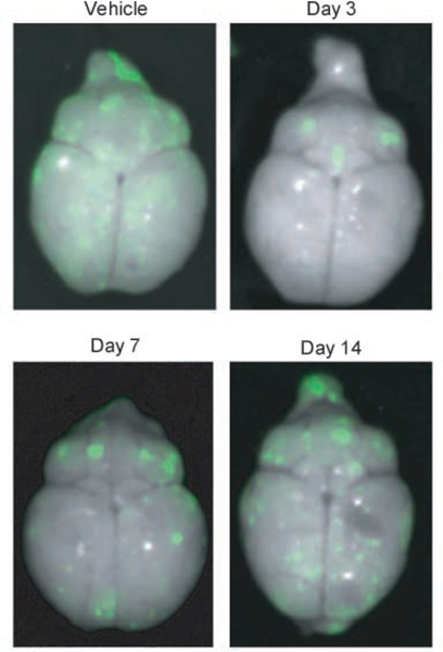
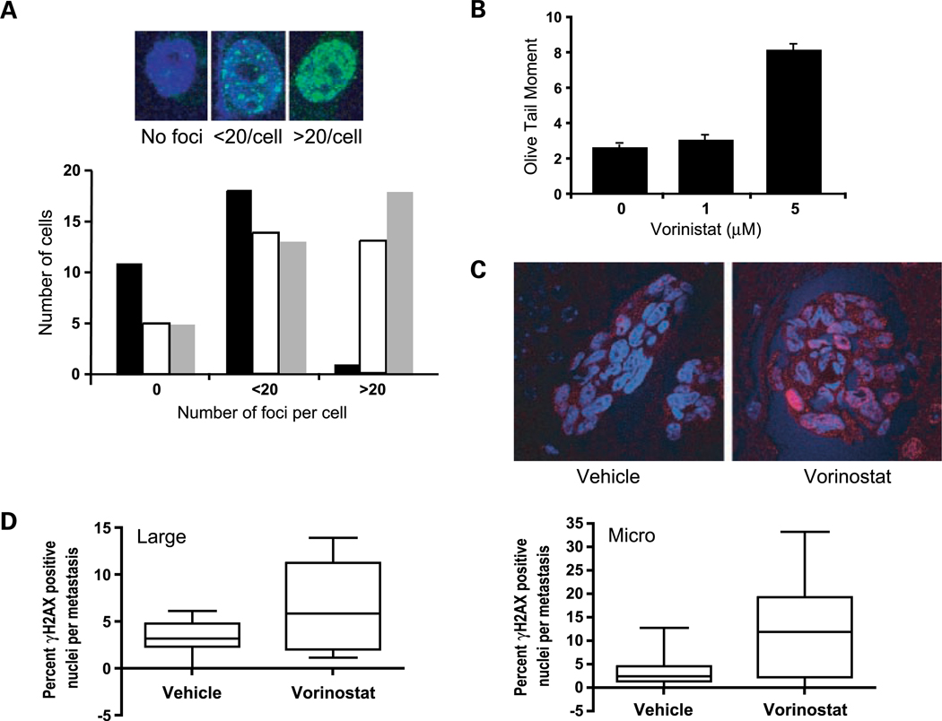
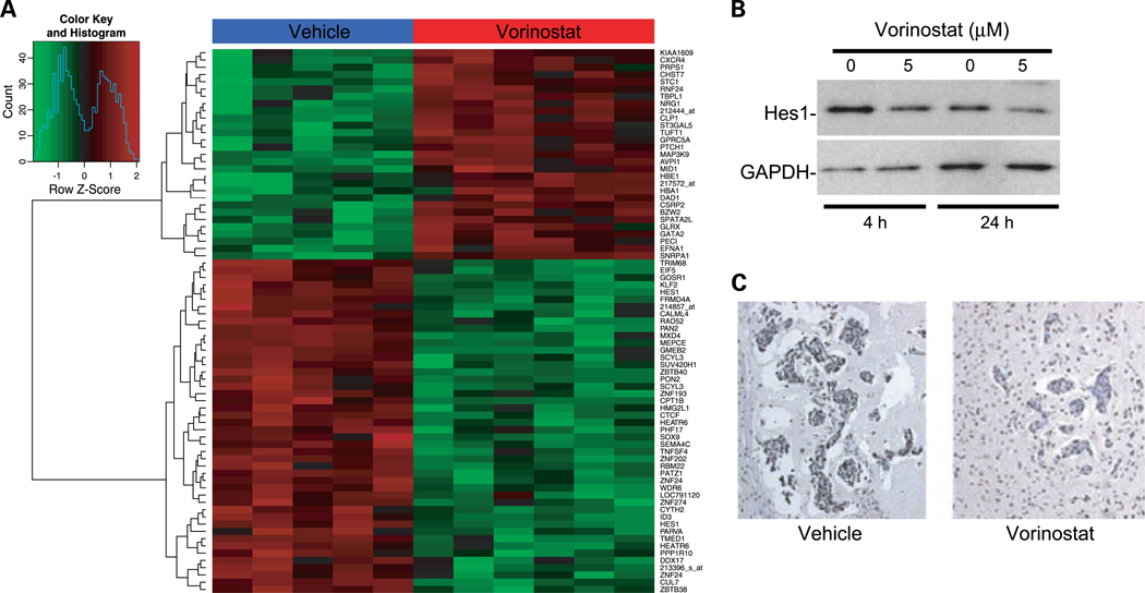
Results: Following systemic administration, uptake of [(14)C]vorinostat was significant into normal rodent brain and accumulation was up to 3-fold higher in a proportion of metastases formed by 231-BR cells. Vorinostat prevented the development of 231-BR micrometastases by 28% (P = 0.017) and large metastases by 62% (P < 0.0001) compared with vehicle-treated mice when treatment was initiated on day 3 post-injection. The inhibitory activity of vorinostat as a single agent was linked to a novel function in vivo: induction of DNA double-strand breaks associated with the down-regulation of the DNA repair gene Rad52.

Conclusions: We report the first preclinical data for the prevention of brain metastasis of triple-negative breast cancer. Vorinostat is brain permeable and can prevent the formation of brain metastases by 62%. Its mechanism of action involves the induction of DNA double-strand breaks, suggesting rational combinations with DNA active drugs or radiation.


Conflict of interest statement

Disclosure of Potential Conflicts of Interest

J. F. Reilly and V. M. Richon, employment, Merck.

Figures

Fig. 1.
Fig. 1.
Pharmacokinetics of vorinostat uptake in normal brain and in experimental brain metastases of breast cancer. A, time course of [3H]vorinostat uptake into normal brain as measured using the in situ brain perfusion technique. Mean ± SE for n = 3 perfusions. The line is the best-fit linear regression to the data. Brain space (mL/g) was calculated as the vascularly corrected brain 3H concentration divided by the [3H]vorinostat concentration of perfusion fluid. B, log BBB PS versus log octanol/water distribution coefficient (logD), a measure of hydrophobicity. The line and black squares are for solutes that cross the BBB by passive diffusion (16). The PS of vorinostat is ∼2 logs below that of normal diffusion. C, a coronal tissue section from a mouse injected intravenously with 150 mg/kg [14C]vorinostat 30 min before death and 1.5 mg 3 kDa Texas Red dextran 10 min before death. Mice received EGFP-transfected 231-BR cells via intracardiac injection 4 wk before the experiment. Green fluorescent metastatic lesions that formed are circled (left). Uptake of the nonspecific 3 kDa Texas Red dextran (red fluorescence; middle) and [14C]vorinostat (autoradiogram; right) for those circled lesions in the same coronal section is shown. Representative images shown for n = 5 mice analyzed.
Fig. 2.
Fig. 2.
Representative dorsal whole-brain images from mice treated with vorinostat. 231-BR-EGFP cells were injected into the left cardiac ventricle of Balb\c mice and vehicle or vorinostat treatment started at days indicated. Brains were dissected at necropsy and imaged using a Maestro 420 Special Imaging System to detect the presence of EGFP-expressing metastases.
Fig. 3.
Fig. 3.
In vitro effects of vorinostat on histone acetylation and apoptosis. A, Western blot analysis of acetylated histone proteins in 231-BR cells treated with increasing concentrations of vorinostat for 24 h. B, apoptosis as measured by the Cell Death Detection ELISAPLUS (Roche). Apoptotic index determined with respect to vehicle control-treated cells given an index of 1 to account for the amount of cell death occurring naturally in a cell population. P < 0.0001, ANOVA, post-hoc Dunnett’s multiple comparison; P = 0.014 for vehicle versus 0.5 μmol/L vorinostat; P = 0.013 for vehicle versus 1 μmol/L vorinostat; P = 0.0001 for vehicle versus 5 μmol/L vorinostat; P < 0.0001 for vehicle versus 10 μmol/L vorinostat. C, clonogenic growth in response to 0.5 μmol/L (P = 0.024) and 1.0 μmol/L (P < 0.0001) vorinostat compared with vehicle control. P values were determined by two-way ANOVA with post-hoc Dunnett’s multiple comparison. D, vorinostat inhibition of cell migration as assessed by Boyden chamber motility experiments. Vorinostat inhibited both unstimulated (bovine serum albumin; P = 0.0007) and fetal bovine serum–stimulated (P < 0.0001) cell migration. P values were determined by three-way ANOVA. Black columns, vehicle control; white columns, 5 μmol/L vorinostat. Representative experiments of at least three conducted (A-D).
Fig. 4.
Fig. 4.
Vorinostat induces DNA damage in vitro and in vivo. A, top, representative immunofluorescent images of γ-H2AX foci (green) in the nucleus of vorinostat-treated 231-BR cells. Cells were treated with 1 μmol/L vorinostat or vehicle for 2 h. γ-H2AX immunofluorescence was done immediately following treatment or 24 h post-treatment. Nuclei were counterstained with 4′,6-diamidino-2-phenylindole. Magnification, ×400. Bottom, quantification of cells from γ-H2AX immunofluorescence. Cells were scored as having no foci, <20 foci per nucleus, or >20 foci per nucleus. Black columns, vehicle; white columns, 1 μmol/L vorinostat for 2 h; gray columns, 24 h after removal of 1 μmol/L vorinostat for 2 h. B, in vitro analysis of DNA DSB using the comet assay. 231-BR cells were treated with vehicle or 1 or 5 μmol/L vorinostat for 24 h. Mean olive tail moment pooled over three experiments. P < 0.0001 (vorinostat effect by ANOVA), post-hoc Dunnett’s multiple comparison; P = 0.34 and P < 0.0001 for vehicle versus 1 and 5 μmol/L, respectively. C, representative images of immunofluorescent γ-H2AX staining in metastatic lesions. Frozen-fixed sections of mouse brains from vehicle- or vorinostat-treated mice were stained for γ-H2AX. Positive foci are visible in pink, and all nuclei were stained with 4′,6-diamidino-2-phenylindole. Magnification, ×400. D, left, quantification of γ-H2AX staining in large metastases. The percentage of positively stained cells per large metastasis was calculated for all large metastases present in one section per mouse. Four or five mice were analyzed per group containing two to four large metastases per section. P = 0.047, weighted ANOVA. Right, quantification of γ-H2AX staining in micrometastases. The percentage of positively stained cells per micrometastasis was calculated. One section per mouse from five mice was analyzed per group with four to six micrometastasis per mouse. P = 0.004, weighted ANOVA.
Fig. 5.
Fig. 5.
Analysis of gene expression changes induced by vorinostat treatment. A, a simple hierarchical clustering of the top 75 genes differentially expressed in the tumor cells of vorinostat-treated mice compared with vehicle controls. Tumor cells were laser capture microdissected from frozen tissue sections. RNA was extracted, amplified, labeled, and applied to Affymetrix Human U133A 2.0 GeneChips. Vorinostat-treated samples are labeled in red and vehicle samples are labeled in blue. B, Western blot validation of decreased Hes1 protein in response to vorinostat treatment. 231-BR cells were treated with 5 μmol/L vorinostat or vehicle for the times indicated. C, immunohistochemical validation of decreased Rad52 protein in vivo. Representative clusters of metastases from vorinostat- and vehicle-treated mice. Magnification, ×200. One section from each of five mice per group was analyzed.

References

    1. Lin N, Bellon J, Winer E. CNS metastases in breast cancer. J Clin Oncol 2004;22:3608–17. - PubMed
    1. Weil R, Palmieri D, Bronder J, Stark A, Steeg P. Breast cancer metastasis to the central nervous system. Am J Pathol 2005;167:913–20. - PMC - PubMed
    1. Bart J, Groen H, Hendrikse N, van der Graaf W, Vaalburg W, deVries E. The blood-brain barrier and oncology: new insights into function and modulation. Cancer Treat Rev 2000;26:449–62. - PubMed
    1. Kniesel U, Wolburg H. Tight junctions of the blood-brain barrier. Cell Mol Neurobiol 1997;20: 57–76. - PMC - PubMed
    1. Deeken JF, Loscher W. The blood-brain barrier and cancer: transporters, treatment, and Trojan horses. Clin Cancer Res 2007;13:1663–74. - PubMed

Publication types

MeSH terms