. 2009 Oct 1;15(19):6128-36.
doi: 10.1158/1078-0432.CCR-08-3180. Epub 2009 Sep 29.

Identification of a novel small molecule HIF-1alpha translation inhibitor

Affiliations

Identification of a novel small molecule HIF-1alpha translation inhibitor

Takuhito Narita et al. Clin Cancer Res. .

Abstract

Purpose: Hypoxia inducible factor-1 (HIF-1), the central mediator of the cellular response to low oxygen, functions as a transcription factor for a broad range of genes that provide adaptive responses to oxygen deprivation. HIF-1 is overexpressed in cancer and has become an important therapeutic target in solid tumors. In this study, a novel HIF-1alpha inhibitor was identified and its molecular mechanism was investigated.

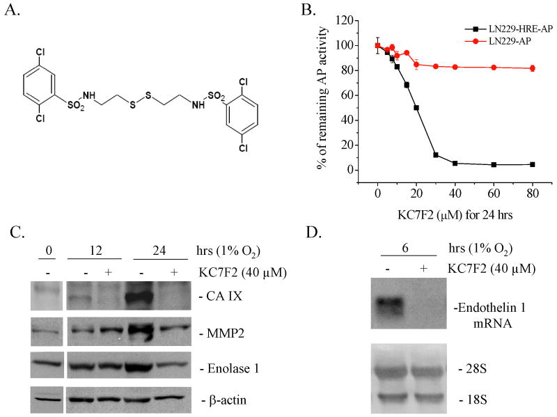
Experimental design: Using a HIF-responsive reporter cell-based assay, a 10,000-member natural product-like chemical compound library was screened to identify novel HIF-1 inhibitors. This led us to discover KC7F2, a lead compound with a central structure of cystamine. The effects of KC7F2 on HIF-1 transcription, translation, and protein degradation processes were analyzed.

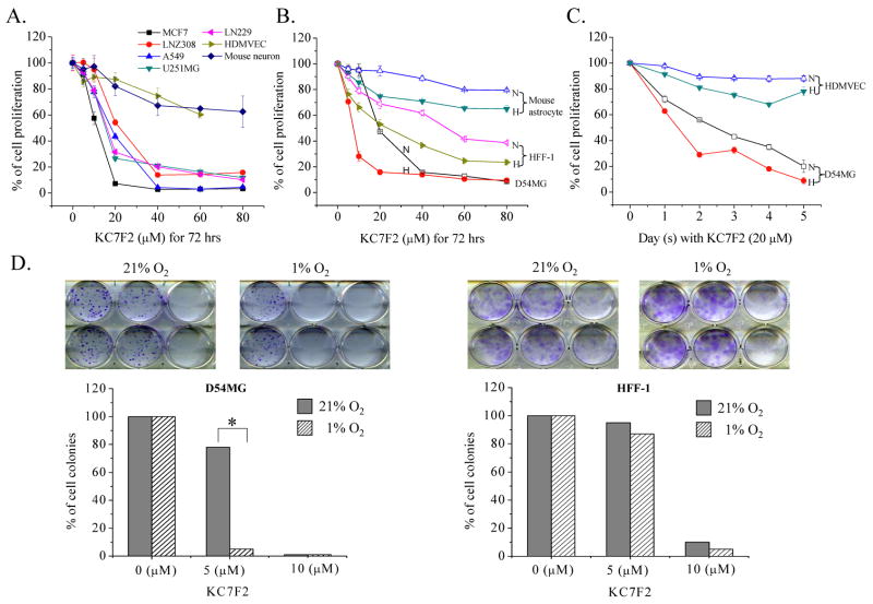
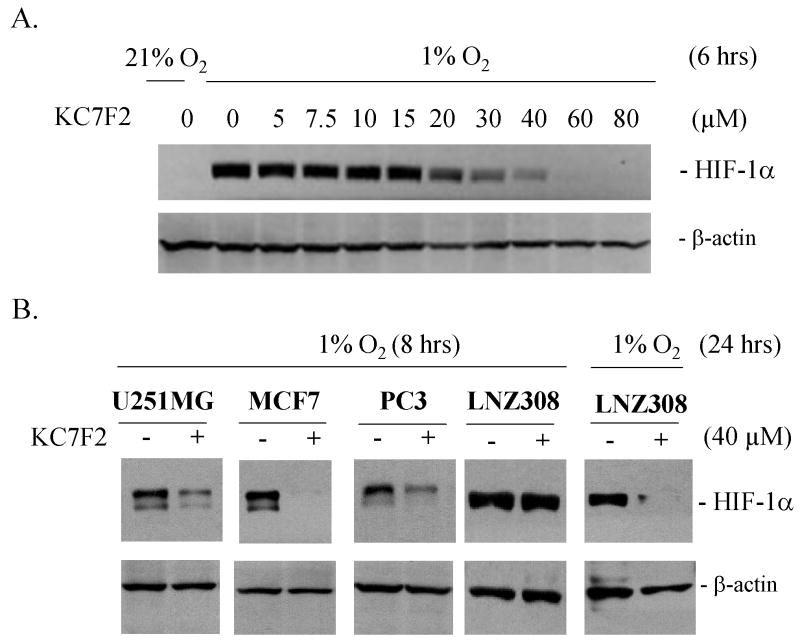
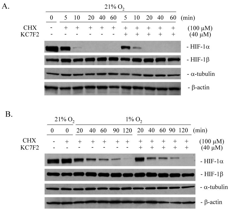
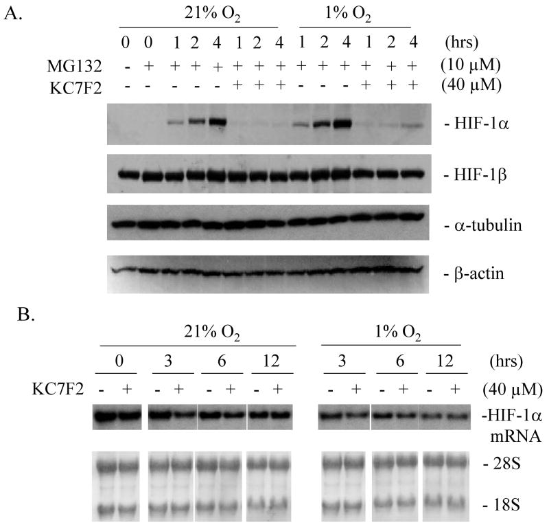
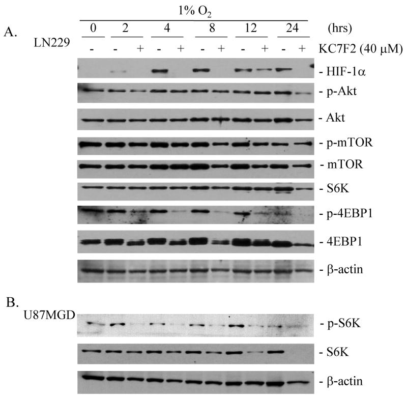
Results: KC7F2 markedly inhibited HIF-mediated transcription in cells derived from different tumor types, including glioma, breast, and prostate cancers, and exhibited enhanced cytotoxicity under hypoxia. KC7F2 prevented the activation of HIF-target genes such as carbonic anhydrase IX, matrix metalloproteinase 2 (MMP2), endothelin 1, and enolase 1. An investigation into the mechanism of action of KC7F2 showed that it worked through the down-regulation of HIF-1alpha protein synthesis, an effect accompanied by the suppression of the phosphorylation of eukaryotic translation initiation factor 4E binding protein 1 and p70 S6 kinase, key regulators of HIF-1alpha protein synthesis.

Conclusion: These results show that KC7F2 is a potent HIF-1 pathway inhibitor and its potential as a cancer therapy agent warrants further study.


Figures

Fig. 1
Fig. 1. KC7F2 inhibits HRE-driven transcription
A, Chemical structure of KC7F2. B, KC7F2 inhibits HRE-mediated alkaline phosphatase (AP) activity under hypoxia (1% O2). LN229-HRE-AP cells were incubated with different concentrations of KC7F2 for 24 hrs. Cells expressing AP constitutively (LN229-AP) were used as a control. The relative remaining AP activities were calculated as the ratios of AP levels in cells treated with KC7F2 versus untreated cells at each concentration. KC7F2 inhibits the transcription of defined HIF-1α target genes in LN229 cells as detected by western blot (C) or northern blot (D).
Fig. 2
Fig. 2. Cytotoxicity analysis in response to KC7F2 treatment
A, Normal cells (HDMVEC and mouse neurons) or cancer cells (MCF7, LNZ308, A549, U251MG and LN229) were exposed for 72 hrs to different doses of KC7F2 under normoxia and their proliferation rates were determined by SRB assay. Cytotoxicity of KC7F2 was more pronounced in tumor cell lines as compared to normal cells. B, Mouse astrocyte, HFF-1 and D54MG cells were exposed to different concentrations of KC7F2 under normoxia or hypoxia for 72 hrs and their proliferation were analyzed by the SRB assay. N stands for normoxia (21% O2, open symbol). H stands for hypoxia (1% O2, filled symbol). C. HDMVEC and D54MG cells were treated with 20 μM KC7F2 under normoxia or hypoxia. The anti-proliferation effects of KC7F2 at different time points were determined by the SRB assay. D. Clonogenic assay of KC7F2 on D54MG and HFF-1 cells under normoxia and hypoxia. The lower panel shows the percent of surviving colonies after treatment.
Fig. 3
Fig. 3. KC7F2 reduces the protein levels of HIF-1α in cancer cell lines of different tissue origin and genetic background
A, LN229 cells were treated with different concentrations of KC7F2 for 6 hrs under hypoxic conditions. Note a strong decrease in HIF-1α levels at concentrations above 20 μM, consistent with the IC50 in the AP reporter assay. B, U251MG, MCF7, PC3, and LNZ308 cells were treated with 40 μM KC7F2 for 8 or 24 hrs under hypoxia and HIF-1α levels analyzed by western blot.
Fig. 4
Fig. 4
KC7F2 does not affect the protein degradation rate of HIF-1α. LN229 cells were treated with 100 μM of cycloheximide (CHX) to inhibit protein synthesis at time zero. The protein levels of HIF-1α, HIF-1β, α-tubulin and β-actin with or without KC7F2 (40 μM) treatment were analyzed under normoxic (A) or hypoxic conditions (B) over a period of 60–120 min. Note that the degradation rate of HIF-1α is slower under hypoxia than normoxia as expected, but neither is changed by KC7F2.
Fig. 5
Fig. 5. KC7F2 inhibits HIF-1α protein synthesis but not its mRNA transcription
A, LN229 cells were treated with 10 μM of MG132 to inhibit protein degradation through the proteasome. The levels of newly synthesized HIF-1α, HIF-1β, α-tubulin and β-actin with or without KC7F2 (40 μM) treatment were determined over a period of 4 hrs. Note that HIF-1α synthesis is dramatically reduced in the presence of KC7F2. B, The levels of HIF-1α mRNA were not affected by KC7F2 (40 μM) in LN229 cells treated for up to 12 hrs, whether under hypoxic or normoxic conditions as measured by northern blot analysis.
Fig. 6
Fig. 6. The inhibition of KC7F2 on HIF-1α protein synthesis involved inhibition of the phosphorylation of 4EBP1 and S6K
A, LN229 cells were pre-treated for one hour with 40 μM of KC7F2 followed by 2–24 hrs of hypoxia. The levels of HIF-1α, and the total and phosphorylated forms of Akt, mTOR, S6K, and 4E binding protein 1 (4EBP1), were examined over time by western blot. The levels of p-Akt, Akt p-mTOR, mTOR, S6K were unchanged or showed only modest change up to 12 hrs in response to KC7F2. In contrast, the phosphorylated 4EBP1 was strongly suppressed by KC7F2 as early as 2 hrs and throughout the 24 hrs incubation under hypoxia. LN229 cells do not express detectable p-S6K as we previously reported (31). B, to examine the effect of KC7F2 on a cell line expressing constitutively p-S6K we treated U87MGD glioma cells for 2–24 hrs with KC7F2 (40 μM) and examined total and phospho-forms of S6K by western blot. Note that phospho-S6K was decreased as early as 2 hrs post-treatment.

Comment in

References

    1. Semenza GL. Hypoxia and cancer. Cancer Metastasis Rev. 2007;26:223–4. - PubMed
    1. Gray LH. Radiobiologic basis of oxygen as a modifying factor in radiation therapy. Am J Roentgenol Radium Ther Nucl Med. 1961;85:803–15. - PubMed
    1. Amellem O, Pettersen EO. Cell inactivation and cell cycle inhibition as induced by extreme hypoxia: the possible role of cell cycle arrest as a protection against hypoxia-induced lethal damage. Cell Proliferation. 1991;24:127–41. - PubMed
    1. Comerford KM, Wallace TJ, Karhausen J, Louis NA, Montalto MC, Colgan SP. Hypoxia-inducible factor-1-dependent regulation of the multidrug resistance (MDR1) gene. Cancer Res. 2002;62:3387–94. - PubMed
    1. Kim H, Peng G, Hicks JM, et al. Engineering human tumor-specific cytotoxic T cells to function in a hypoxic environment. Mol Ther. 2008;16:599–606. - PubMed

Publication types

MeSH terms