. 2009 Sep 30;4(9):e7258.
doi: 10.1371/journal.pone.0007258.

Masitinib (AB1010), a potent and selective tyrosine kinase inhibitor targeting KIT

Affiliations

Masitinib (AB1010), a potent and selective tyrosine kinase inhibitor targeting KIT

Patrice Dubreuil et al. PLoS One. .

Abstract

Background: The stem cell factor receptor, KIT, is a target for the treatment of cancer, mastocytosis, and inflammatory diseases. Here, we characterise the in vitro and in vivo profiles of masitinib (AB1010), a novel phenylaminothiazole-type tyrosine kinase inhibitor that targets KIT.

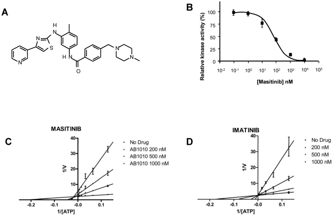
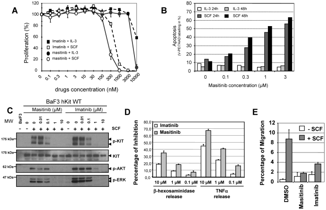
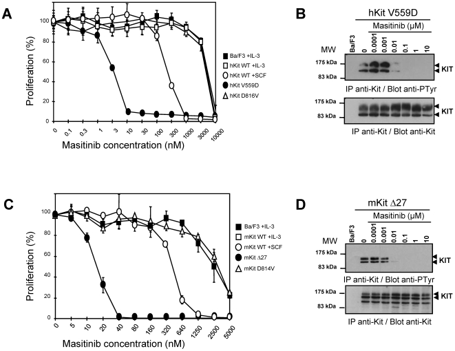
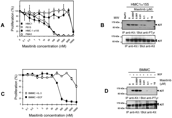
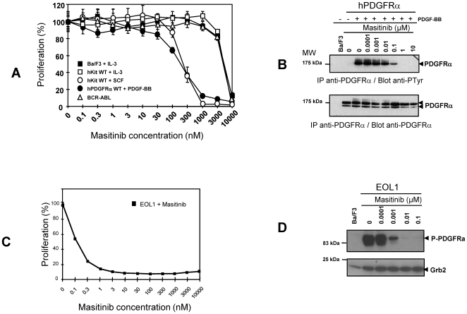
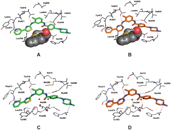
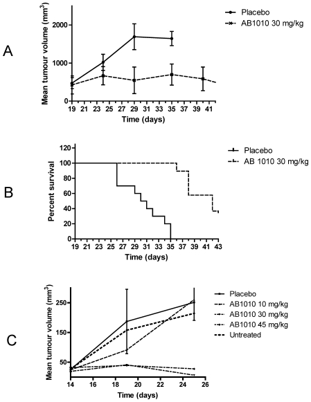
Methodology/principal findings: In vitro, masitinib had greater activity and selectivity against KIT than imatinib, inhibiting recombinant human wild-type KIT with an half inhibitory concentration (IC(50)) of 200+/-40 nM and blocking stem cell factor-induced proliferation and KIT tyrosine phosphorylation with an IC(50) of 150+/-80 nM in Ba/F3 cells expressing human or mouse wild-type KIT. Masitinib also potently inhibited recombinant PDGFR and the intracellular kinase Lyn, and to a lesser extent, fibroblast growth factor receptor 3. In contrast, masitinib demonstrated weak inhibition of ABL and c-Fms and was inactive against a variety of other tyrosine and serine/threonine kinases. This highly selective nature of masitinib suggests that it will exhibit a better safety profile than other tyrosine kinase inhibitors; indeed, masitinib-induced cardiotoxicity or genotoxicity has not been observed in animal studies. Molecular modelling and kinetic analysis suggest a different mode of binding than imatinib, and masitinib more strongly inhibited degranulation, cytokine production, and bone marrow mast cell migration than imatinib. Furthermore, masitinib potently inhibited human and murine KIT with activating mutations in the juxtamembrane domain. In vivo, masitinib blocked tumour growth in mice with subcutaneous grafts of Ba/F3 cells expressing a juxtamembrane KIT mutant.

Conclusions: Masitinib is a potent and selective tyrosine kinase inhibitor targeting KIT that is active, orally bioavailable in vivo, and has low toxicity.


Conflict of interest statement

Competing Interests: This study was financially supported by AB Science, S.A., Paris, France. AB Science is the proprietary holder of masitinib. Masitinib is under clinical development by AB Science. P.D., S.L., M.C., L.G., M.H., N.C., B.H., A.L., C.A., P.S.L., C.D.M. and A.M. are employees of the study sponsor, AB Science. M.C., M.A. and O.H. are scientific advisors to the study sponsor, AB Science. P.D., M.C., L.G., M.H., A.L., A.M. and O.H. are shareholders of the study sponsor, AB Science. M.C., A.L. and A.M. are applicants on numerous patents pertaining to masitinib.

Figures

Figure 1
Figure 1. Masitinib inhibition of recombinant human KIT.
(A) Structure of masitinib. The structure of masitinib is shown without its mesylate counterion. (B) Dose-response of masitinib at 10 µM ATP. Tyrosine phosphorylation by KIT was assayed by measuring the incorporation of phosphate into poly(Glu,Tyr 4∶1). Lineweaver-Burk Plots for masitinib (C) and imatinib (D) with ATP as the varied substrate. Recombinant human KIT tyrosine kinase assays were performed using an ELISA-based assay with poly(Glu,Tyr 4∶1) as a substrate. In (C), the lines intersect to the left of the Y-axis, indicating a mixed mechanism of inhibition for masitinib, whereas in (D), the lines intersect on the Y-axis, indicating a competitive mechanism of inhibition for imatinib.
Figure 2
Figure 2. Masitinib inhibition of KIT in intact cells.
(A) Effect of masitinib and imatinib on SCF and IL-3-stimulated cell proliferation. Ba/F3 cells expressing wild-type (WT) human (hKIT) were incubated for 48 hours with 0.1% conditioned medium from X63-IL-3 cells (IL-3) (filled symbols) or 250 ng/ml murine SCF in the presence of various concentrations of masitinib and imatinib. Cell proliferation was assessed by WST-1 colorimetric assay. (B) Induction of apoptosis by masitinib in Ba/F3 cells expressing wild-type human KIT. Cells were incubated for 24 hours with stem cell factor (SCF) or 0.1% conditioned medium from X63-IL-3 cells (IL-3) in the presence of various concentrations of masitinib. Apoptosis was assessed via Annexin V-phycoerythrin (PE) and 7-amino-actinomycin D (7-AAD) staining, followed by fluorescence-activated cell sorting. A second dataset was acquired for an incubation of 48 hours to verify completeness of the apoptosis process. (C) Effect of masitinib and imatinib on KIT tyrosine phosphorylation in Ba/F3 cells (upper panels) and phosphorylation of the downstream targets AKT and ERK (lower panels). Ba/F3 cells expressing wild-type human KIT (hKIT WT) were incubated for 5 minutes with (+) or without (-) 250 ng/ml murine SCF in the presence of various concentrations of masitinib and imatinib. Tyrosine phosphorylation of KIT, AKT and ERK, were assessed by immunoprecipitation (IP) with the relevant antibody, followed by western blotting (Blot) with anti-phosphotyrosine (pTyr) or anti-KIT molecular weight. Results are representative of at least three independent experiments. MW = molecular weight markers. (D) Comparison of masitinib's and imatinib's ability to inhibit the FcεRI-mediated degranulation and cytokine production in cord blood derived mast cells (CBMC). Left: effect on the release of β-hexosaminidase by IgE-anti IgE activated CBMC after 30 minutes of stimulation. Right: effect on cytokine production by IgE-anti IgE-activated CBMC after 4 hours of simulation via ELISA assessment of TNF-α release. (E) The effect of masitinib and imatinib on the migration of murine BMMCs in response to rmSCF stimulation.
Figure 3
Figure 3. Effect of masitinib on human and mouse KIT mutants.
Effect of masitinib on the proliferation of Ba/F3 cells expressing wild-type (WT) or mutant human (hKIT) (Fig. 3A) or murine (Fig. 3C) KIT (mKIT). Assessment of proliferation was as described for Fig. 2A. Effect of masitinib on tyrosine phosphorylation of KIT mutants in Ba/F3 cells expressing the human V559D mutant (hKIT V559D) (Fig. 3B) or murine Δ27 mutant (mKIT Δ27) (Fig. 3D). KIT tyrosine phosphorylation was assessed as described in Fig. 2B. IP = immunoprecipitation; Blot = western blot; MW = molecular weight markers.
Figure 4
Figure 4. Effect of masitinib on cell proliferation and KIT tyrosine phosphorylation in mastocytoma cell-lines and BMMC.
(A) Effect of masitinib on the proliferation of human (HMC1, HMC-1α155) (filled symbols) and murine (P815, FMA3) mastocytoma cell lines harboring KIT mutants. Cells were incubated for 2 days with the indicated concentrations of masitinib. (B) western blotting analysis of HMC-1α155 tyrosine phosphorylation. (C) Effect of masitinib in the proliferation of BMMCs. BMMCs were incubated for 2 days with 250 ng/ml of stem cell factor (SCF) or 0.1% conditioned medium from X63-IL-3 cells (IL-3) with the indicated concentrations of masitinib. (D) Western blotting analysis of BMMC tyrosine phosphorylation. Cell proliferation was assessed by WST-1 colorimetric assay. Tyrosine phosphorylation of the KIT protein from sensitive cell types in (A) and (C) was analysed by immunoprecipitation (IP) and examined by western blotting (Blot) with antibodies to phosphotyrosine (anti-pTyr) or KIT (anti-Kit). MW = molecular weight.
Figure 5
Figure 5. Effect of masitinib on BCR-ABL and PDGFRα.
(A) Effect of masitinib on the proliferation of Ba/F3 cells expressing human wild-type KIT (hKIT WT), BCR-ABL, human wild-type PDGFRα (hPDGFRα WT). Cells were treated for 48 hours with PDGF-BB, IL-3, or SCF and in the presence of various concentrations of masitinib. Cell growth was assessed by WST-1 colorimetric assay. (B) Ba/F3 cells expressing hPDGFRα were treated for 5 minutes with PDGF-BB and various concentrations of masitinib. Tyrosine phosphorylation of PDGFRα was analysed by immunoprecipitation (IP), followed by western blotting (Blot) with an anti-phosphotyrosine (pTyr) antibody (upper panel) and an anti-PDGFRα antibody (lower panel). Results are representative of two independent experiments. (C) Effect of masitinib on the proliferation of EOL1 cells, a hyperoesinophilic tumour cell line expressing the FIP1L1-PDGFRα chimeric protein. (D) Western blotting analysis of EOL1 tyrosine phosphorylation. MW = molecular weight markers.
Figure 6
Figure 6. Docking of masitinib to human KIT and ABL: comparison with imatinib binding.
(A and B) details of the binding of masitinib (A; green; docking pose) and imatinib (B; orange; X-ray structure 1T46.pdb) to the KIT kinase domain. Masitinib and imatinib interact with the protein via hydrogen bonds involving Glu640, Thr670, Cys673, and His790 and van der Waals interactions with Ala621, Val643, Leu644, Val668, Tyr672, Leu799, Cys809, and Phe811. Cys809 and Phe811, which form a hydrophobic groove for the thiazole and pyrimidine ring, respectively, are shown as space-filling structures. (C and D) Details of the binding of masitinib (C; green; docking pose) and imatinib (right site; orange; X-ray structure 1IEP.pdb) to the ABL kinase domain. Masitinib and imatinib interact with the protein via hydrogen bonds involving Glu286, Thr315, Phe317, and His361 and van der Waals interactions with Tyr252, Ala268, Val289, Met290, Ile313, Phe317, Leu370, and Phe382. In addition, the pyrimidine ring of imatinib is involved in a hydrogen bond network to conserved water molecules around the DFG motif of ABL (shown as red balls).
Figure 7
Figure 7. Masitinib inhibits tumour growth in vivo.
Nude mice were gamma-irradiated and after 24 hours, injected subcutaneously with 1.5×10⁶ Ba/F3 cells expressing the murine Δ27 KIT mutant. (A and B) Effect of intraperitoneal administered masitinib treatment on Δ27 KIT-expressing tumours, with an average pre-treatment tumour volume of 400 mm3 (large tumour experiment). Mice were treated with 30 mg/kg masitinib or a placebo (vehicle control) (n = 10 per group) twice daily for 25 days by intraperitoneal injection. (A) Mean tumour volume assessed every 5 days during the treatment. D19 corresponds to the first day of treatment. (B) Kaplan-Meier survival plot. (C) Effect of oral masitinib treatment on Δ27 KIT-expressing tumours, with an average pre-treatment tumour volume of 40 mm3 (small tumour experiment). Mice were treated twice daily for 11 days with masitinib administered orally at 0 (controls), 10, 30, or 45 mg/kg. D14 corresponds to first day of treatment.

Similar articles

Cited by

References

    1. Roskoski R., Jr Signaling by Kit protein-tyrosine kinase - the stem cell factor receptor. Biochem Biophys Res Commun. 2005;337:1–13. - PubMed
    1. Broudy VC. Stem cell factor and hematopoiesis. Blood. 1997;90:1345–64. - PubMed
    1. Reber L, Da Silva CA, Frossard N. Stem cell factor and its receptor c-Kit as targets for inflammatory diseases. Eur J Pharmacol. 2006;533:327–40. - PubMed
    1. Tests U. Kit mutations in cancer and their treatment with protein kinase inhibitors. Drugs Fut. 2008;33:161–174.
    1. Lennarttson J, Jelacic T, Linnekin D, Shivakrupa R. Normal and Oncogenic Forms of the Receptor Tyrosine Kinase Kit. Stem Cells. 2005;23:16–43. - PubMed

Publication types

LinkOut - more resources