

doi: 10.4103/0972-9941.38908.
Thoracoscopic management of empyema thoracis
Affiliations
- PMID: 19789675
- PMCID: PMC2749197
- DOI: 10.4103/0972-9941.38908
Item in Clipboard
Thoracoscopic management of empyema thoracis
J Minim Access Surg.
2007 Oct.
Abstract
Appropriate management of empyema thoracis is dependent upon a secure diagnosis of the etiology of empyema and the phase of development. Minimal access surgery using video-assisted thoracoscopy (VATS) is one of many useful techniques in treating empyema. Complex empyema requires adjunctive treatment in addition to VATS.
Keywords: Empyema; video-assisted thoracoscopy.
Conflict of interest statement
Figures

Standard AP chest X-ray depicting an elevated diaphragm with loculation of the left pleural space

CT scan of the same patient in Figure 1, demonstrating loculation and septation of the pleural space, indicating a phase II empyema
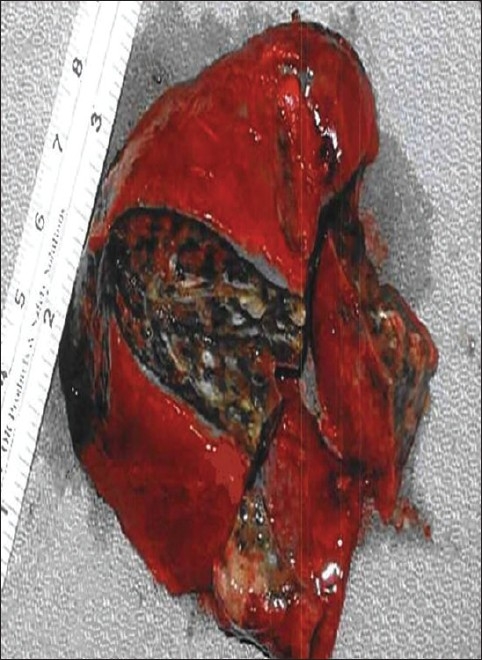
Empyemectomy specimen

Initial thoracoscopic view of VATS decortication of a phase II (fibrinopurulent) empyema

Bronchopleural fistula and empyema following right pneumonectomy for Histoplasmosis and broncholithiasis
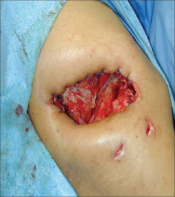
Post-resection empyema treated with Eloesser flap

Wound vacuum placed following Eloesser flap

Right middle lobe lung abscess and pleural empyema

Thoracoscopic view of patient in Figure 8. There is an incidental azygous lobe (double arrows panel a) and an abscess in the right middle lobe (arrow, panel b)
References
-
- Mandal AK, Thadepalli H, Mandal AK, Chettipally U. Outcome of primary empyema thoracis: Therapeutic and microbiologic aspects. Ann Thorac Surg. 1998;66:1782–6. - PubMed
-
- de Souza A, Offner PJ, Moore EE, Biffl WL, Haenel JB, Franciose RJ, et al. Optimal management of complicated empyema. Am J Surg. 2000;180:507–11. - PubMed
-
- Lackner RP, Hughes R, Anderson LA, Sammut PH, Thompson AB. Video-assisted evacuation of empyema is the preferred procedure for management of pleural space infections. Am J Surg. 2000;179:27–30. - PubMed
-
- Meyer DM, Jessen ME, Wait MA, Estrera AS. Early evacuation of traumatic retained hemothoraces using thoracoscopy: A prospective, randomized trial. Ann Thorac Surg. 1997;64:1396–401. - PubMed
-
- Mandal AK, Thadepalli H, Mandal Aloke K, Chettipalli U. Posttraumatic empyema thoracis: A 24-year experience at a major trauma center. J Trauma. 1997;43:764–71. - PubMed
LinkOut - more resources
Full Text Sources