

Obstetric ultrasound scanning by local health workers in a refugee camp on the Thai-Burmese border
- PMID: 19790099
- PMCID: PMC3438883
- DOI: 10.1002/uog.7350
Obstetric ultrasound scanning by local health workers in a refugee camp on the Thai-Burmese border
Abstract
Objectives: Ultrasound examination of the fetus is a powerful tool for assessing gestational age and detecting obstetric problems but is rarely available in developing countries. The aim of this study was to assess the intraobserver and interobserver agreement of fetal biometry by locally trained health workers in a refugee camp on the Thai-Burmese border.
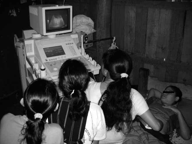
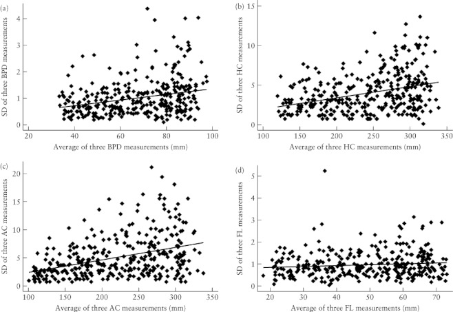
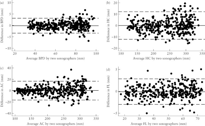
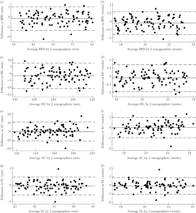
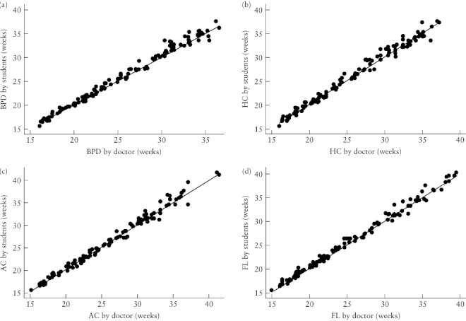
Methods: One expatriate doctor and four local health workers participated in the study, which included examinations performed on every fifth pregnant woman with a singleton pregnancy between 16 and 40 weeks' gestation, and who had undergone an early dating ultrasound scan, attending the antenatal clinic in Maela refugee camp. At each examination, two examiners independently measured biparietal diameter (BPD), head circumference (HC), abdominal circumference (AC) and femur length (FL), with one of the examiners obtaining duplicate measurements of each parameter. Intraobserver measurement error was assessed using the intraclass correlation coefficient (ICC) and interobserver error was assessed by the Bland and Altman 95% limits of agreement method.
Results: A total of 4188 ultrasound measurements (12 per woman) were obtained in 349 pregnancies at a median gestational age of 27 (range, 16-40) weeks in 2008. The ICC for BPD, HC, AC and FL was greater than 0.99 for all four trainees and the doctor (range, 0.996-0.998). For gestational ages between 18 and 24 weeks, interobserver 95% limits of agreement corresponding to differences in estimated gestational age of less than +/- 1 week were calculated for BPD, HC, AC and FL. Measurements by local health workers showed high levels of agreement with those of the expatriate doctor.
Conclusions: Locally trained health workers working in a well organized unit with ongoing quality control can obtain accurate fetal biometry measurements for gestational age estimation. This experience suggests that training of local health workers in developing countries is possible and could allow effective use of obstetric ultrasound imaging.
Figures
References
-
- Ewigman BG, Crane JP, Frigoletto FD, LeFevre ML, Bain RP, McNellis D. Effect of prenatal ultrasound screening on perinatal outcome. RADIUS Study Group. N Engl J Med. 1993;329:821–827. - PubMed
-
- Neilson JP. Ultrasound for fetal assessment in early pregnancy. Cochrane Database Syst Rev. 2000;2:CD000182. - PubMed
-
- Nosten F, McGready R, Mutabingwa T. Case management of malaria in pregnancy. Lancet Infect Dis. 2007;7:118–125. - PubMed
-
- van Dyk B, Motto JA, Buchmann EJ. Routine second-trimester ultrasound for low risk pregnancies in a South African community. Int J Gynaecol Obstet. 2007;98:257–258. - PubMed
-
- Vankayalapati P, Hollis B. Role of ultrasound in obstetrics. Curr Obstet Gynaecol. 2004;14:92–98.
Publication types
MeSH terms
Grants and funding
LinkOut - more resources
Full Text Sources
