

Comparative Study
doi: 10.1016/0888-7543(90)90284-2.
Multipoint linkage analysis and heterogeneity testing in 20 X-linked retinitis pigmentosa families
Affiliations
- PMID: 1979051
- PMCID: PMC6174538
- DOI: 10.1016/0888-7543(90)90284-2
Item in Clipboard
Comparative Study
Multipoint linkage analysis and heterogeneity testing in 20 X-linked retinitis pigmentosa families
Genomics.
1990 Oct.
Abstract
Using multipoint linkage analysis in 20 families segregating for X-linked retinitis pigmentosa (XLRP), the lod scores on a map of eight RFLP loci were obtained. Our results indicate that under the hypothesis of homogeneity the maximal multipoint lod score supports one disease locus located slightly distal to OTC at Xp21.1. Heterogeneity testing for two XLRP loci suggested that a second XLRP locus may be located 8.5 cM proximal to DXS28 at Xp21.3. Further heterogeneity testing for three disease loci failed to detect a third XLRP locus proximal to DXS7 in any of our 20 XLRP families.
Figures
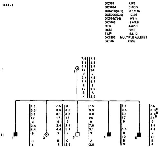
Results of RFLP analysis in GAF-1. Summary of the probes and alleles is given in the upper right table in the same order as the data presented along the sides of the chromosome stick figures. Asterisks (*) denote recombinations. Individual II-5 is recombined for markers distal to DXS84. Data from the most informative marker (+) were used in the calculations when more than one marker at a locus.
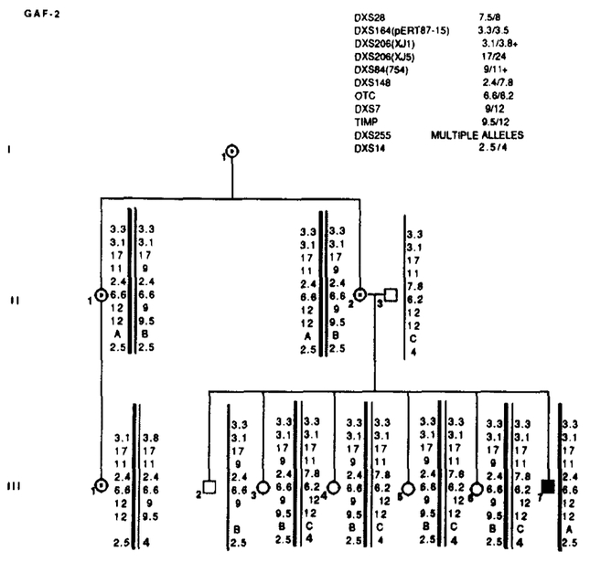
Results of RFLP analysis in GAF-2. Marker loci with their alleles are listed in the upper right table. No recombinations were detected. Data from the most informative marker (+) were used in the calculations when more than one marker at a locus.
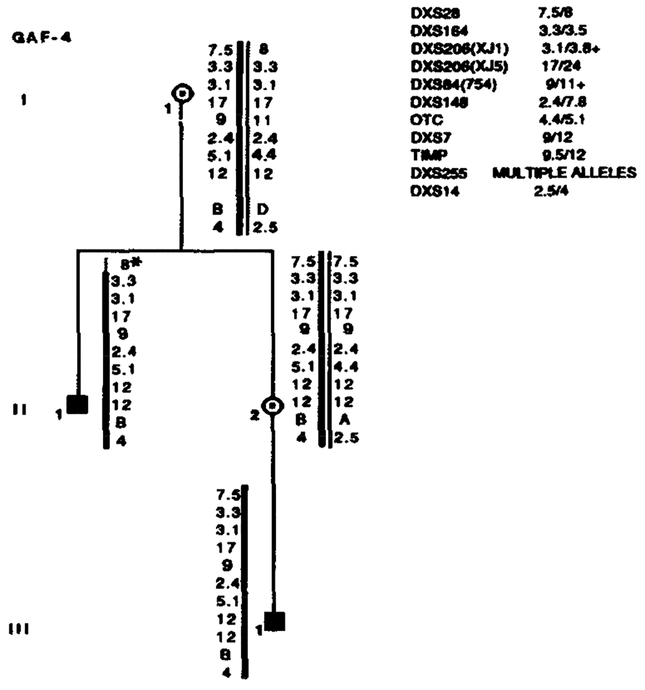
Segregation of RFLP alleles in GAF-4. Marker loci with their alleles are listed in the upper right table. Asterisks (*) denote recombinations. Data from the most informative marker (+) were used in the calculations when more than one marker at a locus.
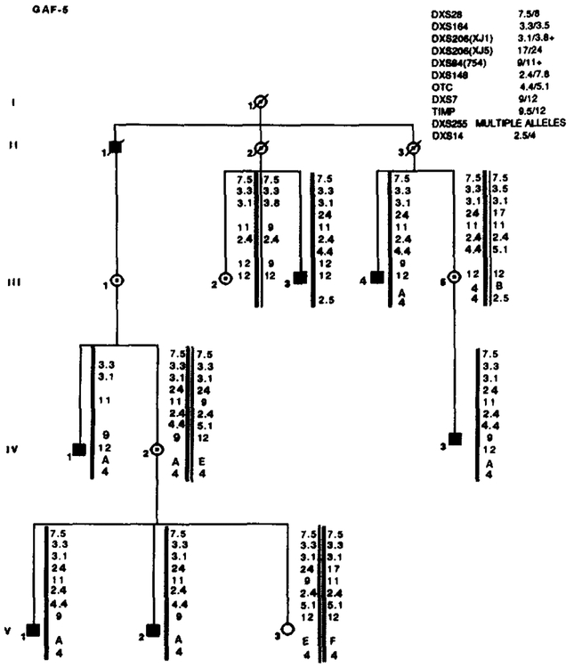
Results of RFLP analysis in GAF-5. Marker loci with their alleles are listed in the upper right table. No recombinations were detected. Data from the most informative marker (+) were used in the calculations when more than one marker at a locus.
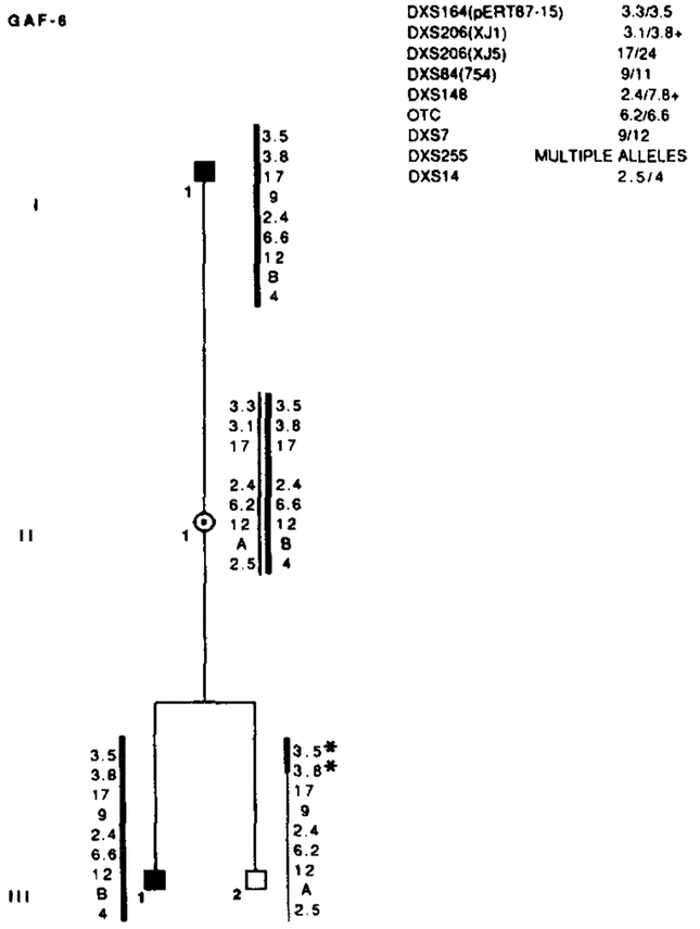
Results of RFLP analysis in GAF-6. Marker loci with their alleles are listed in the upper right table. Asterisks (*) denote recombinations. Data from the most informative marker (+) were used in the calculations when more than one marker at a locus.
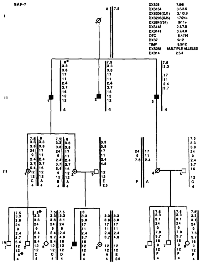
Segregation of RFLP alleles in GAF-7. Marker loci with their alleles are listed in the upper right table. Asterisks (*) denote recombinations. Data from the most informative marker (+) were used in the calculations when more than one marker at a locus.
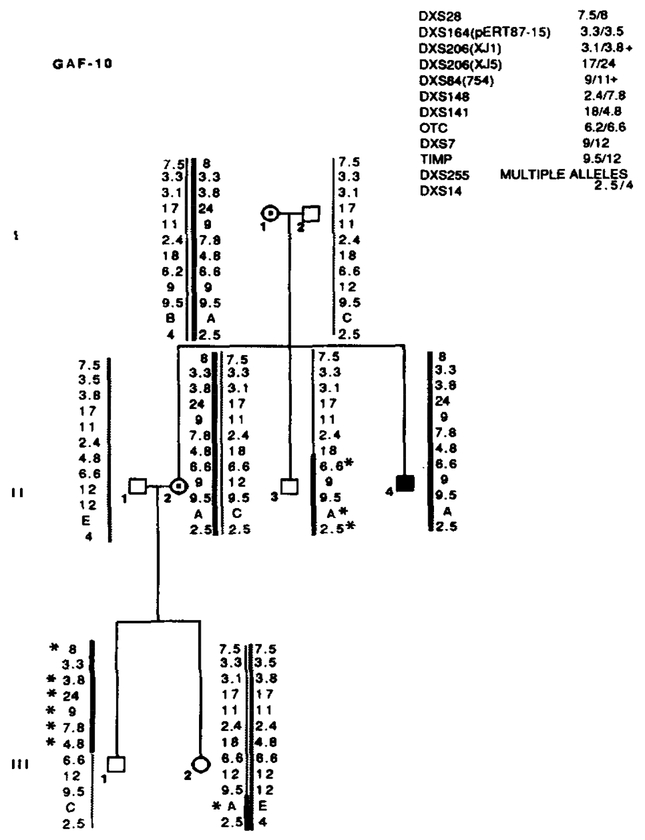
Segregation of RFLP alleles in GAF-10. Marker loci with their alleles are listed in the upper right table. Asterisks (*) denote recombinations. Individual II-3 is recombined for OTC and more proximal markers. His nephew, III-l, is recombined for all markers distal to OTC. Neither male is affected, suggesting that the X L R P mutation is between OTC and DXS141. Data from the most informative marker (+) were used in the calculations when more than one marker at a locus.
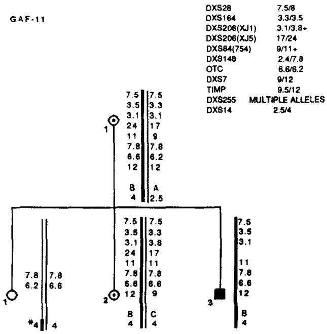
Segregation of RFLP alleles in GAF-11. Marker loci with their alleles are listed in the upper right table. Asterisks (*) denote recombinations. Data from the most informative marker (+) were used in the calculations when more than one marker at a locus.

Results of RFLP analysis in GAF-12. Marker loci with their alleles are listed in the upper right table. Asterisks (*) denote recombinations. Data from the most informative marker (+) were used in the calculations when more than one marker at a locus.
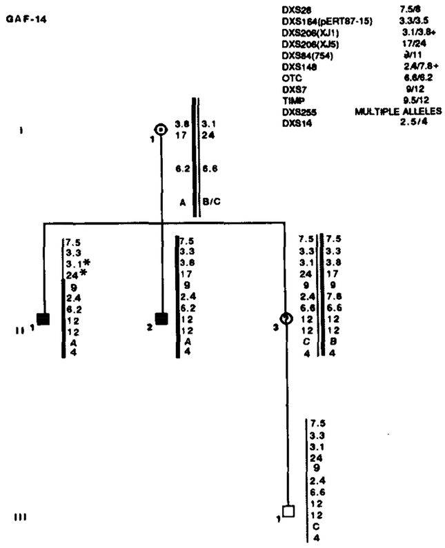
Results of RFLP analysis in GAF-14. Marker loci with their alleles are listed in the upper right table. Asterisks (*) denote recombinations. Data from the most informative marker (+) were used in the calculations when more than one marker at a locus.
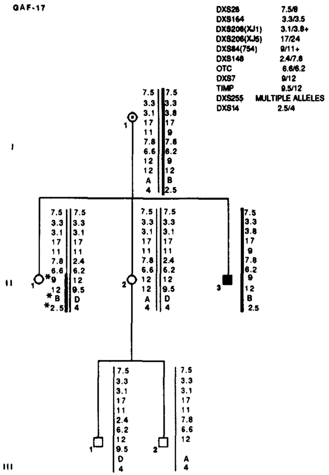
Segregation of RFLP alleles in GAF-17. Marker loci with their alleles are listed in the upper right table. Asterisks (*) denote recombinations. Data from the most informative marker (+) were used in the calculations when more than one marker at a locus.
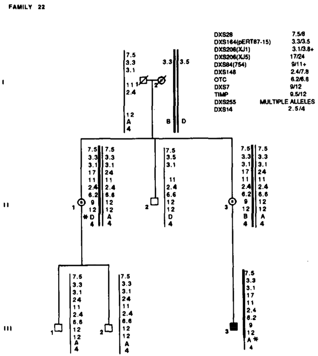
Family 22 segregated with X L R P and RFLP alleles. Marker loci with their alleles are listed in the upper right table. Asterisks (*) denote recombinations. Data from the most informative marker (+) were used in the calculations when more than one marker at a locus.
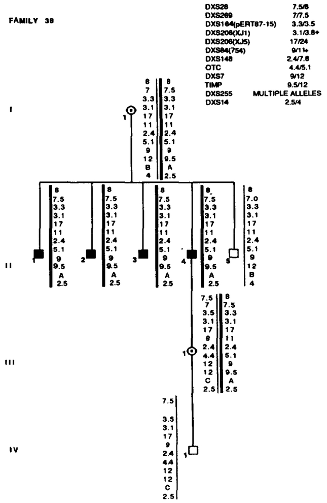
Family 38 segregated with X L R P and RFLP alleles. Marker loci with their alleles are listed in the upper right table. Data from the most informative marker (+) were used in the calculations when more than one marker at a locus.
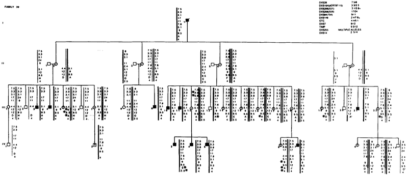
Segregation of RFLP alleles in Family 39. Marker loci with their alleles are listed in the upper right table. Asterisks (*) denote recombinations. Data from the most informative marker (+) were used in the calculations when more than one marker at a locus.
References
-
- Bhattacharya SS, Wright AF, Clayton JF, Price WH, Phillips CI, Mckeown CME, Jay M, Bird AC, Pearson PL, Southern EM, AND Evans HJ (1984). Close genetic linkage between X-linked retinitis pigmentosa and a recombinant DNA probe L1.28. Nature (London) 309:253–255. - PubMed
-
- Denton MJ, Chen JD, Serravalle S, Colley P, Halliday FB, AND Donald J (1988). Analysis of linkage relationships of X-linked retinitis pigmentosa with the following Xp loci: L1.28, OTC, XJ1.1, pERT87, and C7. Hum. Genet 78:60–64. - PubMed
-
- de Saint-Basile G, Bohlek MC, Fischer A, Cartron J, Dufier JL, Griselli C, AND Orkin SH (1988). X p21 D N A microdeletion in a patient with chronic granulomatous disease, retinitis pigmentosa, and the McLeod phenotype. Hum. Genet 80: 85–89. - PubMed
-
- Drayna D, AND White R (1985). The genetic linkage map of the X chromosome. Science 230: 753–758. - PubMed
Publication types
MeSH terms
Substances
Grants and funding
LinkOut - more resources
Full Text Sources
