. 2010 May;20(3):560-70.
doi: 10.1111/j.1750-3639.2009.00333.x. Epub 2009 Sep 10.

Unique molecular characteristics of pediatric myxopapillary ependymoma

Affiliations

Unique molecular characteristics of pediatric myxopapillary ependymoma

Valerie N Barton et al. Brain Pathol. 2010 May.

Abstract

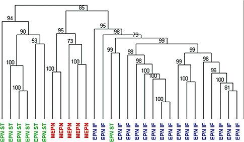
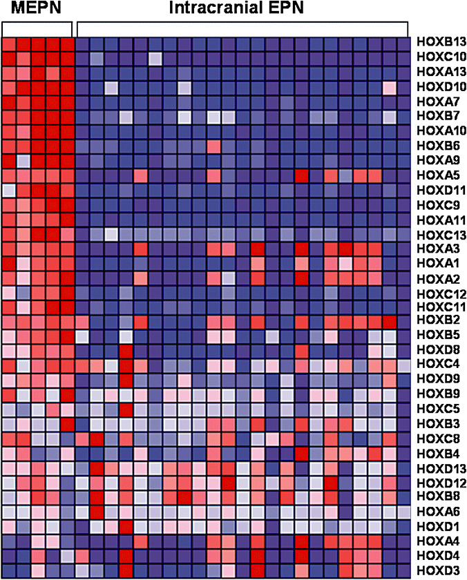
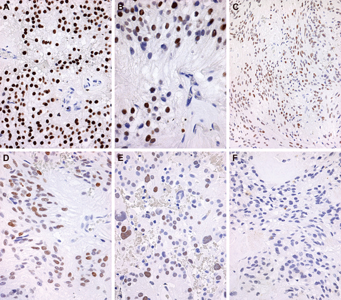
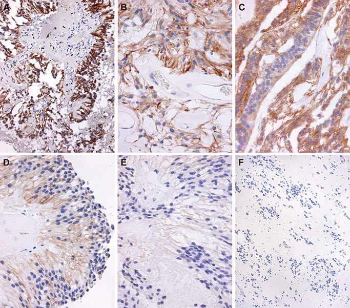
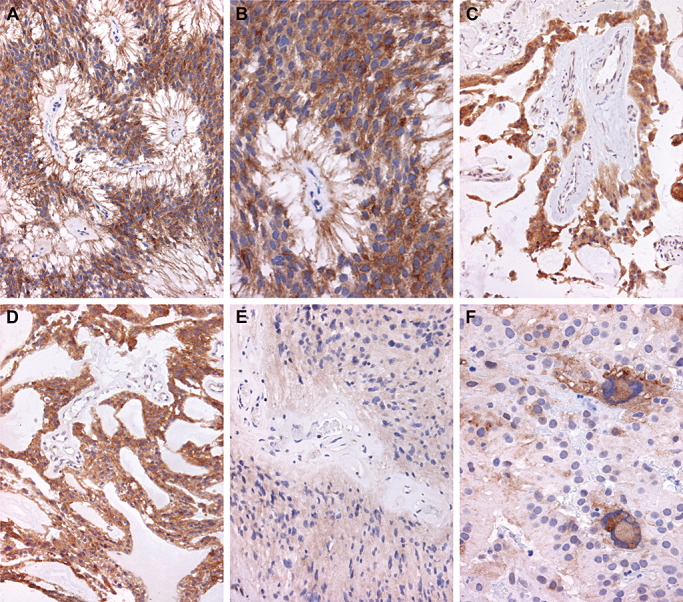
Myxopapillary ependymoma (MEPN) generally can be cured by gross total surgical resection and usually manifest a favorable prognosis. However, surgery is less curative in tumors that are large, multifocal or extend outside the thecal sac. Late recurrences may occur, particularly in pediatric patients. The role of adjuvant therapy is unclear in the clinical management of recurrent tumors. Clinical trial design requires a better understanding of tumor biology. Unique molecular features of MEPN were investigated by using microarray technology to compare the gene expression of five pediatric MEPN to 24 pediatric intracranial ependymoma (EPN). The upregulation of three genes of interest, homeobox B13 (HOXB13), neurofilament, light polypeptide (NEFL) and PDGFR alpha, was further studied by immunohistochemistry in a larger cohort that included adult MEPN and EPN specimens. Protein expression in MEPN was compared to subependymoma, spinal EPN, intracranial EPN and normal fetal and adult ependyma. Immunoreactivity for HOXB13, NEFL and PDGFR alpha was strongest in MEPN and virtually absent in subependymoma. Spinal and intracranial EPN generally expressed weak or focal staining. MEPN manifests unique gene and protein expression patterns compared to other EPNs. Aberrant expression of HOXB13 suggests possible recapitulation of developmental pathways in MEPN tumorigenesis. PDGFR alpha may be a potential therapeutic target in recurrent MEPN.


Figures

Figure 1
Figure 1
Hierarchical clustering analysis of the myxopapillary subgroup of ependymoma. Gene expression profiles of first presentation surgical samples of five pediatric MEPN and 24 pediatric intracranial EPN. Numerical values represent approximately unbiased P values that were computed by multiscale bootstrap resampling. Abbreviations: EPN =  ependymoma; MEPN = myxopapillary ependymoma; IF = infratentorial; ST = supratentorial.
Figure 2
Figure 2
Homeobox family gene expression in pediatric myxopapillary ependymoma compared to pediatric intracranial ependymoma. Data are organized in a heat map format where each row represents a single gene and each column represents an ependymoma sample. Genes are ranked according to difference in expression between MEPN and intracranial EPN. Normalized z‐scores correlating to the abundance of mRNA relative to a common reference are represented by a color scale where red indicates high gene expression and green indicates low gene expression. Abbreviations: MEPN = myxopapillary ependymoma; EPN = ependymoma; HOX = homeobox.
Figure 3
Figure 3
Representative immunohistochemical staining of homeobox B13 (HOXB13). Immunostaining was scored from − to++on a subjective scale. Twenty‐five percent to 100% of cells staining yielded a score of++. Very focal staining in individual cells yielded a score of+. Absence of staining was given a − designation. A. Strong and diffuse nuclear staining of HOXB13, scored as ++, is illustrated in a pediatric myxopapillary ependymoma (MEPN). Immunohistochemistry for HOXB13 with light hematoxylin counterstain, original magnification 400×. B. Higher power magnification of the same pediatric MEPN depicted in A demonstrates a perivascular pseudorosette with absence of HOXB13 staining in endothelial cell nuclei, original magnification 600×. C. Photomicrograph of an adult MEPN with diffuse expression, scored as ++, albeit with slightly less intensity is shown. Immunohistochemistry for HOXB13 with light hematoxylin counterstain, original magnification 200×. D. The same adult MEPN as shown in C is depicted at a higher magnification, original magnification 400×. E. Positive HOXB13 immunostaining reaching a score of ++ was identified only in a single adult spinal ependymoma, as illustrated in this photomicrograph. Immunohistochemistry for HOXB13 with light hematoxylin counterstain, original magnification 400×. F. Photomicrograph of an adult MEPN with negative HOXB13 staining for direct comparison with A–E. Immunohistochemistry for HOXB13 with light hematoxylin counterstain, original magnification 400×.
Figure 4
Figure 4
Representative immunohistochemical staining of neurofilament light polypeptide (NEFL). Immunostaining was scored from − to++on a subjective scale. Twenty‐five percent to 100% of cells staining yielded a score of++. Very focal staining in individual cells yielded a score of+. Absence of staining was given a − designation. A. Photomicrograph of a pediatric myxopapillary ependymoma (MEPN) with strong and diffuse cytoplasmic staining in fibrillary areas of tumor, scored as ++. Immunohistochemistry for NEFL with light hematoxylin counterstain, original magnification 200×. B. Photomicrograph at higher power shows the perivascular, perpendicularly oriented cell processes surrounding hyalinized blood vessels strongly immunoreactive for NEFL; this adult MEPN achieved a ++ score. Immunohistochemistry for NEFL with light hematoxylin counterstain, original magnification 400×. C. Photomicrograph of a pediatric MEPN showing strong, diffuse cytoplasmic staining in areas of the tumor with ependymal, epithelial, non‐fibrillary morphological features; this case scored as ++. Immunohistochemistry for NEFL with light hematoxylin counterstain, original magnification 400×. D. Photomicrograph of an adult MEPN with ++ diffuse immunoreactivity and slightly less intensity. Immunohistochemistry for NEFL with light hematoxylin counterstain, original magnification 400×. E. Focal immunostaining for NEFL in an adult MEPN, yielding a + score. Immunohistochemistry for NEFL with light hematoxylin counterstain, original magnification 400×. F. All subependymomas manifested absence of NEFL staining, as illustrated in this adult example. Immunohistochemistry for NEFL with light hematoxylin counterstain, original magnification 200×.
Figure 5
Figure 5
Representative immunohistochemical staining of platelet‐derived growth factor receptor alpha (PDGFRα). Immunostaining was scored from − to++on a subjective scale. Twenty‐five percent to 100% of cells staining yielded a score of++. Very focal staining in individual cells yielded a score of+. Absence of staining was given a − designation. A. Low power photomicrograph of a pediatric myxopapillary ependymoma (MEPN) with diffuse immunoreactivity for PDGFRα, scored as ++. Immunohistochemistry for PDGFRα with light hematoxylin counterstain, original magnification 200×. B. Photomicrograph at higher power of the same tumor shown in A demonstrates the diffuse cytoplasmic staining throughout all tumor cells of this perivascular pseudorosette. Immunohistochemistry for PDGFRα with light hematoxylin counterstain, original magnification 400×. C. Photomicrograph of a different pediatric MEPN also manifests diffuse immunoreactivity, with diffuse staining scored as ++. Immunohistochemistry for PDGFRα with light hematoxylin counterstain, original magnification 200×. D. Photomicrograph from a different area of the same case as illustrated in C demonstrates that epithelial‐like areas of the MEPN show strong immunoreactivity. Original magnification 200×. E. Photomicrograph of an adult spinal ependymoma (EPN) with diffuse, albeit less intense immunoreactivity, scored as ++. Immunohistochemistry for PDGFRα with light hematoxylin counterstain, original magnification 200×. F. Photomicrograph of a different example of adult spinal EPN shows strong intensity but a lower staining pattern (++, 400×).

References

    1. (2001) FDA Oncology Tools Approval Summary for imatinib mesylate for Accel. Approv., U.S. Food and Drug Administration, Center for Drug Evaluation and Research. Available at: http://www.fda.gov/Drugs/DrugSafety/PostmarketDrugSafetyInformationforPa... (last accessed 15 September 2009).
    1. (2005) FDA Oncology Tools Approval Summary for sorafenib for the treatment of patients with advanced renal cell carcinoma. U.S. Food and Drug Administration Center for Drug Evaluation and Research. Available at: http://www.fda.gov/AboutFDA/CentersOffices/CDER/ucm129234.htm (last accessed 15 September 2009).
    1. (2007) FDA Oncology Tools Approval Summary for sunitinib for the treatment of gastrointestinal stromal tumor after disease progression on or intolerance to imatinib mesylate; for the treatment of advanced renal cell carcinoma. U.S. Food and Drug Administration, Center for Drug Evaluation and Research. Available at: http://www.fda.gov/AboutFDA/CentersOffices/CDER/ucm129255.htm (last accessed 15 September 2009).
    1. Acampora D, D'Esposito M, Faiella A, Pannese M, Migliaccio E, Morelli F et al (1989) The human HOX gene family. Nucleic Acids Res 17:10385–10402 - PMC - PubMed
    1. Bagley CA, Wilson S, Kothbauer KF, Bookland MJ, Epstein F, Jallo GI (2009) Long term outcomes following surgical resection of myxopapillary ependymomas. Neurosurg Rev 32:321–334. - PubMed

Publication types

Substances