

Polyomaviruses of nonhuman primates: implications for research
- PMID: 19793457
- PMCID: PMC2703155
Polyomaviruses of nonhuman primates: implications for research
Abstract
Polyomaviruses are a family of small nonenveloped DNA viruses that infect birds and mammals. At least 7 nonhuman primate polyomaviruses that occur in macaques, African green monkeys, marmosets baboons, and chimpanzees have been described, as well as 4 polyomaviruses that occur in humans. Simian virus 40 (SV40), which infects macaques, was the first nonhuman primate polyomavirus identified as a contaminant of early polio vaccines. Primate polyomaviruses cause inapparent primary infections but persist in the host and can cause severe disease in situations of immunocompromise. This review describes the primate polyomaviruses, and the diseases associated with the viruses of macaques. In macaques, the greatest current concerns are the potential confounding of study results by polyomavirus infections and the zoonotic potential of SV40.
Figures
Similar articles
-
African great apes are naturally infected with polyomaviruses closely related to Merkel cell polyomavirus.J Virol. 2011 Jan;85(2):916-24. doi: 10.1128/JVI.01585-10. Epub 2010 Nov 3. J Virol. 2011. PMID: 21047967 Free PMC article.
-
Genome analysis of non-human primate polyomaviruses.Infect Genet Evol. 2014 Aug;26:283-94. doi: 10.1016/j.meegid.2014.05.030. Epub 2014 Jun 14. Infect Genet Evol. 2014. PMID: 24933462 Review.
-
[New, newer, newest human polyomaviruses: how far?].Mikrobiyol Bul. 2013 Apr;47(2):362-81. doi: 10.5578/mb.5377. Mikrobiyol Bul. 2013. PMID: 23621738 Review. Turkish.
-
Molecular analysis of a novel simian virus 40 (SV40) type in rhesus macaques and evidence for double infections with the classical SV40 type.J Clin Microbiol. 2011 Apr;49(4):1280-6. doi: 10.1128/JCM.01005-10. Epub 2011 Feb 9. J Clin Microbiol. 2011. PMID: 21307214 Free PMC article.
-
Novel polyomavirus detected in the feces of a chimpanzee by nested broad-spectrum PCR.J Virol. 2005 Mar;79(6):3883-7. doi: 10.1128/JVI.79.6.3883-3887.2005. J Virol. 2005. PMID: 15731285 Free PMC article.
Cited by
-
Alterations in cytokines and effects of dexamethasone immunosuppression during subclinical infections of invasive Klebsiella pneumoniae with hypermucoviscosity phenotype in rhesus (Macaca mulatta) and cynomolgus (Macaca fascicularis) macaques.Comp Med. 2010 Feb;60(1):62-70. Comp Med. 2010. PMID: 20158951 Free PMC article.
-
How Simian Virus 40 Hijacks the Intracellular Protein Trafficking Pathway to Its Own Benefit … and Ours.Front Immunol. 2018 May 28;9:1160. doi: 10.3389/fimmu.2018.01160. eCollection 2018. Front Immunol. 2018. PMID: 29892296 Free PMC article. Review.
-
Pathologic characteristics of infectious diseases in macaque monkeys used in biomedical and toxicologic studies.J Toxicol Pathol. 2023 Apr;36(2):95-122. doi: 10.1293/tox.2022-0089. Epub 2023 Feb 13. J Toxicol Pathol. 2023. PMID: 37101957 Free PMC article. Review.
References
-
- Aksamit AJ., Jr 1995. Progressive multifocal leukoencephalopathy: a review of the pathology and pathogenesis. Microsc Res Tech 32:302–311 - PubMed
-
- Ashkenazi A, Melnick JL. 1962. Induced latent infection of monkeys with vacuolating SV40 papovavirus. Virus in kidneys and urine. Proc Soc Exp Biol Med 111:367–372 - PubMed
-
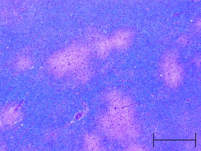
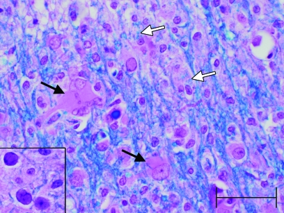
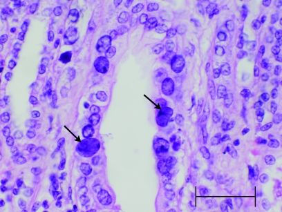
- Axthelm MK, Koralnik IJ, Dang X, Wuthrich C, Rohne D, Stillman IE, Letvin NL. 2004. Meningoencephalitis and demyelination are pathologic manifestations of primary polyomavirus infection in immunosuppressed rhesus monkeys. J Neuropathol Exp Neurol 63:750–758 - PubMed
-
- Berger JR, Houff S. 2006. Progressive multifocal leukoencephalopathy: lessons from AIDS and natalizumab. Neurol Res 28:299–305 - PubMed
Publication types
MeSH terms
LinkOut - more resources
Full Text Sources