. 2008 Feb;58(1):81-7.

Effects of the macrolide drug tylosin on chronic diarrhea in rhesus macaques (Macaca mulatta)

Affiliations

Effects of the macrolide drug tylosin on chronic diarrhea in rhesus macaques (Macaca mulatta)

Rebecca S Blackwood et al. Comp Med. 2008 Feb.

Abstract

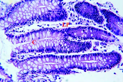
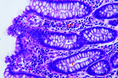
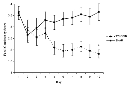
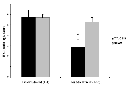
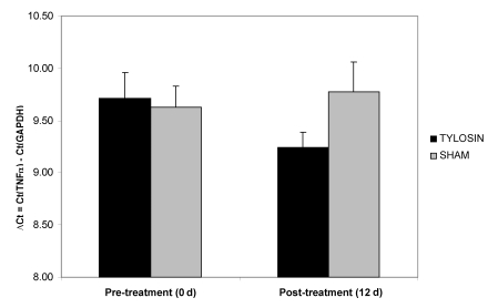
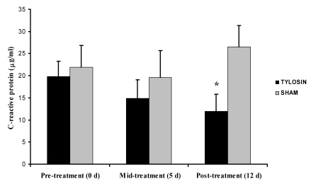
Diarrhea is the gastrointestinal disease most frequently encountered in captive rhesus macaques. The precise pathogenic mechanisms underlying chronic diarrhea in nonhuman primates are not well understood, but a persistent inflammatory component has been implicated strongly. This study evaluated the inflammatory changes in the colon of macaques with diarrhea and assessed the efficacy of a 10-d course of tylosin in a cohort of 21 animals with chronic diarrhea. Stool quality was evaluated daily, and fecal consistency was scored. Colonoscopies were performed; biopsy samples were characterized histologically and assayed for expression of TNFalpha mRNA. Blood samples collected pre-, mid-, and post-treatment were assayed for C-reactive protein (CRP). The results indicated that 63% of the animals receiving tylosin showed improvement in stool quality, compared with 10% in the sham-treated group. Histologically, 82% of animals in the tylosin-treated group had a reduction in the severity of colonic lesions post-treatment, compared with 40% of animals in the sham group. The amount of TNFalpha mRNA before treatment did not differ from that afterward in either tylosin- or sham-treated animals. CRP levels serially decreased in tylosin-treated monkeys; the average post-treatment CRP value for tylosin-treated animals was 11.96 +/- 3.86 microg/ml compared with 26.48 +/- 4.86 microg/ml for sham-treated controls. In conclusion, tylosin significantly improved the fecal consistency score, significantly decreased colonic inflammation, and significantly decreased serum CRP levels post-treatment in rhesus macaques with chronic diarrhea.


Figures

Figure 1.
Figure 1.
Representative histology of colon biopsy specimens. (A) Specimen with a histopathologic score of 2 and histologic diagnosis of mild colitis. The epithelium is intact with mild goblet cell depletion and mild lymphocytic lamina propria (LP) cellular infiltrate. (B) Specimen with a histopathologic score of 9 and histologic diagnosis of moderate colitis. The surface epithelium has moderate attenuation and tufting. There is mild goblet cell depletion, mild crypt (C) branching, and mild crypt abscesses. There is a moderate lymphocytic, plasmacytic lamina propria cellular infiltrate. Hematoxylin and eosin stain; magnification, ×375.
Figure 1.
Figure 1.
Representative histology of colon biopsy specimens. (A) Specimen with a histopathologic score of 2 and histologic diagnosis of mild colitis. The epithelium is intact with mild goblet cell depletion and mild lymphocytic lamina propria (LP) cellular infiltrate. (B) Specimen with a histopathologic score of 9 and histologic diagnosis of moderate colitis. The surface epithelium has moderate attenuation and tufting. There is mild goblet cell depletion, mild crypt (C) branching, and mild crypt abscesses. There is a moderate lymphocytic, plasmacytic lamina propria cellular infiltrate. Hematoxylin and eosin stain; magnification, ×375.
Figure 2.
Figure 2.
Daily mean fecal consistency score for tylosin- and sham-treated groups. Over the 10-d course of tylosin, the score gradually decreased, indicating an improvement in stool quality. Day 10 scores differed significantly (P = 0.002, Wilcoxon rank-sum test) between groups. Data are presented as mean ± SE.
Figure 3.
Figure 3.
Colonic biopsy scores pre- and post-treatment in tylosin- and sham-treated animals. The histopathologic score post-treatment was decreased in the tylosin-treated group and differed significantly (P = 0.001, Wilcoxon rank-sum test) between groups. Data are presented as mean ± SE.
Figure 4.
Figure 4.
Quantification of TNFα mRNA in colonic biopsies pre- and post-treatment in tylosin and sham treated animals. Numerical values are ΔCt (that is, the cycle threshold of the housekeeping gene [GAPDH] subtracted from that of the TNFα gene). TNFα mRNA levels were decreased after treatment with tylosin, but not significantly compared with those of the sham-treated group. Data are expressed as mean ± SE.
Figure 5.
Figure 5.
Serum C-reactive protein levels for tylosin- and sham-treated groups at 0, 5, and 12 d after initiation of therapy. CRP levels did not differ between groups at either 0 or 5 d, but day 12 levels differed significantly (*, P = 0.029, Wilcoxon rank-sum test) between tylosin- and sham-treated animals. Data are presented as mean ± SE.

References

    1. Abel K, Pahar B, Van Rompay KKA, Fritts L, Sin C, Schmidt K, Colon R, McChesney M, Marthas ML. 2006. Rapid virus dissemination in infant macaques after oral simian immunodeficiency virus exposure in the presence of local innate immune responses. J Virol 80:6357–6367 - PMC - PubMed
    1. Blam ME, Stein R, Lichtenstein G. 2001. Integrating anti-tumor necrosis factor therapy in inflammatory bowel disease: current and future prospectives. Am J Gastroenterol 96:1977–1997 - PubMed
    1. Brady A, Morton D. Gastrointestinal system: approach to diarrhea diagnosis and treatment. In: Bennett A, Henrickson, editors Nonhuman primates in biomedical research: diseases San Diego: Academic Press;
    1. Braegger CP, Nicholls S, Murch SH, MacDonald TT, Stephens S. 1992. Tumour necrosis factor α in stool as a marker of intestinal inflammation. Lancet 339:89–91 - PubMed
    1. Cao XY, Dong M, Shen JZ, Wu BB, Wu CM, Du XD, Wang Z, Qi YT, Li BY. 2006. Tilmicosin and tylosin have antiinflammatory properties via modulation of COX-2 and iNOS gene expression and production of cytokines in LPS-induced macrophages and monocytes. Int J Antimicrob Agents 27:431–438 - PubMed

MeSH terms