

NKG2D ligand MICA is retained in the cis-Golgi apparatus by human cytomegalovirus protein UL142
- PMID: 19793804
- PMCID: PMC2786768
- DOI: 10.1128/JVI.01175-09
NKG2D ligand MICA is retained in the cis-Golgi apparatus by human cytomegalovirus protein UL142
Abstract
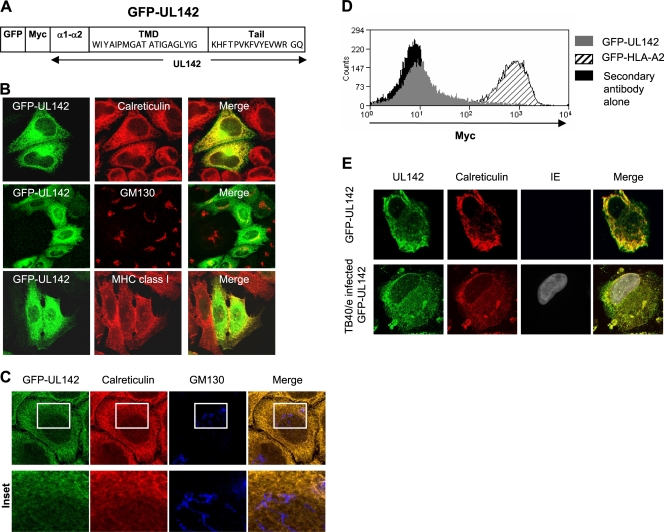
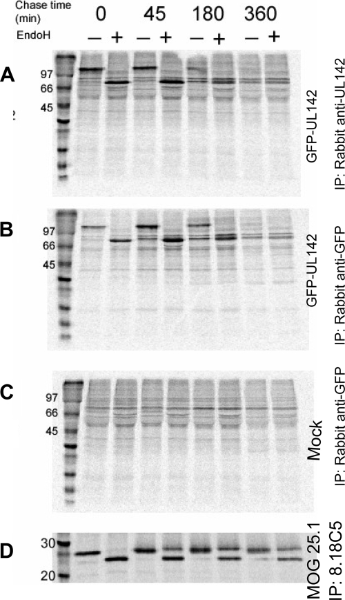
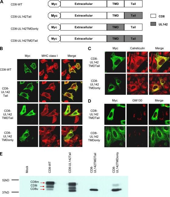
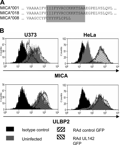
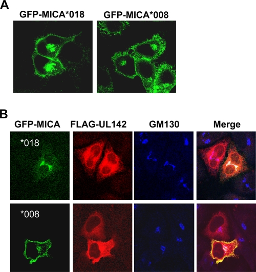
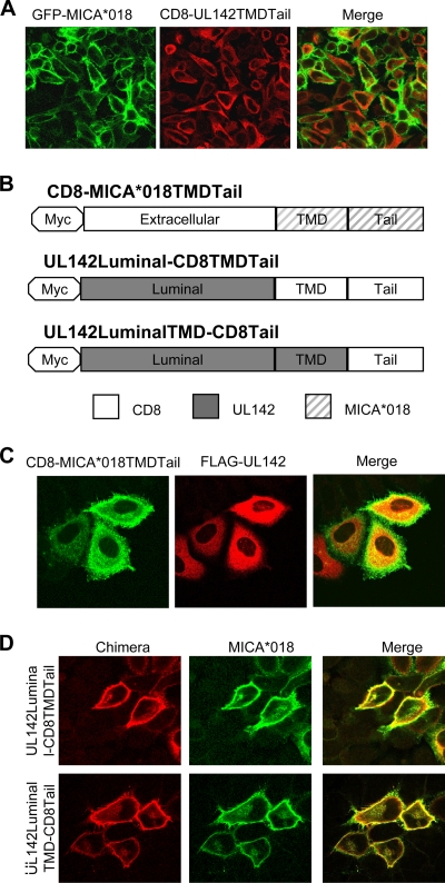
Human cytomegalovirus (HCMV) evades T-cell recognition by down-regulating expression of major histocompatibility complex (MHC) class I and II molecules on the surfaces of infected cells. Contrary to the "missing-self" hypothesis, HCMV-infected cells are refractory to lysis by natural killer (NK) cells. Inhibition of NK cell function is mediated by a number of HCMV immune evasion molecules, which operate by delivering inhibitory signals to NK cells and preventing engagement of activating ligands. One such molecule is UL142, which is an MHC class I-related glycoprotein encoded by clinical isolates and low-passage-number strains of HCMV. UL142 is known to down-modulate surface expression of MHC class I-related chain A (MICA), which is a ligand of the activating NK receptor NKG2D. However, the mechanism by which UL142 interferes with MICA is unknown. Here, we show that UL142 localizes predominantly to the endoplasmic reticulum (ER) and cis-Golgi apparatus. The transmembrane domain of UL142 mediates its ER localization, while we propose that the UL142 luminal domain is involved in its cis-Golgi localization. We also confirm that UL142 down-modulates surface expression of full-length MICA alleles while having no effect on the truncated allele MICA*008. However, we demonstrate for the first time that UL142 retains full-length MICA alleles in the cis-Golgi apparatus. In addition, we propose that UL142 interacts with nascent MICA en route to the cell surface but not mature MICA at the cell surface. Our data also demonstrate that the UL142 luminal and transmembrane domains are involved in recognition and intracellular sequestration of full-length MICA alleles.
Figures
Similar articles
-
Down-regulation of the NKG2D ligand MICA by the human cytomegalovirus glycoprotein UL142.Biochem Biophys Res Commun. 2006 Jul 21;346(1):175-81. doi: 10.1016/j.bbrc.2006.05.092. Epub 2006 May 24. Biochem Biophys Res Commun. 2006. PMID: 16750166
-
Human cytomegalovirus glycoprotein UL16 causes intracellular sequestration of NKG2D ligands, protecting against natural killer cell cytotoxicity.J Exp Med. 2003 Jun 2;197(11):1427-39. doi: 10.1084/jem.20022059. J Exp Med. 2003. PMID: 12782710 Free PMC article.
-
Intracellular retention of the MHC class I-related chain B ligand of NKG2D by the human cytomegalovirus UL16 glycoprotein.J Immunol. 2003 Apr 15;170(8):4196-200. doi: 10.4049/jimmunol.170.8.4196. J Immunol. 2003. PMID: 12682252
-
HCMV-Encoded NK Modulators: Lessons From in vitro and in vivo Genetic Variation.Front Immunol. 2018 Oct 1;9:2214. doi: 10.3389/fimmu.2018.02214. eCollection 2018. Front Immunol. 2018. PMID: 30327650 Free PMC article. Review.
-
The UL16-binding proteins, a novel family of MHC class I-related ligands for NKG2D, activate natural killer cell functions.Immunol Rev. 2001 Jun;181:185-92. doi: 10.1034/j.1600-065x.2001.1810115.x. Immunol Rev. 2001. PMID: 11513139 Review.
Cited by
-
Varicella-Zoster Virus and Herpes Simplex Virus 1 Differentially Modulate NKG2D Ligand Expression during Productive Infection.J Virol. 2015 Aug;89(15):7932-43. doi: 10.1128/JVI.00292-15. Epub 2015 May 20. J Virol. 2015. PMID: 25995251 Free PMC article.
-
The signal sequence of type II porcine reproductive and respiratory syndrome virus glycoprotein 3 is sufficient for endoplasmic reticulum retention.J Vet Sci. 2013;14(3):307-13. doi: 10.4142/jvs.2013.14.3.307. Epub 2013 Jun 30. J Vet Sci. 2013. PMID: 23820208 Free PMC article.
-
Classical and non-classical MHC I molecule manipulation by human cytomegalovirus: so many targets—but how many arrows in the quiver?Cell Mol Immunol. 2015 Mar;12(2):139-53. doi: 10.1038/cmi.2014.105. Epub 2014 Nov 24. Cell Mol Immunol. 2015. PMID: 25418469 Free PMC article. Review.
-
Diverse cytomegalovirus US11 antagonism and MHC-A evasion strategies reveal a tit-for-tat coevolutionary arms race in hominids.Proc Natl Acad Sci U S A. 2024 Feb 27;121(9):e2315985121. doi: 10.1073/pnas.2315985121. Epub 2024 Feb 20. Proc Natl Acad Sci U S A. 2024. PMID: 38377192 Free PMC article.
-
Role of Polymorphisms of NKG2D Receptor and Its Ligands in Acute Myeloid Leukemia and Human Stem Cell Transplantation.Front Immunol. 2021 Mar 30;12:651751. doi: 10.3389/fimmu.2021.651751. eCollection 2021. Front Immunol. 2021. PMID: 33868289 Free PMC article. Review.
References
-
- Anthony Nolan Trust. 2008. MICA nucleotide sequence alignments. Anthony Nolan Research Institute, London, United Kingdom. http://hla.alleles.org/data/mica.html.
-
- Arnon, T. I., H. Achdout, O. Levi, G. Markel, N. Saleh, G. Katz, R. Gazit, T. Gonen-Gross, J. Hanna, E. Nahari, A. Porgador, A. Honigman, B. Plachter, D. Mevorach, D. G. Wolf, and O. Mandelboim. 2005. Inhibition of the NKp30 activating receptor by pp65 of human cytomegalovirus. Nat. Immunol. 6:515-523. - PubMed
-
- Bahram, S., H. Inoko, T. Shiina, and M. Radosavljevic. 2005. MIC and other NKG2D ligands: from none to too many. Curr. Opin. Immunol. 17:505-509. - PubMed
-
- Barnes, P. D., and J. E. Grundy. 1992. Down-regulation of the class I HLA heterodimer and beta 2-microglobulin on the surface of cells infected with cytomegalovirus. J. Gen. Virol. 73(Pt. 9):2395-2403. - PubMed
Publication types
MeSH terms
Substances
Grants and funding
LinkOut - more resources
Full Text Sources
Other Literature Sources
Molecular Biology Databases
Research Materials
