. 2010 Jan;84(1):59-75.
doi: 10.1128/JVI.00899-09.

The cytoplasmic domain of human immunodeficiency virus type 1 transmembrane protein gp41 harbors lipid raft association determinants

Affiliations

The cytoplasmic domain of human immunodeficiency virus type 1 transmembrane protein gp41 harbors lipid raft association determinants

Polung Yang et al. J Virol. 2010 Jan.

Abstract

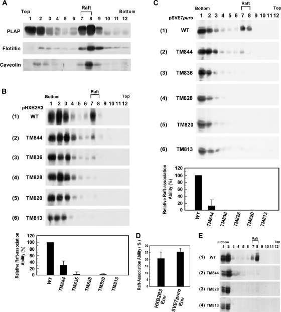
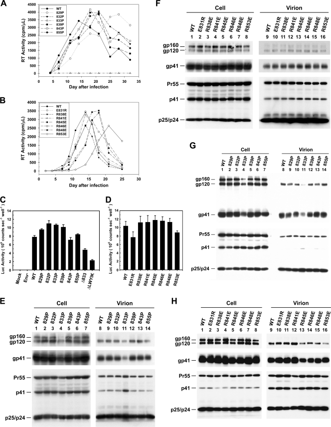
The molecular basis for localization of the human immunodeficiency virus type 1 envelope glycoprotein (Env) in detergent-resistant membranes (DRMs), also called lipid rafts, still remains unclear. The C-terminal cytoplasmic tail of gp41 contains three membrane-interacting, amphipathic alpha-helical sequences, termed lentivirus lytic peptide 2 (LLP-2), LLP-3, and LLP-1, in that order. Here we identify determinants in the cytoplasmic tail which are crucial for Env's association with Triton X-100-resistant rafts. Truncations of LLP-1 greatly reduced Env localization in lipid rafts, and the property of Gag-independent gp41 localization in rafts was conserved among different strains. Analyses of mutants containing single deletions or substitutions in LLP-1 showed that the alpha-helical structure of the LLP-1 hydrophobic face has a more-critical role in Env-raft associations than that of the hydrophilic face. With the exception of a Pro substitution for Val-833, all Pro substitution and charge-inverting mutants showed wild-type virus-like one-cycle viral infectivity, replication kinetics, and Env incorporation into the virus. The intracellular localization and cell surface expression of mutants not localized in lipid rafts, such as the TM844, TM813, 829P, and 843P mutants, were apparently normal compared to those of wild-type Env. Cytoplasmic subdomain targeting analyses revealed that the sequence spanning LLP-3 and LLP-1 could target a cytoplasmic reporter protein to DRMs. Mutations of LLP-1 that affected Env association with lipid rafts also disrupted the DRM-targeting ability of the LLP-3/LLP-1 sequence. Our results clearly demonstrate that LLP motifs located in the C-terminal cytoplasmic tail of gp41 harbor Triton X-100-resistant raft association determinants.


Figures

FIG. 1.
FIG. 1.
(A) Schematic representation of the gp41 cytoplasmic domain and truncation mutants examined in this study. The cytoplasmic tail of gp41 contains a tyrosine-based endocytic YSPL signal located at residue 712, a hydrophilic region, a diaromatic YW motif, and three amphipathic α-helices, termed LLP-2, LLP-3, and LLP-1, at its C terminus. The amino acid sequence from residues 806 to 856 of the WT HXB2 Env is presented in single amino acid code, and the C-terminal dileucine motif is underlined in the sequence. Truncation mutants (TMs) generating stop codons immediately downstream of the indicated amino acids and their respective sequences are also shown. (B) pHXB2R3-based mutant proviruses used in this study. All mutants were generated by a PCR overlap cloning strategy, and the mutation sites are indicated. A dash or dot indicates that the residue in that position of the mutant provirus sequence is identical to or absent from that of the WT provirus sequence, respectively. The substituted amino acids in the mutant proviruses are also indicated.
FIG. 2.
FIG. 2.
Analyses of Env cytoplasmic tail truncation mutants for lipid raft localization. (A) Subconfluent 293T cell monolayers were transfected with pHXBn-PLAP-IRES-N+ or mock transfected by a standard calcium phosphate coprecipitation method. Cells were lysed with 1% Triton X-100 in PBS on ice and subjected to sucrose density gradient ultracentrifugation followed by SDS-PAGE and Western blotting analysis with antibodies against PLAP, flotillin-1, and caveolin-1. (B) 293T cell monolayers were transfected with each of the WT or mutant proviral clones, and cold Triton X-100-extracted cell lysates were subjected to sucrose density gradient ultracentrifugation followed by Western blotting analysis with the Chessie 8 MAb. (Top) The distribution patterns of gp41 from a representative set of data are shown. (Bottom) The percentages of the levels of WT and mutant gp41 proteins fractionated into DRMs relative to the total gp41 levels were calculated by quantifying ECL images. The percentages of association with DRMs of mutant proteins relative to that of the WT protein were quantified from three independent studies, with the means ± standard deviations shown. (C) (Top) 293T cells were cotransfected with pIIIextat together with each of the WT and mutant pSVE7puro plasmids, and the gp41 distribution profiles from a set of representative data are shown. (Bottom) The relative DRM association ability of the mutant proteins was expressed as a percentage of that of WT gp41. The diagram represents the results from three independent experiments (means ± standard deviations). (D) The absolute raft association ability of Env was quantified from three separate raft flotation analyses, and the mean ± standard deviation is indicated. (E) CEM-SS cells were infected with VSV G trans-complemented WT and mutant viruses containing 106 cpm of RT activity. The cold Triton X-100-extracted cell lysates were analyzed by sucrose density gradient centrifugation, and the profiles of gp41 partitioning into the DRMs are shown.
FIG. 3.
FIG. 3.
Analyses of WT and mutant NL4-3 viruses. (A) 293T cells were transfected with WT, 12LE, or 30LE Gag mutant NL4-3 proviruses in the presence of pHCMV-VSV G. Cell-free, VSV G trans-complemented WT and mutant viruses containing equal amounts of RT activity were used to challenge the T-cell lymphoma SupT1 cell line, and cell and virion lysates were subjected to SDS-PAGE followed by Western immunoblotting analyses using 902 and Chessie 8 MAbs (top and middle panels, respectively) and MAb 183 (bottom panel). (B) 293T cells transfected with pHXB2R3 or with WT, 12LE, or 30LE mutant pNL4-3 clones were subjected to a lipid raft flotation assay. The gp41 distribution profile for each transfection is shown. (C) 293T cells were transfected with pHXBn-PLAP-IRES-N+ or mock transfected, lysed with 0.5% Triton X-100 in PBS, and then subjected to Clapham's multilayer step gradient centrifugation. Proteins in each fraction were then analyzed by Western immunoblotting with PLAP, flotillin-1, and caveolin-1 antibodies. (D) 293T cells transfected with the indicated proviral constructs were lysed with Triton X-100 and subjected to multilayer lipid raft flotation analysis. (E) CEM-SS cells were infected with VSV G trans-complemented HXB2 and NL4-3 viruses, and the cold Triton X-100-extracted cell lysates were analyzed by the conventional three-layer lipid raft flotation assay.
FIG. 4.
FIG. 4.
Analyses of lipid raft association ability of Env proteins. (A) 293T cells transfected with WT pHXB2R3 (top), the Tat-independent pCX-Env plasmid (middle), or the Tat-dependent pSVE7puro plasmid (bottom) were processed for multilayer lipid raft flotation analysis. (B) 293T cells expressing each of the Env proteins derived from HIV-1 LTR-directed HXB2, NL4-3, and ConB clones in the absence of Gag coexpression were assessed by a three-layer density gradient lipid raft flotation analysis. For both panels, two independent studies were performed, with similar results obtained. The data from a representative set are thus shown.
FIG. 5.
FIG. 5.
Effects of cholesterol on Env association with lipid rafts. (A) 293T cells transfected with WT pHXB2R3 were treated with β-MCD at the indicated concentrations in serum-free DMEM at 37°C for 30 min prior to being lysed with cold Triton X-100. The resultant cell extracts were then subjected to a lipid raft flotation assay. (B) 293T cells cotransfected with WT pSVE7puro and pIIIextat were incubated in serum-free DMEM (top), with 20 mM β-MCD in serum-free DMEM (middle), or with 20 mM β-MCD followed by replenishment with 100 μg/ml of cholesterol prepared as a cholesterol-β-MCD complex (bottom) prior to cold detergent lysis. Cell lysates were then subjected to sucrose gradient lipid raft flotation analysis.
FIG. 6.
FIG. 6.
Site-directed mutational analysis of LLP-1 for Env localization in lipid rafts. 293T cells transfected with WT or mutant pHXB2R3 proviruses with point deletions (A), Pro substitutions (B), Ala, Ser, or Pro substitutions for Val-832 and Val-833 (C), or charge-switching residues (D), as indicated, were analyzed for gp41 association with lipid rafts. In each case, the relative raft association properties of mutant proteins were expressed as percentages of that of the WT Env, and diagrams representing the results from three independent experiments (means ± standard deviations) are shown.
FIG. 7.
FIG. 7.
Characterization of mutant viruses. (A and B) Viral replication analyses. Cell-free viruses obtained from 293T cells transfected with the WT and Pro-substituted mutants (A) or charge-reversing mutants (B) were normalized by RT activity prior to challenge with CEM-SS cells, and culture supernatants were measured for RT activity at different days postinfection. The studies were performed at least three times, with similar results obtained. Thus, a representative set of data for each group of infection is shown. (C and D) One-cycle viral infectivity assay. LuSIV cells were infected with WT or mutant viruses, and infected cells were lysed and assayed for luciferase activity as indicated in Materials and Methods. The background luciferase activity detected in cells mock infected or challenged with virus produced from env-defective pHXBCATΔBgl, marked as Env, was used as a negative control. The diagrams represent the results of luciferase activities of WT and mutant viruses from three independent experiments (means ± standard deviations). (E and F) Viral protein expression in 293T cells. 293T cells were transfected with WT or mutant proviruses as indicated. Two days after transfection, equal volumes of cell and virion lysates were subjected to SDS-PAGE followed by Western blotting analysis using 902, Chessie 8, and 183 MAbs. (G and H) Viral protein expression in SupT1 cells. VSV G trans-complemented WT and mutant viruses were normalized by their RT activity and then used to challenge SupT1 cells. Two days after infection, cell cultures were harvested, and cell and virion lysates were resolved by Western blotting as indicated in panels E and F.
FIG. 8.
FIG. 8.
Analysis of subcellular localization of mutant viruses. (A) Intracellular localization of Env proteins. HeLa cells grown on coverslips coated with gelatin were transfected with WT or mutant proviruses, as indicated. Two days after transfection, cells were processed for immunostaining, followed by confocal microscopy. (B and C) Analyses of total and cell surface expression of Env proteins. (B) 293T cells were transfected with WT or mutant proviruses, and 2 days after transfection, cells were harvested and examined for total and cell surface expression of Env by flow cytometry. Transfection with pHXBCATΔBgl, marked as Env, was used as a negative control. The study was performed three times, with similar results obtained. Data from a representative set are shown. (C) The specific levels of total and surface Env of the WT and mutant viruses were obtained by subtracting the background level from each value obtained by flow cytometry. The relative total and surface levels of mutants, which were expressed as percentages of those of WT Env, were quantitated from three separate analyses, with the means ± standard deviations shown.
FIG. 9.
FIG. 9.
Analyses of lipid raft-targeting ability of cytoplasmic subdomains. (A) 293T cells were transfected with pcDNA3-β-gal chimeras encoding β-Gal or the indicated gp41 cytoplasmic tail fragments fused to the C terminus of β-Gal. Cold 1% Triton X-100-extracted cell lysates were subjected to lipid raft sucrose density gradient centrifugation followed by Western blotting analysis with a β-Gal MAb. A set of data from a representative experiment is shown in panels 1 to 9, whereas the average values from two separate analyses are shown in the bottom graph. (B) 293T cells were transfected with either β-Gal or WT or mutant β-Gal-786-856 chimeras, as indicated. Cell lysates were subjected to a lipid raft flotation analysis followed by Western blotting using a β-Gal MAb. Results from a representative set are shown in panels 1 to 6. Also, the average values from two independent analyses are shown in the bottom graph.
FIG. 10.
FIG. 10.
Heptagonal representation of the LLP-1 α-helix. (A) The amino acid sequence of LLP-1 (amino acids 828 to 856) was plotted in a seven-point wheel format to represent the helical configuration of selected point deletions. Blue, positively charged residue; red, negatively charged residue; purple, glutamine; green, histidine. The WT sequence maintains an amphipathic helical arrangement with segregated hydrophilic (charged) and hydrophobic residues. In all deletion mutants, all but the Δ854 mutant altered the hydrophilic/hydrophobic orientation of the α-helix. (B) Positions of residues replaced by Pro are mapped in the heptagonal representation of the LLP-1 helix. Red, mutations that disrupted Env-raft associations; green, mutations that did not disrupt Env-raft associations. (C) Positions of Arg/Glu mutations in the LLP-1 α-helix. None of the charge conversion mutations in the hydrophilic face affected the lipid raft localization ability of the Env protein, and they are therefore marked in green.

Similar articles

Cited by

References

    1. Anderson, R. G. 1998. The caveolae membrane system. Annu. Rev. Biochem. 67:199-225. - PubMed
    1. Arreaza, G., and D. A. Brown. 1995. Sorting and intracellular trafficking of a glycosylphosphatidylinositol-anchored protein and two hybrid transmembrane proteins with the same ectodomain in Madin-Darby canine kidney epithelial cells. J. Biol. Chem. 270:23641-23647. - PubMed
    1. Bartz, S. R., M. E. Rogel, and M. Emerman. 1996. Human immunodeficiency virus type 1 cell cycle control: Vpr is cytostatic and mediates G2 accumulation by a mechanism which differs from DNA damage checkpoint control. J. Virol. 70:2324-2331. - PMC - PubMed
    1. Berlioz-Torrent, C., B. L. Shacklett, L. Erdtmann, L. Delamarre, I. Bouchaert, P. Sonigo, M. C. Dokhelar, and R. Benarous. 1999. Interactions of the cytoplasmic domains of human and simian retroviral transmembrane proteins with components of the clathrin adaptor complexes modulate intracellular and cell surface expression of envelope glycoproteins. J. Virol. 73:1350-1361. - PMC - PubMed
    1. Berman, P. W., W. M. Nunes, and O. K. Haffar. 1988. Expression of membrane-associated and secreted variants of gp160 of human immunodeficiency virus type 1 in vitro and in continuous cell lines. J. Virol. 62:3135-3142. - PMC - PubMed

Publication types

Substances

LinkOut - more resources