

The amino terminus of herpes simplex virus type 1 glycoprotein K (gK) modulates gB-mediated virus-induced cell fusion and virion egress
- PMID: 19793812
- PMCID: PMC2786757
- DOI: 10.1128/JVI.01329-09
The amino terminus of herpes simplex virus type 1 glycoprotein K (gK) modulates gB-mediated virus-induced cell fusion and virion egress
Abstract
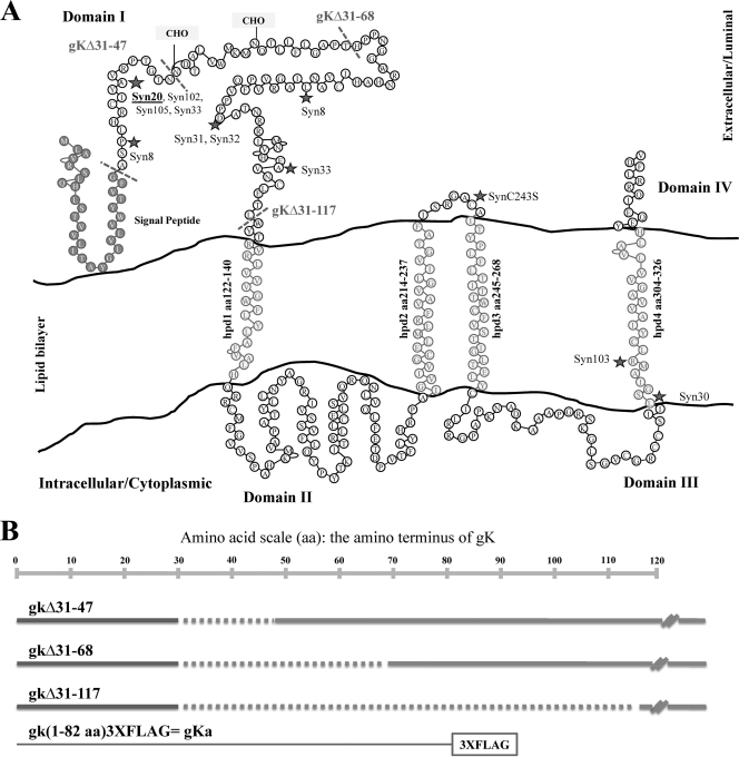
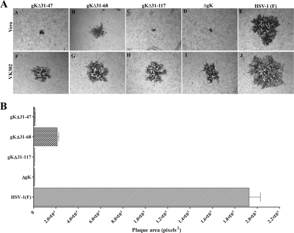
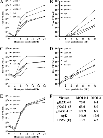
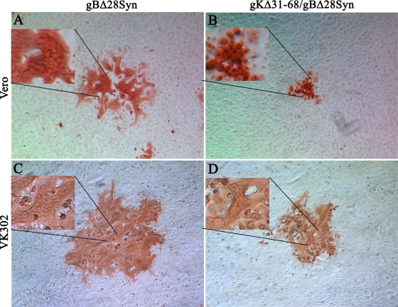
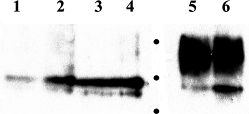
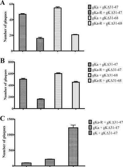
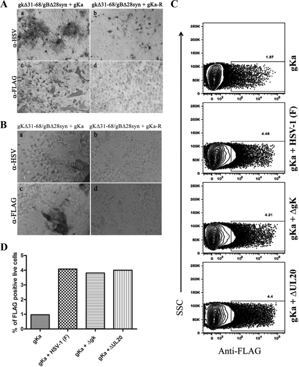
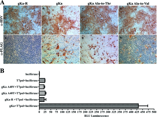
Herpes simplex virus type 1 (HSV-1)-induced cell fusion is mediated by viral glycoproteins and other membrane proteins expressed on infected cell surfaces. Certain mutations in the carboxyl terminus of HSV-1 glycoprotein B (gB) and in the amino terminus of gK cause extensive virus-induced cell fusion. Although gB is known to be a fusogenic glycoprotein, the mechanism by which gK is involved in virus-induced cell fusion remains elusive. To delineate the amino-terminal domains of gK involved in virus-induced cell fusion, the recombinant viruses gKDelta31-47, gKDelta31-68, and gKDelta31-117, expressing gK carrying in-frame deletions spanning the amino terminus of gK immediately after the gK signal sequence (amino acids [aa] 1 to 30), were constructed. Mutant viruses gKDelta31-47 and gKDelta31-117 exhibited a gK-null (DeltagK) phenotype characterized by the formation of very small viral plaques and up to a 2-log reduction in the production of infectious virus in comparison to that for the parental HSV-1(F) wild-type virus. The gKDelta31-68 mutant virus formed substantially larger plaques and produced 1-log-higher titers than the gKDelta31-47 and gKDelta31-117 mutant virions at low multiplicities of infection. Deletion of 28 aa from the carboxyl terminus of gB (gBDelta28syn) caused extensive virus-induced cell fusion. However, the gBDelta28syn mutation was unable to cause virus-induced cell fusion in the presence of the gKDelta31-68 mutation. Transient expression of a peptide composed of the amino-terminal 82 aa of gK (gKa) produced a glycosylated peptide that was efficiently expressed on cell surfaces only after infection with the HSV-1(F), gKDelta31-68, DeltagK, or UL20-null virus. The gKa peptide complemented the gKDelta31-47 and gKDelta31-68 mutant viruses for infectious-virus production and for gKDelta31-68/gBDelta28syn-mediated cell fusion. These data show that the amino terminus of gK modulates gB-mediated virus-induced cell fusion and virion egress.
Figures
Similar articles
-
The amino terminus of herpes simplex virus 1 glycoprotein K is required for virion entry via the paired immunoglobulin-like type-2 receptor alpha.J Virol. 2013 Mar;87(6):3305-13. doi: 10.1128/JVI.02982-12. Epub 2013 Jan 9. J Virol. 2013. PMID: 23302878 Free PMC article.
-
The Amino Terminus of Herpes Simplex Virus 1 Glycoprotein K (gK) Is Required for gB Binding to Akt, Release of Intracellular Calcium, and Fusion of the Viral Envelope with Plasma Membranes.J Virol. 2018 Feb 26;92(6):e01842-17. doi: 10.1128/JVI.01842-17. Print 2018 Mar 15. J Virol. 2018. PMID: 29321326 Free PMC article.
-
The herpes simplex virus type 1 UL20 protein and the amino terminus of glycoprotein K (gK) physically interact with gB.J Virol. 2010 Sep;84(17):8596-606. doi: 10.1128/JVI.00298-10. Epub 2010 Jun 23. J Virol. 2010. PMID: 20573833 Free PMC article.
-
Two Sides to Every Story: Herpes Simplex Type-1 Viral Glycoproteins gB, gD, gH/gL, gK, and Cellular Receptors Function as Key Players in Membrane Fusion.Viruses. 2021 Sep 16;13(9):1849. doi: 10.3390/v13091849. Viruses. 2021. PMID: 34578430 Free PMC article. Review.
-
Glycoprotein K of herpes simplex virus: a transmembrane protein encoded by the UL53 gene which regulates membrane fusion.Virus Genes. 1999;18(1):81-90. doi: 10.1023/a:1008025520655. Virus Genes. 1999. PMID: 10334040 Review.
Cited by
-
Cysteines and N-Glycosylation Sites Conserved among All Alphaherpesviruses Regulate Membrane Fusion in Herpes Simplex Virus 1 Infection.J Virol. 2017 Oct 13;91(21):e00873-17. doi: 10.1128/JVI.00873-17. Print 2017 Nov 1. J Virol. 2017. PMID: 28835497 Free PMC article.
-
Intramuscular Vaccination With the HSV-1(VC2) Live-Attenuated Vaccine Strain Confers Protection Against Viral Ocular Immunopathogenesis Associated With γδT Cell Intracorneal Infiltration.Front Immunol. 2021 Nov 15;12:789454. doi: 10.3389/fimmu.2021.789454. eCollection 2021. Front Immunol. 2021. PMID: 34868077 Free PMC article.
-
Deletion of a Predicted β-Sheet Domain within the Amino Terminus of Herpes Simplex Virus Glycoprotein K Conserved among Alphaherpesviruses Prevents Virus Entry into Neuronal Axons.J Virol. 2015 Dec 9;90(5):2230-9. doi: 10.1128/JVI.02468-15. J Virol. 2015. PMID: 26656706 Free PMC article.
-
Phenylalanine residues at the carboxyl terminus of the herpes simplex virus 1 UL20 membrane protein regulate cytoplasmic virion envelopment and infectious virus production.J Virol. 2014 Jul;88(13):7618-27. doi: 10.1128/JVI.00657-14. Epub 2014 Apr 23. J Virol. 2014. PMID: 24760889 Free PMC article.
-
Novel Oncolytic Herpes Simplex Virus 1 VC2 Promotes Long-Lasting, Systemic Anti-melanoma Tumor Immune Responses and Increased Survival in an Immunocompetent B16F10-Derived Mouse Melanoma Model.J Virol. 2021 Jan 13;95(3):e01359-20. doi: 10.1128/JVI.01359-20. Print 2021 Jan 13. J Virol. 2021. PMID: 33177208 Free PMC article.
References
Publication types
MeSH terms
Substances
Grants and funding
LinkOut - more resources
Full Text Sources
Other Literature Sources
