. 2009 Dec;83(24):12895-906.
doi: 10.1128/JVI.00942-09. Epub 2009 Sep 30.

Crystal structure of a novel conformational state of the flavivirus NS3 protein: implications for polyprotein processing and viral replication

Affiliations

Crystal structure of a novel conformational state of the flavivirus NS3 protein: implications for polyprotein processing and viral replication

René Assenberg et al. J Virol. 2009 Dec.

Abstract

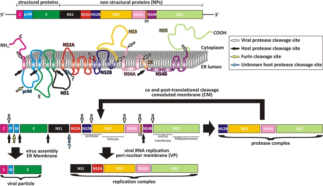
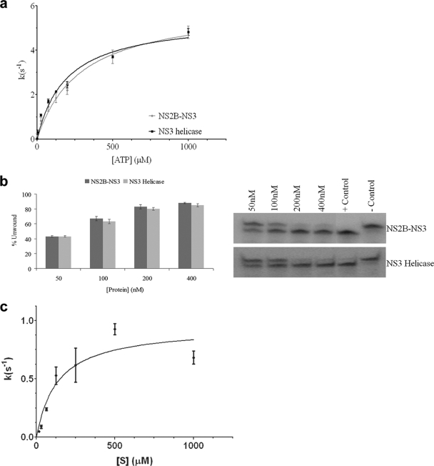
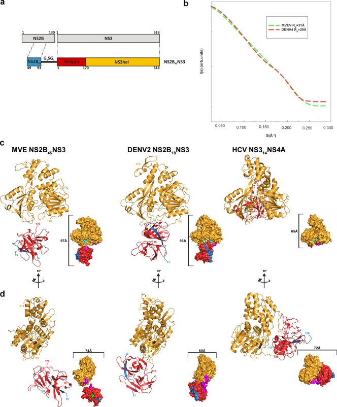
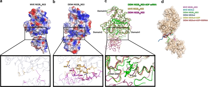
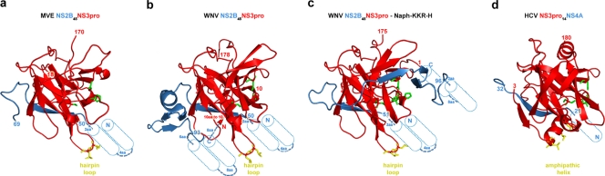
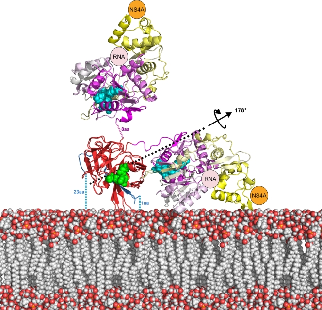
The flavivirus genome comprises a single strand of positive-sense RNA, which is translated into a polyprotein and cleaved by a combination of viral and host proteases to yield functional proteins. One of these, nonstructural protein 3 (NS3), is an enzyme with both serine protease and NTPase/helicase activities. NS3 plays a central role in the flavivirus life cycle: the NS3 N-terminal serine protease together with its essential cofactor NS2B is involved in the processing of the polyprotein, whereas the NS3 C-terminal NTPase/helicase is responsible for ATP-dependent RNA strand separation during replication. An unresolved question remains regarding why NS3 appears to encode two apparently disconnected functionalities within one protein. Here we report the 2.75-A-resolution crystal structure of full-length Murray Valley encephalitis virus NS3 fused with the protease activation peptide of NS2B. The biochemical characterization of this construct suggests that the protease has little influence on the helicase activity and vice versa. This finding is in agreement with the structural data, revealing a single protein with two essentially segregated globular domains. Comparison of the structure with that of dengue virus type 4 NS2B-NS3 reveals a relative orientation of the two domains that is radically different between the two structures. Our analysis suggests that the relative domain-domain orientation in NS3 is highly variable and dictated by a flexible interdomain linker. The possible implications of this conformational flexibility for the function of NS3 are discussed.


Figures

FIG. 1.
FIG. 1.
Schematic diagram of flavivirus polyprotein organization and processing. (Top) Linear organization of the structural and nonstructural proteins within the polyprotein. (Middle) Putative membrane topology of the polyprotein predicted from biochemical and cellular analyses, which is then processed by cellular and viral proteases (indicated by arrows). (Bottom) Different complexes that are thought to arise in different cellular compartments during and following polyprotein processing.
FIG. 2.
FIG. 2.
Catalytic activity studies of MVEV NS2B45NS3. (a) Comparison of the ATPase activities of MVEV NS2B45NS3 (light gray) and MVEV NS3171-618 (dark gray). The ATPase assay was carried out with 5 nM of enzyme in the presence of the indicated concentrations of ATP. The amount of inorganic phosphate released during catalysis was measured with malachite green. (b) Helicase activities of MVEV NS2B45NS3 (dark gray) and MVEV NS3171-618 (light gray). Unwinding activity was measured by using a radiolabeled double-stranded RNA substrate. Control lanes are included (positive control [heat denatured duplex] and negative control [in the absence of enzyme]). Enzyme concentrations are indicated. The values represent average data from three experiments. (c) Assay of the protease activity of NS2B45NS3 was carried out with 5 nM of enzyme in the presence of the indicated concentrations of peptide (see Materials and Methods). The amount of AMC released during proteolysis was detected by excitation at 354 nm and emission at 442 nm using a SpectraFluorPlus reader (Tecan).
FIG. 3.
FIG. 3.
Overall structure of MVEV NS2B45NS. (a) Diagram of the MVEV NS2B and NS3 protein organization and of the NS2B45NS3 synthetic construct used for crystallization. (b) Superimposition of the scattering from an ab initio model of MVEV NS2B45NS3 and DENV4 NS2B18NS3 computed by use of the program CRYSOL (50). As a result of within-experiment errors, the two curves are indistinguishable. Rg is the radius of gyration. (c) Comparison of the structures of MVEV (MVE) NS2B45NS3, DENV4 NS2B18NS3, and HCVNS314NS4A. A cartoon diagram (main) and surface representation (inset) of equivalent views of the three synthetic constructs are shown. Color coding is the same as that in panel a: NS2B (and NS4A) is blue, the NS3pro domain is red, NS3hel is yellow, the NS3pro catalytic pocket is green, the NS3hel catalytic pocket is cyan, and the interdomain linker loop is magenta. (d) Orthogonal view of panel c.
FIG. 4.
FIG. 4.
Structural analysis of protease-helicase interdomain interactions. (a) Electrostatic surface view of MVEV NS2B45NS3 and ribbon diagram of the protease (magenta)-helicase (yellow) interdomain region, with residues at the interacting interface represented as sticks (inset). Residues involved in hydrogen bonds and salt bridges are shown in boldface type, and interatomic distances are indicated. (b) DENV4 NS2B18NS3. (c) Structural superimposition of MVEV NS2B45NS3 (this study) (yellow), DENV4 NS2B18NS3 (PDB accession number 2vbc) (magenta), and DENV4NS3hel+ADP+ssRNA (PDB accession number 2jlz) (green). In DENV4, protease domain residue S68 contacts the catalytically relevant N329 in the helicase domain (which is blocked in a catalytically unfavorable conformation). E66 of the protease domain in particular is likely to block access to the ATP base, as its side chain sits directly in front of the binding pocket and appears to orientate R418 and K201 via long-range electrostatic interactions. In addition, D175 and E177 of the linker region contact R202 of the P loop, and D175 further contacts G80 of the protease domain. Indeed, superimposition of helicase domain 1 of DENV4 NS2B-NS3 and the ADP and ssRNA-bound helicase domain of DENV4 (PDB accession number 1jlv [34]), which represents the product-bound form, shows that domain 2 movement would cause a significant clash with the protease domain. (d) Analysis of the linker orientations in different flavivirus NS3 crystal structures. Shown is a ribbon diagram and transparent surface representation of the structure of MVEV NS2B45NS3. Shown is the superimposition of the interdomain linker region (residues 169 to 181) of MVEV NS2B45NS3 (magenta) (this study) with those of the MVEV NS3hel structure (cyan) (PDB accession number 1v80), DENV4 NS2B18NS3 (green) (PDB accession number 2vbc), and a number of DENV4 NS3hel structures solved in the presence and absence of cofactors (DENV4 NS3hel [blue] [PDB accession number 2jlq], DENV4 NS3hel+ADP [yellow] [PDB accession number 2jls], and DENV4NS3hel+ADP+ssRNA [red] [PDB accession number 2jlz]).
FIG. 5.
FIG. 5.
Structural comparison of cofactor-bound Flaviviridae NS3 protease domains. (a) Cartoon representation of MVEV NS2B45NS3pro (this study). (b) WNV NS2B45NS3pro (PDB accession number 2ggv). (c) WNV NS2B45NS3pro bound to Naph-KKR-H (PDB accession number 3e90). (d) HCV NS3pro18NS4A (PDB accession number 1cu1). Blue, NS2B and NS4A cofactors; red, NS3pro; green, stick representation of the NS3pro catalytic triad (His-Asp-Ser); yellow, hydrophobic residues in its membrane-inserted region. Putative transmembrane helices of the cofactors NS2B and NS4A are shown as cylinders, together with their distance to the nearest visualized residue in the atomic structure.
FIG. 6.
FIG. 6.
Model for the structural organization of MVEV NS2B45NS3 and DENV4 NS2B18NS3 at the cellular membrane. Positioning on the membrane is based upon the tight anchoring of NS2B-NS3 at three points, at the NS2B N- and C-terminal transmembrane helices and at the NS3pro hydrophobic helix, as predicted by the structure of WNV NS2B45NS3pro-Naph-KKR-H and as shown in Fig. 5c. After positioning of WNV NS2B45NS3pro-Naph-KKR-H on the membrane, MVEV NS2B45NS3 and DENV4 NS2B18NS3 were superimposed based on the protease domain alone. A model for the membrane is shown as van der Waals balls and, atomic structures are shown in a cartoon representation and color coded according to the following convention: NS2B (blue), NS3pro (red), and NS3hel by subdomains (domain 1, magenta; domain 2, white; domain 3, yellow). NS4A (shown schematically in orange) was positioned at the NS3 C terminus (domain 3), and the RNA (shown schematically in pink) is positioned in the ssRNA binding groove (shown to be bound in DENV4NS3hel+ADP+ssRNA [PDB accession number 2jlz]). The NS3pro (green) and NS3hel catalytic pockets are shown in a ball representation. The position of hinge axis is shown. aa, amino acids.

References

    1. Adams, P. D., R. W. Grosse-Kunstleve, L. W. Hung, T. R. Ioerger, A. J. McCoy, N. W. Moriarty, R. J. Read, J. C. Sacchettini, N. K. Sauter, and T. C. Terwilliger. 2002. PHENIX: building new software for automated crystallographic structure determination. Acta Crystallogr. D Biol. Crystallogr. 58:1948-1954. - PubMed
    1. Aleshin, A. E., S. A. Shiryaev, A. Y. Strongin, and R. C. Liddington. 2007. Structural evidence for regulation and specificity of flaviviral proteases and evolution of the Flaviviridae fold. Protein Sci. 16:795-806. - PMC - PubMed
    1. Arias, C. F., F. Preugschat, and J. H. Strauss. 1993. Dengue 2 virus NS2B and NS3 form a stable complex that can cleave NS3 within the helicase domain. Virology 193:888-899. - PubMed
    1. Assenberg, R., J. Ren, A. Verma, T. S. Walter, D. Alderton, R. J. Hurrelbrink, S. D. Fuller, S. Bressanelli, R. J. Owens, D. I. Stuart, and J. M. Grimes. 2007. Crystal structure of the Murray Valley encephalitis virus NS5 methyltransferase domain in complex with cap analogues. J. Gen. Virol. 88:2228-2236. - PubMed
    1. Benarroch, D., B. Selisko, G. A. Locatelli, G. Maga, J. L. Romette, and B. Canard. 2004. The RNA helicase, nucleotide 5′-triphosphatase, and RNA 5′-triphosphatase activities of dengue virus protein NS3 are Mg2+-dependent and require a functional Walker B motif in the helicase catalytic core. Virology 328:208-218. - PubMed

Publication types

MeSH terms