. 2009 Dec;83(24):12680-91.
doi: 10.1128/JVI.01476-09. Epub 2009 Sep 30.

Apolipoprotein E but not B is required for the formation of infectious hepatitis C virus particles

Affiliations

Apolipoprotein E but not B is required for the formation of infectious hepatitis C virus particles

Jieyun Jiang et al. J Virol. 2009 Dec.

Erratum in

  • J Virol. 2010 May;84(9):4864

Abstract

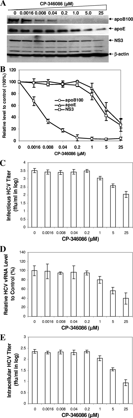
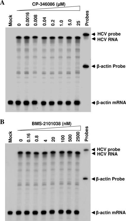
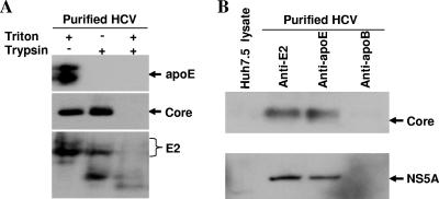
Our previous studies have found that hepatitis C virus (HCV) particles are enriched in apolipoprotein E (apoE) and that apoE is required for HCV infectivity and production. Studies by others, however, suggested that both microsomal transfer protein (MTP) and apoB are important for HCV production. To define the roles of apoB and apoE in the HCV life cycle, we developed a single-cycle HCV growth assay to determine the correlation of HCV assembly with apoB and apoE expression, as well as the influence of MTP inhibitors on the formation of HCV particles. The small interfering RNA (siRNA)-mediated knockdown of apoE expression remarkably suppressed the formation of HCV particles. However, apoE expressed ectopically could restore the defect of HCV production posed by the siRNA-mediated knockdown of endogenous apoE expression. In contrast, apoB-specific antibodies and siRNAs had no significant effect on HCV infectivity and production, respectively, suggesting that apoB does not play a significant role in the HCV life cycle. Additionally, two MTP inhibitors, CP-346086 and BMS-2101038, efficiently blocked secretion of apoB-containing lipoproteins but did not affect HCV production unless apoE expression and secretion were inhibited. At higher concentrations, however, MTP inhibitors blocked apoE expression and secretion and consequently suppressed the formation of HCV particles. Furthermore, apoE was found to be sensitive to trypsin digestion and to interact with NS5A in purified HCV particles and HCV-infected cells, as demonstrated by coimmunoprecipitation. Collectively, these findings demonstrate that apoE but not apoB is required for HCV assembly, probably via a specific interaction with NS5A.


Figures

FIG. 1.
FIG. 1.
Effect of apoB-specific antibodies on HCV infectivity. HCV was incubated with increasing concentrations (0, 0.4, 2.0, 10, and 50 μg/ml) of normal mouse IgG, apoB polyclonal antibodies (rabbit and goat anti-apoB [α-ApoB]) or MAbs (464B1B3, apoB MAb3, and apoB MAb4), and apoE MAb23 at room temperature for 1 h, followed by infection of Huh7.5 cells. At 2 h p.i., the antibody-HCV mixture was removed, and the HCV-infected cells were washed twice with PBS and then incubated with DMEM containing 10% FBS. At 3 days p.i., the level of HCV NS3 protein in the cells was determined by Western blotting (A) and the titer of infectious HCV in the medium was determined by serial dilution and immunofluorescence staining for NS3-positive cell foci (B).
FIG. 2.
FIG. 2.
Knockdown of apoB and apoE expression by specific siRNAs. (A) Determination of apoB and apoE secretion by Western blotting. Huh7.5 cells were infected with HCV and then transfected with apoB, apoE, or NSC siRNA at various concentrations, using Lipofectamine RNAiMax (Invitrogen) following the manufacturer's instructions. At 24 h p.i., the levels of apoB and apoE secreted into the medium were quantified by Western blotting using apoB (Biodesign) and apoE antibodies, respectively. (B) Correlation of apoB-100 secretion and siRNA concentration. The levels of apoB-100 relative to the control (without siRNA transfection) were converted to percentages of the control (100%). The relative levels of apoB-100 (y axis) were plotted against siRNA concentrations (x axis). (C) Correlation of apoE level and siRNA concentration. The relative levels of apoE were plotted (y axis) against siRNA concentrations (x axis). The relative levels of apoB and apoE shown in panels B and C are average values for three independent experiments.
FIG. 3.
FIG. 3.
Effects of siRNA-mediated knockdown of apoB and apoE expression on HCV replication and production. Huh7.5 cells were infected with HCV at an MOI of 5 and then transfected with apoB, apoE, or NSC siRNA as described in the legend to Fig. 2. At 24 h p.i., the medium was collected and cells were lysed in RIPA buffer. (A) Detection of NS3 protein in HCV-infected and siRNA-transfected cells by Western blotting using an NS3-specific MAb. (B and C) Influence of apoB and apoE siRNAs on HCV production. HCV in the medium of HCV-infected and siRNA-transfected cells was used to infect naïve Huh7.5 cells. The levels of NS3 protein (B) and positive-strand HCV RNA (C) were determined by Western blotting and RPA, respectively, as described in Materials and Methods. (D) Quantification of infectious HCV by serial dilution and IFA. HCV in the medium was serially diluted and used to infect naïve Huh7.5 cells on coverslips. The titers of infectious HCV were determined in FFU/ml as described for Fig. 1B. The titers of infectious HCV were plotted against siRNA concentrations. (E) Correlation of HCV vRNA level in the medium with siRNA concentration. HCV vRNA in the medium was extracted with Trizol reagent and quantified by real-time RT-PCR. The HCV vRNA level was calculated as a percentage of the control level (without siRNA). (F) Correlation of intracellular HCV titers with siRNA concentrations. Intracellular HCV particles were prepared from HCV-infected and siRNA-transfected Huh7.5 cells as described in Materials and Methods. Intracellular HCV titers were determined in the same way as for panel D. The titers of intracellular HCV were plotted against siRNA concentrations. The means ± standard deviations derived from three independent experiments were used for panels D to F. White bars, NSC siRNA; gray bars, apoB siRNA; black bars, apoE siRNA.
FIG. 4.
FIG. 4.
Ectopic expression of apoE restored HCV production, which was otherwise blocked by siRNA-mediated knockdown of endogenous apoE expression. Huh7.5 cells in 12-well plates were transfected with 50 nM of apoE or NSC siRNA. At 12 h posttransfection, pCMV6XL5/mApoE or vector DNA was transfected into cells with DMRIE-C (Invitrogen). After 12 h of incubation, Huh7.5 cells were infected with HCV at an MOI of approximately 5. At 24 h p.i., the medium was collected and cells were lysed in RIPA buffer. The medium was used for determinations of apoE levels and infectivity (HCV production). (A) Determination of levels of apoE secretion in the medium and of NS3 protein in the cells by Western blotting. Western blot analysis of apoE and HCV NS3 was performed as described in the legends to Fig. 2A and 3, respectively. (B and C) Restoration of HCV production by ectopic expression of apoE. HCV in the medium of siRNA-transfected or both siRNA- and apoE cDNA-transfected cells was used to infect naïve Huh7.5 cells. At 3 days p.i., the levels of NS3 protein (B) and positive-strand HCV RNA (C) were determined by Western blotting and RPA, respectively. The levels of positive-strand HCV RNA were quantified by phosphorimager analysis and converted into percentages of the control level, considering the level of HCV RNA without siRNA and apoE-expressing DNA as 100%. Numbers at the top of panels A and B and below panel C indicate the concentration of siRNA and the amount of DNA used in experiments.
FIG. 5.
FIG. 5.
Effect of CP-346086 at low concentrations on apoB and apoE secretion and HCV production in multiple-cycle HCV growth assays. Huh7.5 cells were infected with HCV at an MOI of 0.3 and then incubated with DMEM containing CP-346086 at concentrations varying from 0.008 to 1 μM. After 3 days p.i., the medium was collected for determination of apoB and apoE secretion and HCV production. (A) Determination of apoB and apoE secretion by Western blotting. (B) Determination of infectious HCV by infectivity assay. HCV in the medium was used to infect naïve Huh7.5 cells. At 3 days p.i., the level of HCV NS3 protein was determined by Western blotting. (C) Correlation of apoB-100 and apoE secretion with HCV production. The levels of apoB-100, apoE, and NS3 in panels A and B were quantified and converted to percentages of the control levels, considering the levels of apoB-100, apoE, and NS3 in the absence of CP-346086 as 100%. Relative levels of apoB-100, apoE, and NS3 were plotted against concentrations of CP-346086. (D) Determination of infectious HCV titers by IFA. Infectious HCV in the medium of HCV-infected and inhibitor-treated cells was titrated and quantified by IFA as FFU/ml.
FIG. 6.
FIG. 6.
Suppression of apoE secretion and HCV production by CP-346086 at high concentrations in multiple-cycle HCV growth assay. Huh7.5 cells were infected with HCV at an MOI of 0.3 and then treated with CP-346086 at various concentrations from 3.125 to 25 μM. After 3 days p.i., the medium was collected for determination of the levels of apoB-100 and apoE secretion and HCV production. (A) Levels of apoB-100 and apoE secretion. ApoB and apoE in the medium were detected by Western blotting. (B) Determination of infectious HCV by infectivity assay. HCV in the medium was used to infect naïve Huh7.5 cells. At 3 days p.i., NS3 was detected by Western blotting. (C) Correlation of apoB and apoE with HCV production. The data shown in panels A and B were quantified and converted to relative levels of apoB, apoE, and NS3 as percentages of the control values. Values are means ± standard deviations derived from three experiments. (D) Determination of infectious HCV titers by IFA. Infectious HCV titers (log FFU/ml) were plotted against CP-340686 concentrations (μM).
FIG. 7.
FIG. 7.
Effect of CP-346086 on HCV production in single-cycle HCV growth assays. Huh7.5 cells were infected with HCV at an MOI of 5 at 37°C for 2 h and then treated with CP-346086 at concentrations varying from 0.0016 to 25 μM. At 24 h p.i., the medium was collected for determination of the levels of apoB, apoE, and infectious HCV, while cells were harvested for preparation of intracellular HCV particles. (A) Effect of CP-346086 on apoB and apoE secretion and production of infectious HCV. ApoB-100 and apoE in the medium were detected by Western blotting. Infectious HCV in the medium was determined by infectivity assay, as described for Fig. 3 to 6. HCV NS3 protein in the subsequently infected Huh7.5 cells was detected by Western blotting. (B) Correlation of apoB-100 and apoE secretion with level of infectious HCV. The levels of apoB-100, apoE, and NS3 represent the means ± standard deviations derived from three different experiments, as shown in panel A. Relative levels of apoB-100, apoE, and NS3 were calculated as percentages of the control values. (C) Determination of infectious HCV titers by IFA. (D) HCV vRNA level relative to control level in the medium. The extraction and quantification of HCV vRNA in the medium were performed as described in the legend to Fig. 3. (E) Infectious titers of intracellular HCV particles determined by IFA. Intracellular HCV titers were plotted against CP-346086 concentrations (μM).
FIG. 8.
FIG. 8.
Effect of BMS-2101038 on HCV production in single-cycle HCV growth assays. Huh7.5 cells were infected with HCV at an MOI of 5 and then incubated with DMEM containing various concentrations of BMS-2101038. At 24 h p.i., the medium was collected and used to determine the levels of apoB-100 and apoE secretion and HCV production. (A) Levels of apoB and apoE determined by Western blotting. ApoB-100 and apoE in the medium were detected by Western blotting using apoB and apoE MAbs, respectively. HCV NS3 in Huh7.5 cells that were infected with medium derived from HCV-infected and BMS-2101038-treated cells was detected by Western blot analysis. (B) Correlation of apoB-100 and apoE secretion with HCV production. The levels of apoB-100, apoE, and NS3 relative to the control levels (without inhibitor treatment) were converted from the data shown in panel A and plotted against concentrations of BMS-2101038.
FIG. 9.
FIG. 9.
Effects of MTP inhibitors on HCV RNA replication. HCV infection and treatment with MTP inhibitors were the same as those described in the legends to Fig. 7 and 8. At 24 h p.i., total RNAs in the HCV-infected and MTP inhibitor-treated Huh7.5 cells were extracted with Trizol reagent. The levels of positive-strand HCV RNA were determined by RPA, using a radiolabeled RNA probe containing the negative-sense HCV 3′ UTR as described in Materials and Methods. The level of β-actin mRNA was used as a control for normalization of the amounts of total RNA used between samples. The concentrations of MTP inhibitors are indicated at the top. RNA probes and products are highlighted by arrows on the right. Mock, naive Huh7.5 cells without HCV infection.
FIG. 10.
FIG. 10.
Biochemical analysis of apoE in HCV particles. (A) Sensitivity of apoE to trypsin digestion. The preparation and purification of HCV particles were described in Materials and Methods. Purified HCV was treated with 4 μg/ml of trypsin (Sigma) in the absence or presence of 1% Triton X-100 at 37°C for 1 h. Trypsin reactions were terminated by the addition of 1 mM phenylmethylsulfonyl fluoride and a 1/100 volume of protease inhibitor cocktail (Roche). HCV C, E2, and apoE were detected by Western blotting using an ECL substrate (Pierce) and apoE-, C-, and E2-specific MAbs. (B) IP of HCV particles. HCV E2-, apoB-, and apoE-specific MAbs were individually coupled to AminoLink Plus coupling resin (Pierce). The antibody-conjugated resin was then incubated with purified HCV particles at 4°C overnight. Upon elution, proteins of HCV particles were separated by 10% SDS-PAGE, followed by transfer of proteins onto a PVDF membrane. HCV core and NS5A proteins were detected by Western blotting.
FIG. 11.
FIG. 11.
Determination of apoE and NS5A interaction by co-IP. (A) Co-IP of NS5A with apoE in HCV particles. Purified HCV particles were lysed by treatment with M-PER protein extraction reagent (Pierce). The resulting HCV lysate was subjected to co-IP using apoE-, E2-, and apoB-specific MAbs as described in Materials and Methods. (B) Co-IP of NS5A with apoE in HCV-infected cells. A lysate of naïve or HCV-infected Huh7.5 cells was incubated with protein G-conjugated agarose beads (Invitrogen) bound with normal mouse IgG or apoB- or apoE-specific MAb. Upon extensive washing, precipitated proteins were separated by 10% SDS-PAGE and then transferred onto a PVDF membrane. HCV NS5A protein was subsequently detected by Western blotting using an NS5A-specific MAb.

Similar articles

Cited by

References

    1. Alter, M. J. 2006. Epidemiology of viral hepatitis and HIV co-infection. J. Hepatol. 44:S6-S9. - PubMed
    1. Andre, P., F. Komurian-Pradel, S. Deforges, M. Perret, J. L. Berland, M. Sodoyer, S. Pol, C. Brechot, G. Paranhos-Baccala, and V. Lotteau. 2002. Characterization of low- and very-low-density hepatitis C virus RNA-containing particles. J. Virol. 76:6919-6928. - PMC - PubMed
    1. Andre, P., G. Perlemuter, A. Budkowska, C. Brechot, and V. Lotteau. 2005. Hepatitis C virus particles and lipoprotein metabolism. Semin. Liver Dis. 25:93-104. - PubMed
    1. Appel, N., M. Zayas, S. Miller, J. Krijnse-Locker, T. Schaller, P. Friebe, S. Kallis, U. Engel, and R. Bartenschlager. 2008. Essential role of domain III of nonstructural protein 5A for hepatitis C virus infectious particle assembly. PLoS Pathog. 4:e1000035. - PMC - PubMed
    1. Blight, K. J., A. A. Kolykhalov, and C. M. Rice. 2000. Efficient initiation of HCV RNA replication in cell culture. Science 290:1972-1975. - PubMed

Publication types

MeSH terms

LinkOut - more resources