. 2009 Dec;83(23):12290-300.
doi: 10.1128/JVI.01333-09. Epub 2009 Sep 30.

The African swine fever virus virion membrane protein pE248R is required for virus infectivity and an early postentry event

Affiliations

The African swine fever virus virion membrane protein pE248R is required for virus infectivity and an early postentry event

Irene Rodríguez et al. J Virol. 2009 Dec.

Abstract

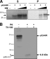
The African swine fever virus (ASFV) protein pE248R, encoded by the gene E248R, is a late structural component of the virus particle. The protein contains intramolecular disulfide bonds and has been previously identified as a substrate of the ASFV-encoded redox system. Its amino acid sequence contains a putative myristoylation site and a hydrophobic transmembrane region near its carboxy terminus. We show here that the protein pE248R is myristoylated during infection and associates with the membrane fraction in infected cells, behaving as an integral membrane protein. Furthermore, the protein localizes at the inner envelope of the virus particles in the cytoplasmic factories. The function of the protein pE248R in ASFV replication was investigated by using a recombinant virus that inducibly expresses the gene E248R. Under repressive conditions, the ASFV polyproteins pp220 and pp62 are normally processed and virus particles with morphology indistinguishable from that of those produced in a wild-type infection or under permissive conditions are generated. Moreover, the mutant virus particles can exit the cell as does the parental virus. However, the infectivity of the pE248R-deficient virions was reduced at least 100-fold. An investigation of the defect of the mutant virus indicated that neither virus binding nor internalization was affected by the absence of the protein pE248R, but a cytopathic effect was not induced and early and late gene expression was impaired, indicating that the protein is required for some early postentry event.


Figures
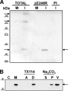
FIG. 1.
FIG. 1.
Myristoylation and membrane association of protein pE248R. (A) Mock-infected (M) and ASFV-infected (I) Vero cells were labeled with [3H]myristic acid for 3 h at 16 hpi, and cell extracts were analyzed by electrophoresis in SDS-12% polyacrylamide gels before (TOTAL) and after immunoprecipitation with anti-pE248R antibody (αE248R) or preimmune serum (PI). Molecular mass markers are indicated on the left (in kDa). The labeled bands are indicated by arrows. (B) Subcellular distribution of protein pE248R in ASFV-infected cells. Infected cells were disrupted at 20 hpi and fractionated into cytosolic (C) and membrane/particulate (M) fractions, as described in Materials and Methods. The membrane fraction was treated with sodium carbonate, and after centrifugation, equivalent amounts of the supernatant (S) and sediment (P), along with 2 μg of purified ASFV particles (V), were analyzed by immunoblotting using the anti-pE248R antibody. Alternatively, the membrane fraction was extracted with Triton X-114 and subjected to temperature-induced phase separation. After centrifugation, the aqueous (A) and detergent-rich (D) phases were analyzed as before. The band corresponding to the protein pE248R is indicated by an arrow. Molecular mass markers are indicated on the left (in kDa).
FIG. 2.
FIG. 2.
Topology of membrane-associated pE248R. (A) The protein pE248R was translated in vitro as indicated in Materials and Methods in the absence (−) or presence of 2 and 4 μl of canine microsomal membranes. Soluble (S) and membrane-associated (P) fractions were analyzed by electrophoresis in SDS-12 to 20% polyacrylamide gels under reducing conditions. (B) After in vitro translation, the membrane fraction was treated (+) or not (−) with proteinase K in the absence (−) or presence (+) of Triton X-100, and the samples were analyzed by SDS-PAGE under reducing conditions. The position of pE248R and that of the protected fragment are indicated by arrows. Molecular mass markers are indicated on the left (in kDa).
FIG. 3.
FIG. 3.
Immunofluorescence microscopy analysis of BA71V-infected Vero cells. Mock-infected (Mock) or BA71V-infected (BA71V) cells were fixed at 16 hpi and double labeled with anti-E248R antibody detected with Alexa 488 goat anti-rabbit IgG (pE248R) and with anti-p54 antibody detected with Alexa 594 goat anti-rat IgG (p54). Cells were counterstained with 4′,6-diamidino-2-phenylindole to visualize cellular and viral DNA. Arrowheads indicate the positions of some viral factories.
FIG. 4.
FIG. 4.
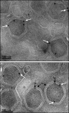
Localization of protein pE248R in virus particles. BA71V-infected cells were fixed at 24 hpi and processed for cryosectioning. Ultrathin sections were incubated with anti-pE248R antibody followed by protein A-gold (10-nm diameter). White arrows indicate labeling on the inner envelope of the virus particles. The capsid (c) and inner envelope (ie) layers are indicated by black arrows in the lower panel. Bars, 100 nm.
FIG. 5.
FIG. 5.
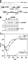
(A) Genomic structure of ASFV recombinant vE248Ri. The recombinant virus vE248Ri was obtained from vGUSREP, a BA71V-derived recombinant virus, which contains the lac repressor-encoding gene lacI inserted in the nonessential thymidine kinase locus (26). In the vE248Ri virus, the promoter of the gene E248R was replaced by an inducible promoter, p72.I*, which is composed of a strong late promoter (p72.4) and the operator sequence O1 from the E. coli lac operon. (B) Inducible expression of protein pE248R. COS-7 cells were either mock infected (M) or infected with parental BA71V (WT) or recombinant vE248Ri virus in the presence (+) or absence (−) of 250 μM IPTG. At 12 hpi, the cells were lysed and analyzed, along with 2 μg of purified ASFV particles (V), by immunoblotting with antibodies against the proteins pE248R and p72 (loading control). The positions of the detected proteins are indicated. Molecular mass markers are indicated on the left (in kDa). (C) One-step growth curves of vE248Ri virus. COS-7 cells were infected with 5 PFU of vE248Ri virus per cell in the presence or absence of 250 μM IPTG. At the indicated times of infection, the total virus titer of each sample was determined by plaque assay on Vero cells in the presence of the inducer. Parental BA71V infections were also titrated as a control.
FIG. 6.
FIG. 6.
Polyprotein processing in cells infected with vE248Ri virus. COS-7 cells were mock infected (M) or infected with parental BA71V (WT) or recombinant vE248Ri virus in the presence (+) or absence (−) of IPTG. At 12 hpi, the cells were lysed and analyzed by immunoblotting with antibodies against the polyprotein pp220 and its mature products p150 and p37 or the polyprotein pp62 and its mature product p35. A sample of 2 μg of highly purified ASFV (V) was also analyzed. The positions of the detected proteins are indicated. Molecular mass markers are indicated on the left (in kDa).
FIG. 7.
FIG. 7.
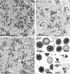
Electron microscopy of vE248Ri-infected cells. COS-7 cells infected with parental BA71V (A) or with vE248Ri virus in the presence (B) or in the absence (C) of IPTG were fixed at 24 hpi and processed for conventional electron microscopy. The overall appearance of the viral factories is similar under all conditions, with the presence of numerous precursor viral membranes (arrowheads), as well as immature (open-head arrows) and mature (filled-head arrows) virions. As shown at a higher magnification, the morphology of the “full” vE248Ri virions generated in the absence of the inducer (F) is indistinguishable from that of the recombinant virus formed under permissive conditions (E) or of parental BA71V virus (D). Panels G and H show the budding and release of vE248Ri virus produced in the presence and absence of IPTG, respectively. Arrows indicate budding and released virus.
FIG. 8.
FIG. 8.
Polypeptide composition of vE248Ri(−) virus. (A) 35S-labeled BA71V and vE248Ri virus grown in the absence of IPTG [vE248Ri(−)] were purified by Percoll gradient centrifugation (18) and analyzed by SDS-PAGE followed by fluorography. The two main labeled bands, corresponding to the proteins p150 and p72, are indicated by arrows. Molecular mass markers (in kDa) are indicated on the left. (B) The presence of the proteins p72, p37, pE248R, and p17 was analyzed by Western blotting in BA71V, vE248Ri(+), and vE248Ri(−) virus. Molecular mass markers (in kDa) are indicated on the left.
FIG. 9.
FIG. 9.
Lack of cytopathic effect following infection with vE248Ri(−) virus produced in the absence of IPTG. COS-7 cells were mock infected (Mock) or infected with parental BA71V (BA71V), vE248Ri(+) virions grown in the presence of IPTG [vE248Ri(+)], or vE248Ri(−) virions grown in the absence of IPTG [vE248Ri(−)]. At 48 hpi, cells were examined by phase-contrast microscopy.
FIG. 10.
FIG. 10.
Binding and internalization of vE248Ri virus. Vero cells were incubated with 5 PFU/cell of BA71V or equivalent amounts of vE248Ri(+) or vE248Ri(−) virus, as indicated. The cells were fixed after 2 h at 4°C (0 hpi) or after shifting to 37°C and further incubating for 4 h (4 hpi). The cells were double labeled with anti-p72 monoclonal antibody detected with Alexa Fluor 488 goat anti-mouse IgG (green) and anti-p37 detected with Alexa Fluor 594 goat anti-rabbit IgG (red). Cells were counterstained with TO-PRO-3 to visualize DNA. The arrows point to the red signal, corresponding to p37.
FIG. 11.
FIG. 11.
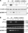
Analysis of early and late gene expression in cells infected with parental BA71V and vE248Ri virus. Vero cells were mock infected (M) or infected with 5 PFU/cell of BA71V (WT) or equivalent amounts of vE248Ri(+) or vE248Ri(−) virus. AraC (40 μg/ml) was added or not as indicated, and the cells were collected at 18 hpi. (A) Cell extracts were analyzed by Western blotting using antibodies against the early protein pE296R or the late protein p72. As a loading control, an anti-β-actin antibody was used. The sizes (in kDa) of molecular mass markers are indicated on the right. (B) Total RNA was extracted, and mRNA for p72, pE296R, and β-actin was analyzed by RT-PCR as indicated in Materials and Methods. Size markers (in nucleotides) are indicated on the right.

Similar articles

Cited by

References

    1. Aguado, B., E. Viñuela, and A. Alcamí. 1991. African swine fever virus fatty acid acylated proteins. Virology 185:942-945. - PubMed
    1. Alcamí, A., A. L. Carrascosa, and E. Viñuela. 1989. Saturable binding sites mediate the entry of African swine fever virus into Vero cells. Virology 168:393-398. - PubMed
    1. Alcamí, A., A. L. Carrascosa, and E. Viñuela. 1989. The entry of African swine fever virus into Vero cells. Virology 171:68-75. - PubMed
    1. Alcamí, A., A. L. Carrascosa, and E. Viñuela. 1990. Interaction of African swine fever virus with macrophages. Virus Res. 17:93-104. - PubMed
    1. Alejo, A., G. Andrés, and M. L. Salas. 2003. African swine fever virus proteinase is essential for core maturation and infectivity. J. Virol. 77:5571-5577. - PMC - PubMed

Publication types

MeSH terms

Substances