. 2009 Dec;83(23):12355-67.
doi: 10.1128/JVI.01593-09. Epub 2009 Sep 30.

Heavily isotype-dependent protective activities of human antibodies against vaccinia virus extracellular virion antigen B5

Affiliations

Heavily isotype-dependent protective activities of human antibodies against vaccinia virus extracellular virion antigen B5

Mohammed Rafii-El-Idrissi Benhnia et al. J Virol. 2009 Dec.

Abstract

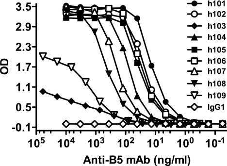
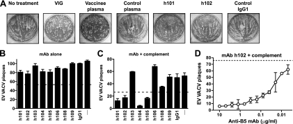
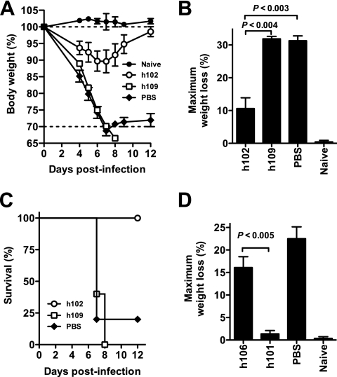
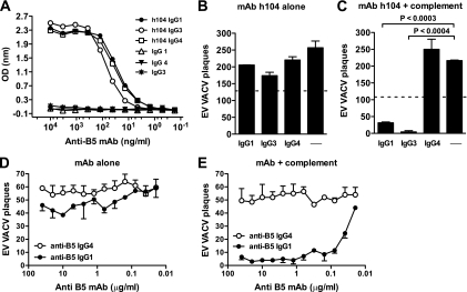
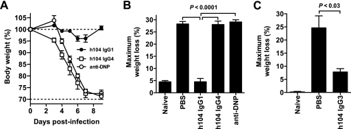
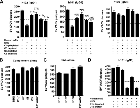
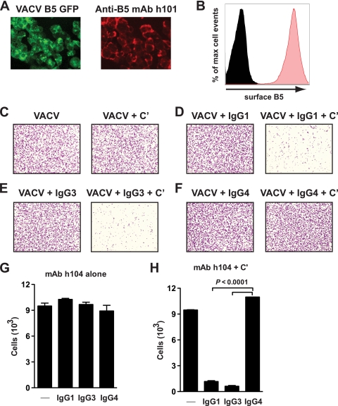
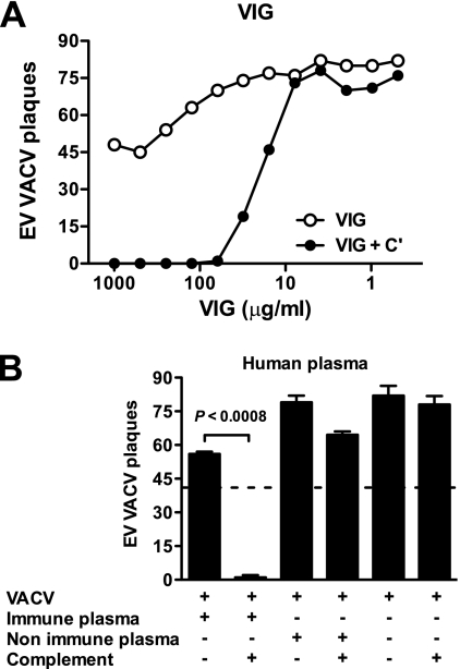
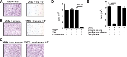
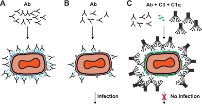
Antibodies against the extracellular virion (EV or EEV) form of vaccinia virus are an important component of protective immunity in animal models and likely contribute to the protection of immunized humans against poxviruses. Using fully human monoclonal antibodies (MAbs), we now have shown that the protective attributes of the human anti-B5 antibody response to the smallpox vaccine (vaccinia virus) are heavily dependent on effector functions. By switching Fc domains of a single MAb, we have definitively shown that neutralization in vitro--and protection in vivo in a mouse model--by the human anti-B5 immunoglobulin G MAbs is isotype dependent, thereby demonstrating that efficient protection by these antibodies is not simply dependent on binding an appropriate vaccinia virion antigen with high affinity but in fact requires antibody effector function. The complement components C3 and C1q, but not C5, were required for neutralization. We also have demonstrated that human MAbs against B5 can potently direct complement-dependent cytotoxicity of vaccinia virus-infected cells. Each of these results was then extended to the polyclonal human antibody response to the smallpox vaccine. A model is proposed to explain the mechanism of EV neutralization. Altogether these findings enhance our understanding of the central protective activities of smallpox vaccine-elicited antibodies in immunized humans.


Figures

FIG. 1.
FIG. 1.
Binding affinities of human anti-B5 MAbs. Titration of anti-B5 MAbs was done using rB5 ELISA. Data are representative of three independent experiments. OD, optical density.
FIG. 2.
FIG. 2.
Complement-dependent human MAb neutralization of VACV EV. (A) Human anti-B5 MAbs, VIG, and immune human plasma exhibited comet tail inhibition activity in vitro. Vero E6 cells were infected with VACVIHDJ and then cultured in the absence of antibody (no treatment), with human VIG (100 μg/ml), with human plasma samples from a smallpox vaccine recipient or unvaccinated people (1/100 dilution), with anti-B5 MAbs (20 μg/ml; h101 or h102), or with irrelevant MAb (20 μg/ml; IgG1). (B to D) VACV EV virion neutralization by human anti-B5 MAbs is dependent on complement. VACV EV neutralization activity of the full panel of monoclonal antibodies against B5 at 10 μg/ml in the absence (B) or the presence (C) of 10% rabbit complement. VACV EV alone (—) and an irrelevant human IgG1 MAb plus EV (IgG1) were negative controls. The dashed line indicates 50% neutralization based on VACV EV alone (B) or in the presence of complement without antibody (C). (D) Titrated VACV EV neutralization activity of human anti-B5 MAb h102 (IgG1) in the presence of 10% of rabbit complement. The dashed line indicates the plaque number of VACV EV in the presence of complement without antibody. All data are representative of three or more experiments.
FIG. 3.
FIG. 3.
Protection against a lethal VACVWR infection. High-affinity human anti-B5 MAb (h102) exhibited protection in vivo. (A to C) BALB/c mice were inoculated i.p. with 100 μg of anti-B5 MAb (h102 or h109) or an equivalent volume of PBS. One day later, mice were challenged intranasally with 5 × 10⁴ PFU of purified VACVWR. Naive mice were unchallenged (no infection; “Naive”). (A) Body weight was tracked daily. Dotted lines indicate starting body weight (upper line) or terminal body weight (bottom). (B) Weight loss nadirs for each group are quantitated. (C) Survival after intranasal challenge. H102 MAb-treated mice were protected from death compared to results for mock-treated mice (P < 0.02, MAb h102 versus PBS) and compared to results with low-affinity MAb h109 (P < 0.003). Group average ± SEM from one of three independent experiments is shown. (D) BALB/c mice were inoculated i.p. with a high-affinity anti-B5 MAb (h101 or h106) or PBS at day −1 and challenged intranasally with 5 × 10⁴ PFU of purified VACVWR at day 0. Maximum weight loss (%) is shown. H101-treated mice exhibited no significant weight loss compared to results for uninfected mice (P ≫ 0.05) and were significantly better protected than mice treated with h106 or given PBS treatment (P < 0.005 or P < 0.002, respectively; n = 5/group).
FIG. 4.
FIG. 4.
Isotype and complement-dependent human antibody neutralization of VACV EV. (A) Titration of anti-B5 MAb h104 IgG1, IgG3, or IgG4 isotype by rB5 ELISA. (B to C) VACV EV neutralization activity with or without different MAb h104 isotypes at 10 μg/ml in the absence (B) or the presence (C) of 10% of rabbit complement. VACV EV alone (B) or plus complement (C) (—) was a negative control. The dashed line indicates 50% neutralization based on VACV EV alone (B) or in the presence of complement without antibody (C). (D and E) Titrated VACV EV neutralization activity of human anti-B5 MAbs in the absence (D) or the presence (E) of 10% of rabbit complement. All data are representative of three or more experiments.
FIG. 5.
FIG. 5.
Anti-B5 protection in vivo is antibody isotype dependent. Anti-B5 MAbs h104 IgG1 and h104 IgG3 were protective in vivo, while IgG4 was not protective. (A and B) BALB/c mice were inoculated i.p. with 100 μg of anti-B5 MAb h104 IgG1, h104 IgG4, a negative control human MAb (anti-DNP IgG1) or PBS at day −1 and challenged intranasally with 5 × 10⁴ PFU of purified VACVWR at day 0. Naive mice were unchallenged (no infection; “Naive”). Weight loss kinetics for each group (A) and maximum weight loss (weight nadir) (B) are shown. Dotted lines indicated the starting body weight (upper line) or terminal body weight (bottom line). H104 IgG1 isotype-treated mice exhibited no significant weight loss compared to results for uninfected mice (P ≫ 0.05) and were significantly better protected than mice treated with the h104 IgG4 isotype, human anti-DNP, or PBS (P < 0.0001; n = 6/group). The group average ± SEM from one of two independent experiments is shown. (C) In an independent experiment, BALB/c mice were inoculated i.p. with 100 μg of the anti-B5 MAb h104 IgG3 isotype or PBS at day −1 and challenged intranasally with 5 × 10⁴ PFU of purified VACVWR at day 0. Maximum weight loss (weight nadir) is shown. H104 IgG3 isotype-treated mice were significantly better protected than mice treated with PBS (P < 0.03; n = 6/group). The group average ± SEM from one of three independent experiments is shown.
FIG. 6.
FIG. 6.
. Complement activation pathways. (A to C) VACV EV neutralization by the anti-B5 MAb at 10 μg/ml (h101 IgG1, h102 IgG1, or h106 IgG4 isotype) in the presence of human complement (NHS) or C1q-, C3-, fB- or C5-depleted human sera (10%). The dashed line indicates 50% neutralization based on that for VACV EV alone without antibody and complement. Significant decreases in anti-B5 MAb (h101 or h102 IgG1 isotype) VACV EV neutralization were observed when the complement component C1q, C3, C5, or fB was depleted. Percent EV neutralization activity loss in the absence of C1q or C3 was higher than that in the absence of fB or C5. (B) EV neutralization by human complement (NHS) or C1q-, C3-, fB-, or C5-depleted sera alone. (C) Anti-B5 MAb h101 IgG1, h102 IgG1, or h106 IgG4 neutralization in the absence of complement. (D) EV neutralization by anti-B5 MAb (h101 IgG1) at 10 μg/ml in the presence of human complement (NHS), C1q-depleted sera, or C1q-purified protein. The addition of the C1q protein (10 μg/ml) to C1q-depleted sera restored EV neutralization activity. Data are representative of three experiments. Error bars indicate the SEM in each group.
FIG. 7.
FIG. 7.
Human IgG1 or IgG3 anti-B5 antibodies are able to direct complement lysis of VACV-infected cells. (A) Vero E6 cell monolayers were infected with VACVWR B5-GFP (green, multiplicity of infection = 8), and surface expression of B5 (red) was determined 12 h postinfection by surface staining with anti-B5 MAb h101 and immunofluorescence microscopy. (B) Surface expression of B5 was tested (red filled curve) after infection with VACVWR by surface staining of infected cells with anti-B5 MAb and performance of flow cytometry. Uninfected cells, negative control (black filled curve). (C to H) Anti-B5-directed complement lysis of infected cells. Virus-infected cells stained with crystal violet are shown at magnification ×40. VACV-infected cells were treated with media (C) (left) or 20% rabbit complement (+ C′) (C to F) (right) in the absence (C) or presence (D to F) of anti-B5 MAb h104 isotypes. (G and H) Quantitation of live cell numbers (cells/image field) from experiment shown in panels C to F. Destruction of VACV-infected cells was statistically significant in the presence of complement plus anti-B5 IgG1 or IgG3 isotype versus results for anti-B5 IgG1 or IgG3 alone (P < 0.0001), complement alone (P < 0.001), or complement plus anti-B5 IgG4 (P < 0.0001). No killing was observed for anti-B5 IgG4 in the absence or presence of complement (P ≫ 0.05). Error bars indicate the SEM in each group. One of two independent experiments is shown.
FIG. 8.
FIG. 8.
EV neutralization by antibodies from human vaccinees is complement dependent. VACV EV neutralization activity of VIG (A) or human plasma (B) in the presence or absence of complement. (A) Titrated VACV EV neutralization activity of VIG in the absence (VIG) or the presence of 10% of rabbit complement (VIG + C′). VIG possessed strong EV-neutralizing activity in the presence of complement. (B) Plasma samples used are from a single human vaccinee (“immune”) or nonimmune human plasma. The dashed line indicates 50% neutralization based on results for VACV EV alone without plasma samples and complement. EV neutralization was highly statistically significant in the presence of immune human plasma plus complement (P < 0.0008). Error bars indicate SEM in each group. One of two (A) or one of three (B) independent experiments is shown.
FIG. 9.
FIG. 9.
VIG or plasma from individual human vaccinees directs complement lysis of VACV-infected cells. (A to C) Virus-infected Vero E6 cells stained with crystal violet at magnification ×40. VACV-infected cells were treated with VIG, plasma from a single human vaccinee (immune), or nonimmune human plasma alone (A to C, left) or with complement (+ C′) (A to C, right). (D and E) Quantitation of live cell numbers (cells/image field) after cell lysis in the presence of VIG (D) or human plasma (E). Destruction of VACV-infected cells was highly statistically significant in the presence of VIG plus complement (P < 0.02) or immune human plasma plus complement (P < 0.0006). No killing was observed for nonimmune human plasma in the absence or presence of complement (P ≫ 0.05; not significant). Error bars indicate the SEM for each group. One of two independent experiments is shown.
FIG. 10.
FIG. 10.
Model of VACV EV neutralization. Schematic diagrams of potential virion neutralization pathways. B5 is drawn in blue. Another representative EV surface antigen is drawn in gray. Ab, anti-B5 antibody. (A) Basic occupancy model. Antibodies against B5 could completely coat the virion surface and thereby neutralize the virus. This model failed. (B) Model 2. VACV EV escape neutralization by anti-B5 antibody binding due to low density of the B5 protein on the surface of EV. Direct occupancy of B5 with anti-B5 IgG is insufficient to block infection of target cells. (C) Complement-assisted coating of VACV EV (opsonization). Antibody-mediated protection against VACV EV is dependent on antibody recruitment of complement C1q and covalent attachment of C3 to the surface of the virus. See Discussion for details.

Comment in

Similar articles

Cited by

References

    1. Aldaz-Carroll, L., J. C. Whitbeck, M. Ponce de Leon, H. Lou, L. Hirao, S. N. Isaacs, B. Moss, R. J. Eisenberg, and G. H. Cohen. 2005. Epitope-mapping studies define two major neutralization sites on the vaccinia virus extracellular enveloped virus glycoprotein B5R. J. Virol. 79:6260-6271. - PMC - PubMed
    1. Aldaz-Carroll, L., Y. Xiao, J. C. Whitbeck, M. P. de Leon, H. Lou, M. Kim, J. Yu, E. L. Reinherz, S. N. Isaacs, R. J. Eisenberg, and G. H. Cohen. 2007. Major neutralizing sites on vaccinia virus glycoprotein B5 are exposed differently on variola virus ortholog B6. J. Virol. 81:8131-8139. - PMC - PubMed
    1. Amanna, I. J., M. K. Slifka, and S. Crotty. 2006. Immunity and immunological memory following smallpox vaccination. Immunol. Rev. 211:320-337. - PubMed
    1. Angal, S., D. J. King, M. W. Bodmer, A. Turner, A. D. Lawson, G. Roberts, B. Pedley, and J. R. Adair. 1993. A single amino acid substitution abolishes the heterogeneity of chimeric mouse/human (IgG4) antibody. Mol. Immunol. 30:105-108. - PubMed
    1. Bell, E., M. Shamim, J. C. Whitbeck, G. Sfyroera, J. D. Lambris, and S. N. Isaacs. 2004. Antibodies against the extracellular enveloped virus B5R protein are mainly responsible for the EEV neutralizing capacity of vaccinia immune globulin. Virology 325:425-431. - PubMed

Publication types