. 2009 Nov;136(21):3567-74.
doi: 10.1242/dev.039214. Epub 2009 Sep 30.

Pancreatic neurogenin 3-expressing cells are unipotent islet precursors

Affiliations

Pancreatic neurogenin 3-expressing cells are unipotent islet precursors

Renaud Desgraz et al. Development. 2009 Nov.

Abstract

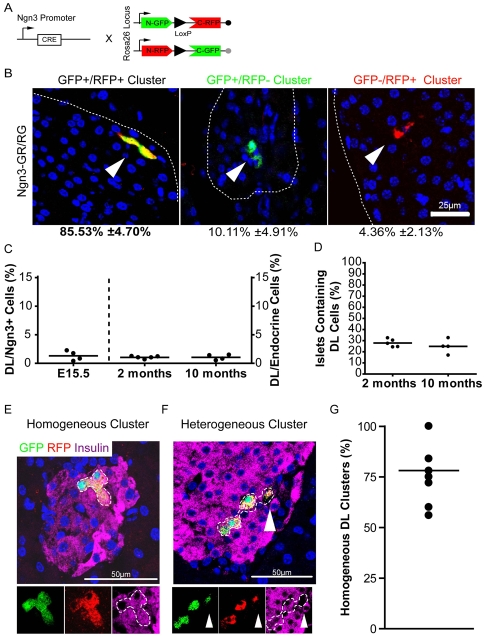
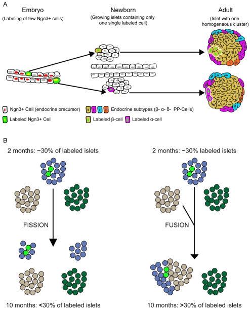
Pancreatic islet endocrine cells arise during development from precursors expressing neurogenin 3 (Ngn3). As a population, Ngn3(+) cells produce all islet cell types, but the potential of individual Ngn3(+) cells, an issue central to organogenesis in general and to in vitro differentiation towards cell-based therapies, has not been addressed. We performed in vivo clonal analyses in mice to study the proliferation and differentiation of very large numbers of single Ngn3(+) cells using MADM, a genetic system in which a Cre-dependent chromosomal translocation labels, at extremely low mosaic efficiency, a small number of Ngn3(+) cells. We scored large numbers of progeny arising from single Ngn3(+) cells. In newborns, labeled islets frequently contained just a single tagged endocrine cell, indicating for the first time that each Ngn3(+) cell is the precursor of a single endocrine cell. In adults, small clusters of two to three Ngn3(+) progeny were detected, but all expressed the same hormone, indicating a low rate of replication from birth to adult stages. We propose a model whereby Ngn3(+) cells are monotypic (i.e. unipotent) precursors, and use this paradigm to refocus ideas on how cell number and type must be regulated in building complete islets of Langerhans.


Figures

Fig. 1.
Fig. 1.
In vivo clonal analysis to determine the differentiation potency of Ngn3+ cells. (A) Ngn3+ cells can be multipotent or unipotent. (B) Clonal analysis of Ngn3+ cells. A small fraction of Ngn3+ cells are genetically labeled during pancreatic development, such that the clonal progeny of individual labeled cells can be followed from embryogenesis to adulthood. If the labeled endocrine precursor is multipotent, its labeled clonal progeny in the adult islet should express different hormone genes. Conversely, if the labeled endocrine precursor is unipotent, then its descendants will only produce one hormone.
Fig. 2.
Fig. 2.
Ngn3+ tagged cells principally form homogeneous clusters of doubly labeled (DL) endocrine cells. (A) The Ngn3-Cre and Rosa26GR/RG transgenes used to generate Ngn3-Cre; Rosa26GR/RG (Ngn3-GR/RG) mice by backcrossing. (B) Confocal micrographs of 2-month-old Ngn3-GR/RG islets stained for EGFP (green) and RFP (red), showing the proportions of DL (EGFP+ RFP+) cells and of cells positive for EGFP or RFP (n=5 mice; 426 clusters of labeled cells scored). Islets are outlined. (C) The proportion of DL cells relative to Ngn3+ cells (shown on the left scale; E15.5 embryos, n=4, 600 cells scored per embryo) or to total endocrine cells (right scale; adults, n=4-5 mice, 60,000 and 150,000 cells scored at 2 and 10 months, respectively). About 1% of cells are labeled; this proportion remains constant at any stage analyzed (ANOVA, P>0.05). Note that Ngn3 expression has ceased when cells become DL (see Fig. S2B in the supplementary material). (D) Proportion of islets containing DL cells in 2- and 10-month-old mice (800-1200 islets scored per group). (E,F) Confocal micrographs of islets from Ngn3-GR/RG mice containing clusters of cells stained for EGFP (green), RFP (red) and insulin (purple). Clusters of clonal DL cells are outlined. The cluster in E is formed by three β-cells and is therefore homogeneous, whereas that in F is heterogeneous because it is formed by three β-cells and one non-β unidentified endocrine cell (arrowhead). (G) The proportion of homogenous clusters of DL cells in 2-month-old adult islets (each dot represents one individual; n=6 mice, 465 islets scored).
Fig. 3.
Fig. 3.
DL islet cells are found as isolated single cells at birth. (A) Expected recombination events predicted by the Poisson distribution (left) and recombination events per islet observed at birth (right). White, gray and black areas represent the proportion of islets containing one, two or three DL cells per islet (n=4, 150 DL cells), respectively. (B) Proportion of homogenous clusters of DL cells, calculated per islet cell type with the knowledge that 22% of islets contain more than one cluster of DL cells. Expected and observed proportions of homogeneous clusters were compared using Fisher's exact test. No statistical difference was found. This indicates that the observed frequencies of homogeneous clusters correspond to those expected if labeled Ngn3+ cells were unipotent. n=4 mice per group, 100-200 islets per group; NS, not significant. (C) Mean cross-section diameter of homogeneous and heterogeneous clusters. Heterogeneous clusters are more dispersed (15 clusters per group, from four mice, *P<0.05). (D) Size (number of cells) of homogeneous and heterogeneous clusters at 2 and 10 months. Note the increased size of homogeneous clusters between 2 and 10 months of age. At 10 months, heterogeneous clusters are twice the size of homogeneous clusters (n=4-5 mice, greater than 50 clusters per group, ***P<0.001). (E) Number of DL cells per cluster in adult islets. The prevailing size of DL cell clusters changes with age in 2- and 10-month-old transgenic mice. Diamonds indicate the median cluster size; n=4-5 mice per age group; 220 and 340 clusters of DL cells were scored in 800 and 1200 islets, respectively. (F) DL cluster size and islet size are not correlated. The average number of cells per cluster is the same in small and large islets (coefficient of determination: R2=0.036; 223 labeled islets scored of all sizes, from five mice). (G-I) Paraffin sections of P0, 2- and 10-month-old islets from Ngn3-GR/RG mice stained for EGFP (DAB) and counterstained with Hemalun. At birth (G), one isolated DL cell is shown (arrowhead), whereas one cluster of two adjacent DL cells is seen at 2 months (H), and another of four DL cells at 10 months (I). Scale bars: 25 μm in G; 50 μm in H,I.
Fig. 4.
Fig. 4.
Islets are larger in aged mice. (A) The number of islets per pancreas is constant throughout life (n=3 mice per group, with 250-550 islets sampled per group; see Materials and methods). (B) Average islet volume increases with age (n=4-5 mice per group, with 800-1200 islets scored per group; *P<0.05). (C) Frequency distribution of islet size from 2- and 10-month-old mice (n=5 mice per group; 60,000-150,000 islet cells, with 800-1200 islets scored per group). (D) Average number of cells per homogeneous cluster of DL cells relative to cell composition and age. Homogenous clusters composed of β-cells (white bars) are larger than clusters of non-β-cells (gray bars) at 2 and 10 months [44-132 clusters scored (bar chart, **P<0.01); n=4-5 mice per group (red dots, P=0.057)]. The size of the β-cell clusters increases with age.
Fig. 5.
Fig. 5.
An overview of islet development and homeostasis. (A) One Ngn3+ cell becomes one endocrine cell at birth. Soon after Ngn3 expression, labeled endocrine precursor cells differentiate into hormone-expressing cells, without division. Individual, labeled, hormone-expressing cells present at birth within early islets proliferate during postnatal life. β-cells proliferate more than non-β-cells, resulting in the formation of larger homogeneous clusters of labeled β-cells. (B) Normal islet homeostasis does not involve islet fission or fusion. In the absence of islet cell neogenesis (i.e. the proportion of labeled cells is constant), islet fission would result in a decreased proportion of labeled islets over time (from one-third to one-quarter in the figure, left panel). Conversely, islet fusion would result in an increased proportion of labeled islets with age (from one-third to a half, right panel).

Similar articles

Cited by

References

    1. Bonal, C. and Herrera, P. L. (2008). Genes controlling pancreas ontogeny. Int. J. Dev. Biol. 52, 823-835. - PubMed
    1. Bonal, C., Thorel, F., Ait-Lounis, A., Reith, W., Trumpp, A. and Herrera, P. L. (2009). Pancreatic inactivation of c-Myc decreases acinar mass and transdifferentiates acinar cells into adipocytes in mice. Gastroenterology 136, 309-319. - PubMed
    1. Brennand, K., Huangfu, D. and Melton, D. (2007). All beta cells contribute equally to islet growth and maintenance. PLoS Biol. 5, e163. - PMC - PubMed
    1. D'Amour, K. A., Bang, A. G., Eliazer, S., Kelly, O. G., Agulnick, A. D., Smart, N. G., Moorman, M. A., Kroon, E., Carpenter, M. K. and Baetge, E. E. (2006). Production of pancreatic hormone-expressing endocrine cells from human embryonic stem cells. Nat. Biotechnol. 24, 1392-1401. - PubMed
    1. Delacour, A., Nepote, V., Trumpp, A. and Herrera, P. L. (2004). Nestin expression in pancreatic exocrine cell lineages. Mech. Dev. 121, 3-14. - PubMed

Publication types

MeSH terms

Substances