. 2009 Nov;136(21):3575-84.
doi: 10.1242/dev.038133. Epub 2009 Sep 30.

BMP signaling regulates sympathetic nervous system development through Smad4-dependent and -independent pathways

Affiliations

BMP signaling regulates sympathetic nervous system development through Smad4-dependent and -independent pathways

Yuka Morikawa et al. Development. 2009 Nov.

Abstract

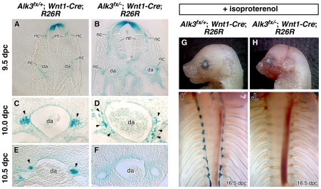
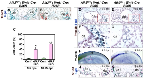
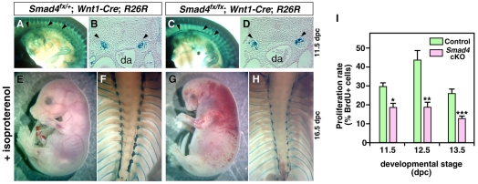
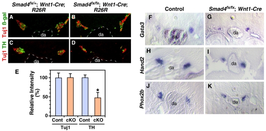
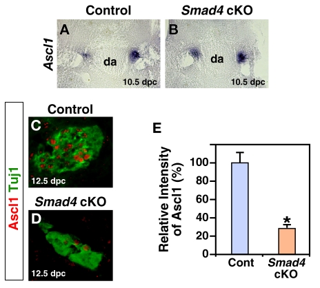
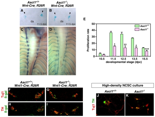
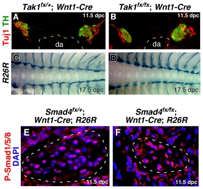
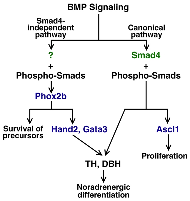
Induction of the sympathetic nervous system (SNS) from its neural crest (NC) precursors is dependent on BMP signaling from the dorsal aorta. To determine the roles of BMP signaling and the pathways involved in SNS development, we conditionally knocked out components of the BMP pathways. To determine if BMP signaling is a cell-autonomous requirement of SNS development, the Alk3 (BMP receptor IA) was deleted in the NC lineage. The loss of Alk3 does not prevent NC cell migration, but the cells die immediately after reaching the dorsal aorta. The paired homeodomain factor Phox2b, known to be essential for survival of SNS precursors, is downregulated, suggesting that Phox2b is a target of BMP signaling. To determine if Alk3 signals through the canonical BMP pathway, Smad4 was deleted in the NC lineage. Loss of Smad4 does not affect neurogenesis and ganglia formation; however, proliferation and noradrenergic differentiation are reduced. Analysis of transcription factors regulating SNS development shows that the basic helix-loop-helix factor Ascl1 is downregulated by loss of Smad4 and that Ascl1 regulates SNS proliferation but not noradrenergic differentiation. To determine if the BMP-activated Tak1 (Map3k7) pathway plays a role in SNS development, Tak1 was deleted in the NC lineage. We show that Tak1 is not involved in SNS development. Taken together, our results suggest multiple roles for BMP signaling during SNS development. The Smad4-independent pathway acts through the activation of Phox2b to regulate survival of SNS precursors, whereas the Smad4-dependent pathway controls noradrenergic differentiation and regulates proliferation by maintaining Ascl1 expression.


Figures

Fig. 1.
Fig. 1.
BMP signaling is required for formation of sympathetic ganglia. Alk3 was deleted in the NC lineage using Wnt1-Cre, and NC cells were simultaneously traced by genetically marking by β-galactosidase expression from the R26R knock-in allele. (A-F) β-Galactosidase expression in Alk3fx/+; Wnt1-Cre; R26R/+ which are phenotypically wild-type (A,C,E), and Alk3fx/-; Wnt1-Cre; R26R/+ cKO (B,D,F) embryos at 9.5 dpc (A,B), 10.0 dpc (C,D), and 10.5 dpc (E,F). The loss of Alk3 in the NC lineage (arrowheads) shows that the cells reach the dorsal aorta (B,D) but fail to form sympathetic ganglia in Alk3fx/-; Wnt1-Cre; R26R/+ cKO embryos (F). (G-J) The loss of Alk3 in the NC lineage results in craniofacial defects and the absence of the SNS. The early lethality caused by the lack of embryonic synthesis of norepinephrine was overcome by administering dams with isoproterenol. At 16.5 dpc, relative to control littermates (G), Alk3 cKOs (H,J) exhibit severe craniofacial defects (H) and complete loss of the SNS (J). In panels I and J, organs were removed before photographing from the ventral side. da, dorsal aorta; nc, neural crest; nt, neural tube.
Fig. 2.
Fig. 2.
BMP signaling is required for the survival of sympathetic precursors and for the expression of Phox2b. To determine if sympathetic precursor cells were dying by apoptosis, 9.5 dpc embryos were analyzed by TUNEL. (A-C) Immunohistochemical analysis of apoptosis in Alk3fx/+; Wnt1-Cre; R26R/+ (A) and Alk3fx/-; Wnt1-Cre; R26R/+ cKO (B) embryos at 9.5 dpc. TUNEL-positive cells are highlighted with red arrowheads. The number of TUNEL-positive sympathetic precursor cells were quantified at 9.5 and 10.25 dpc (C). *, P≤0.022; **, P≤0.000014. (D-G) The expression of Phox2b in β-galactosidase-stained sympathetic precursor cells was analyzed in 10.0 dpc embryos by immunohistochemistry using a Phox2b-specific antibody. F and G show higher magnification of the regions enclosed by the red box in panels D and E. In control sections (D,F), Phox2b is expressed in cells of the developing SNS, whereas expression of Phox2b protein could not be detected in Alk3 cKO embryos (E,G). (H-K) Onset of Phox2b expression is not dependent on BMP signaling. The expression of Phox2b was examined by whole mount in situ hybridization in control (H) and Alk3fx/-; Wnt1-Cre (I) embryos at 9.5 dpc. Phox2b is expressed in the SNS forming at the rostral region of control embryos but is not expressed in mutant embryos. Immunohistochemical analysis of the NC marker Sox10 was used to identify cells forming the SNS of control (J) and Alk3fx/-; Wnt1-Cre; R26R/+ cKO (K) embryos at 9.5 dpc. Sox10 is expressed in both control (J) and cKO (K) β-galactosidase-expressing cells showing that loss of Phox2b expression in mutant embryos is not due to the absence of SNS progenitors. Purple arrowheads point to the forming SNS. da, dorsal aorta; nt, neural tube.
Fig. 3.
Fig. 3.
Smad4 is not required for formation of the SNS but is required for proliferation. (A-D) Smad4 was deleted in the NC lineage using Wnt1-Cre, and the NC lineage was simultaneously traced using the R26R allele. NC-derived cells in Smad4fx/+; Wnt1-Cre; R26R/+ (A,B), and Smad4fx/fx; Wnt1-Cre; R26R/+ cKO (C,D) embryos at 11.5 dpc were stained for β-galactosidase activity and cleared (A,C) or sectioned (B,D). The sympathetic ganglia (arrowheads), DRG, and enteric nervous system form in the absence of Smad4. (E-H) Pharmacological rescue of Smad4 cKO embryos. Dams were administered isoproterenol, and Smad4fx/+; Wnt1-Cre; R26R/+ (E,F) and Smad4fx/fx; Wnt1-Cre; R26R/+ cKO (G,H) embryos were collected at 16.5 dpc. Smad4 cKO embryos showed gross morphological defects of the head (G) and a hypoplastic SNS (H). (I) The proliferation rate of cells in the SNS was quantified by BrdU incorporation from 11.5 to 13.5 dpc of development, showing that loss of Smad4 results in reduced proliferation. *, P≤0.03; **, P≤0.01; ***, P≤0.007. da, dorsal aorta.
Fig. 4.
Fig. 4.
The Smad4-dependent pathway is required for noradrenergic differentiation but not for neurogenesis. (A-E) Immunofluorescent analysis of Smad4fx/+; Wnt1-Cre; R26R/+ (A,C, control) and Smad4fx/fx; Wnt1-Cre; R26R/+ cKO (B,D) embryos at 12.5 dpc for expression of the pan-neuronal marker Tuj1 (A,B) and the noradrenergic marker TH (C,D). The levels of Tuj1 were quantified by the level of immunofluorescence relative to β-galactosidase, and the levels of TH were quantified relative to Tuj1 immunofluorescence per ganglia (E). (F-K) Effect of Smad4 loss on expression of transcriptional factors regulating TH expression. In situ hybridization of control (F,H,J) and Smad4fx/fx; Wnt1-Cre cKO (G,I,K) embryos at 12.5 dpc. Zinc finger factor Gata3 (F,G), bHLH factor Hand2 (H,I) and homeodomain factor Phox2b (J,K) were not affected by loss of Smad4. *, P≤0.031.
Fig. 5.
Fig. 5.
Continued Ascl1 expression depends on Smad4. (A,B) Expression of Ascl1 in control (A) and Smad4fx/fx; Wnt1-Cre cKO (B) embryos was analyzed at 10.5 dpc by in situ hybridization. The activation of Ascl1 expression is not affected by loss of Smad4. (C-E) Immunofluorescent analysis of control (C) and Smad4fx/fx; Wnt1-Cre cKO (D) embryos at 12.5 dpc shows that Ascl1 expression decreases in the absence of Smad4. The expression of Ascl1 was quantified relative to the expression of the pan-neuronal marker Tuj1 (E). Continued expression of Ascl1 depends on expression of Samd4. *, P≤0.043. da, dorsal aorta.
Fig. 6.
Fig. 6.
Ascl1 regulates proliferation of sympathetic neuroblasts. (A-D) Sympathetic ganglia (arrowheads) were traced using Wnt1-Cre and the R26R locus in Wnt1-Cre; R26R (A,C) and in Ascl1-/-; Wnt1-Cre; R26R (B,D) embryos at 11.5 dpc (A,B) and 16.5 dpc (C,D) embryos. The sizes of ganglia were comparable between control and mutant embryos at 11.5 dpc but were dramatically reduced by the loss of Ascl1 by 16.5 dpc. (E) To determine if a lower neuroblast proliferation rate accounted for the decreased size of the SNS in Ascl1-/- embryos, the proliferation rate was quantified by BrdU incorporation. (F-K) Ascl1 is not required for neurogenesis or noradrenergic differentiation. Immunofluorescent analysis of control (F,H) and Ascl1-/- (G,I) embryos at 12.5 dpc shows that Tuj1 (F,G) and TH (H,I) expression in the mutant is comparable to that in the control. Sympathetic ganglia were delineated using Wnt1-Cre to activate β-galactosidase expression from the R26R locus and visualizing using immunofluorescent analysis. To analyze the potential of Ascl1 null NCSCs to differentiate in vitro, NCSCs from control (J) and Ascl1-/- (K) embryos were isolated and plated at high density. Neuronal differentiation was determined by expression of the pan-neuronal marker Tuj1, and noradrenergic differentiation by TH expression. The NCSCs from Ascl1-/- embryos are able to differentiate into noradrenergic neurons in vitro. *, P≤0.0003; **, P≤0.007; ***, P≤0.03. da, dorsal aorta.
Fig. 7.
Fig. 7.
BMP signaling is Tak1 independent, but R-Smads are nuclear localized during SNS development. (A-D) The Tak1 pathway does not play a role in SNS development. Differentiation of the SNS was analyzed by expression of TH and Tuj1 in control (A) and Tak1fx/fx; Wnt1-Cre (B) embryos at 11.5 dpc. Expression of TH and Tuj1 was not affected by the loss of Tak1. NC-derived cells in Tak1fx/+; Wnt1-Cre; R26R/+ (C), and Smad4fx/fx; Wnt1-Cre; R26R/+ cKO (D) embryos at 17.5 dpc were stained for β-galactosidase activity. Development of sympathetic ganglia was not affected by loss of Tak1. (E,F) Nuclear localization of phopho-Smad1/5/8 in the SNS was Smad4 independent. The distribution of phospho-Smad1/5/8 was examined by immunofluorescent analysis in Smad4fx/+; Wnt1-Cre; R26R/+ (E) and Smad4fx/fx; Wnt1-Cre; R26R/+ cKO (F) embryos at 11.5 dpc. The SNS was identified by β-galactosidase staining and is delineated by the white dashed lines. Loss of Smad4 does not affect the ability of activated R-Smads to enter the nucleus. da, dorsal aorta.
Fig. 8.
Fig. 8.
The roles of BMP signaling pathways during SNS development. Both Smad4-dependent and -independent pathways are involved in SNS development. The Smad4-independent pathway activates the paired homeobox factor Phox2b, which regulates the survival of precursors and the expressions of bHLH factor Hand2 and zinc finger factor Gata3. The Smad4-dependent pathway is not required for Ascl1 gene activation but is required for continued expression. Ascl1 regulates proliferation of neuroblasts but not their noradrenergic differentiation. Both pathways converge to regulate noradrenergic differentiation of sympathetic neurons.

Similar articles

Cited by

References

    1. Chalazonitis, A., D'Autreaux, F., Guha, U., Pham, T. D., Faure, C., Chen, J. J., Roman, D., Kan, L., Rothman, T. P., Kessler, J. A. et al. (2004). Bone morphogenetic protein-2 and -4 limit the number of enteric neurons but promote development of a TrkC-expressing neurotrophin-3-dependent subset. J. Neurosci. 24, 4266-4282. - PMC - PubMed
    1. Chalazonitis, A., Pham, T. D., Li, Z., Roman, D., Guha, U., Gomes, W., Kan, L., Kessler, J. A. and Gershon, M. D. (2008). Bone morphogenetic protein regulation of enteric neuronal phenotypic diversity: relationship to timing of cell cycle exit. J. Comp. Neurol. 509, 474-492. - PMC - PubMed
    1. D'Autreaux, F., Morikawa, Y., Cserjesi, P. and Gershon, M. D. (2007). Hand2 is necessary for terminal differentiation of enteric neurons from crest-derived precursors but not for their migration into the gut or for formation of glia. Development 134, 2237-2249. - PubMed
    1. Dai, Y. S., Hao, J., Bonin, C., Morikawa, Y. and Cserjesi, P. (2004). JAB1 enhances HAND2 transcriptional activity by regulating HAND2 DNA binding. J. Neurosci. Res. 76, 613-622. - PubMed
    1. Danielian, P. S., Muccino, D., Rowitch, D. H., Michael, S. K. and McMahon, A. P. (1998). Modification of gene activity in mouse embryos in utero by a tamoxifen-inducible form of Cre recombinase. Curr. Biol. 8, 1323-1326. - PubMed

Publication types

MeSH terms