. 2009 Nov;136(21):3597-606.
doi: 10.1242/dev.035857. Epub 2009 Sep 30.

Talin 1 and 2 are required for myoblast fusion, sarcomere assembly and the maintenance of myotendinous junctions

Affiliations

Talin 1 and 2 are required for myoblast fusion, sarcomere assembly and the maintenance of myotendinous junctions

Francesco J Conti et al. Development. 2009 Nov.

Abstract

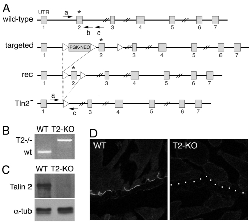
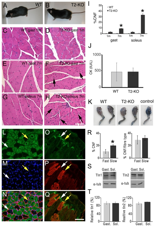
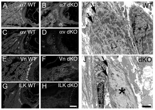
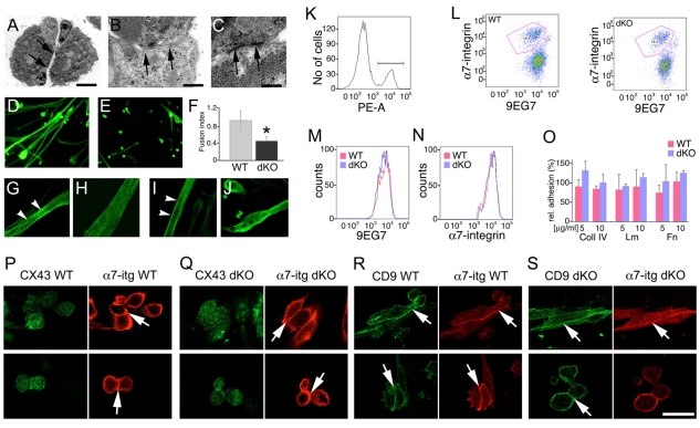
Talin 1 and 2 connect integrins to the actin cytoskeleton and regulate the affinity of integrins for ligands. In skeletal muscle, talin 1 regulates the stability of myotendinous junctions (MTJs), but the function of talin 2 in skeletal muscle is not known. Here we show that MTJ integrity is affected in talin 2-deficient mice. Concomitant ablation of talin 1 and 2 leads to defects in myoblast fusion and sarcomere assembly, resembling defects in muscle lacking beta1 integrins. Talin 1/2-deficient myoblasts express functionally active beta1 integrins, suggesting that defects in muscle development are not primarily caused by defects in ligand binding, but rather by disruptions of the interaction of integrins with the cytoskeleton. Consistent with this finding, assembly of integrin adhesion complexes is perturbed in the remaining muscle fibers of talin 1/2-deficient mice. We conclude that talin 1 and 2 are crucial for skeletal muscle development, where they regulate myoblast fusion, sarcomere assembly and the maintenance of MTJs.


Figures

Fig. 1.
Fig. 1.
Generation of Tln2-KO mice. (A) Schematic representation of the targeting strategy. (B) PCR result from genotyping using the primers in (A) with DNA from 1-month-old wild-type (WT) and Tln2-KO mice (T2-KO). 325-bp and 569-bp bands indicative of wild-type and Tln2-alleles, respectively, were observed. (C) Protein extracts from 1-month old gastrocnemius muscle were analyzed by western blot. Talin 2 expression was ablated in Tln2-KO mice. Membranes were probed with α-tubulin as a loading control. (D) Longitudinal sections of 1-month-old gastrocnemius muscle were stained with antibodies against talin 2. Talin 2 was undetectable in Tln2-KO muscle. Scale bar: 50 μm.
Fig. 2.
Fig. 2.
Tln2-KO mice develop a myopathy with centrally nucleated fibers. (A,B) Normal appearance of Tln2-KO mice. (C-H) Sections of gastrocnemius muscle were stained with H&E. Centrally nucleated fibers were evident in gastrocnemius (C-F) and soleus muscles (G,H) of mice that were 1 (C,D) and 7 (E-H) months old. (I) Quantification of centrally nucleated fibers (CNF) in gastrocnemius and soleus muscles. The number of affected fibers increased with the age of Tln2-KO mice (P=0.006, gastrocnemius; *, P=0.0007, soleus). (J) Serum creatine kinase (CK) levels were normal in 5-month-old Tln2-KO mice (n=4-7 per genotype). (K) EBD was injected into 5-month-old mice. No dye incorporation was noted in muscles of Tln2-KO mice. Occasionally, dye incorporation was observed irrespective of genotype (control), validating the experimental set up (n=4-7 per genotype). (L-N,R) Co-immunostaining of Tln2-KO muscle for MHCf (green) and DAPI (blue) revealed that fast and slow fibers were affected; laminin staining (red) highlights muscle fiber contours. (O-Q) Co-immunostaining for MHCf (red) and talin 2 (green) revealed that talin 2 was expressed at the MTJs of fast and slow fibers. Yellow and white arrows point to fast and slow fibers, respectively. (R) Quantification of the distribution of central nuclei (CNF) in slow and fast fibers (*, P=0.02). (S,T) Expression of talin 1 and talin 2 was evaluated by western blot. Equivalent expression levels were observed in soleus and gastrocnemius muscles. Scale bars: 100 μm.
Fig. 3.
Fig. 3.
Talin 2 is required for MTJ integrity but not for sarcomere organization. (A-C) EM micrographs of gastrocnemius (A,B) and soleus (C) isolated from 3-month-old mice. Disorganization was evident to varying degrees in Tln2-KO muscle fibers, which accumulated necrotic material. Muscle fibers appeared contracted, but the Z-line and A-band were evident (white arrows and asterisks, respectively). (D-I) Electron micrographs of MTJ from soleus (D,E) and gastrocnemius (G-I) of 3-month-old mice. In wild-type mice, myofilaments reached the end of muscle fibers (D,G, arrowhead). In Tln2-KO mice, myofilaments were detached from the MTJ, and necrotic material accumulated in the gaps (E,H,I, asterisks). Lateral detachment of the cytoskeleton from the sarcolemma was occasionally noted (F, arrowheads). (J-Q) Longitudinal sections of gastrocnemius muscle were immunostained with antibodies to α7 integrin (J,K), ILK (L,M), vinculin (Vn) (N,O) and talin 1 (Tln1) (P,Q). All proteins were localized at MTJs, but talin 1 staining was increased in the mutants (arrow in Q). Scale bars: 2 μm in A-C; 5 μm in D-I; 100 μm in J-Q.
Fig. 4.
Fig. 4.
Tln1/2-dKO mice die at birth with skeletal muscle defects. (A) Tln1/2-dKO embryos (dKO) had a contracted posture compared with that of wild-type (WT) embryos. (B,C) Immunostaining of sections from E18.5 wild-type and Tln1/2-dKO muscle showed that talin 2 was effectively ablated from costameres and MTJs (white arrows). (D-K) Sections showing intracostal muscle in E18.5 wild-type (D,F) and Tln1/2-dKO embryos (E,G,H-K) stained with H&E. Myofibers from Tln1/2-dKO mice had abnormal morphology and variation in fiber size (black arrows in G-K). Scale bars: 50 μm in B,C; 100 μm in D-E; 50 μm in F-K.
Fig. 5.
Fig. 5.
Defective sarcomere assembly in Tln1/2-dKO muscle. (A-F) Sections from E18.5 embryos were immunostained with antibodies against vinculin (Vn) (A,B), laminin (Lm) (C,D) and tenascin C (Tn C) (E,F). Expression levels of vinculin were reduced in Tln1/2-dKO embryos. Laminin localized around myofibers but appeared disorganized in mutants (arrow in D). Tenascin C was exclusively localized at the MTJ and in periosteum in controls (E) but was expressed in Tln1/2-dKOs in extrajunctional areas as well (arrow in F). (G-I) Electron micrographs of intercostal muscles from wild-type (G) and Tln1/2-dKO embryos (H,I). The cytoskeletal structure appeared immature and disorganized in Tln1/2-dKO embryos. Disorganized filamentous material accumulated throughout the myofiber, and Z-bands appeared to be incompletely assembled (arrows in H,I) compared with those of controls (arrow in G). Scale bars: 25 μm in A-D; 50 μm in G,H; 2 μm in I.
Fig. 6.
Fig. 6.
Compromised assembly of integrin complexes in Tln1/2-dKO mice. (A-H) Sections from E18.5 embryos were immunostained with antibodies against α7 and αv-integrins and to vinculin (Vn) and ILK. The localization of integrins and their effectors at MTJs was disrupted in Tln1/2-dKO muscle. Dotted lines highlight the location of the MTJ in Tln1/2-dKO muscle. (I,J) Electron micrographs of the MTJ of intercostal muscles from E18.5 wild-type (I, arrows) and Tln1/2-dKO (J, arrows) embryos. Muscle fibers close to MTJs in Tln1/2-dKO were disorganized (asterisk in J). Scale bars: 50 μm in A-H; 2 μm in I,J.
Fig. 7.
Fig. 7.
Defective fusion but normal integrin activation in myoblasts from Tln1/2-dKO mice. (A-C) Electron micrographs of muscle from E18.5 Tln1/2-dKO embryos revealed myoblasts at intermediate stages of fusion. Plasma membranes were aligned (arrows in A,B) and electron-dense adhesion plaques were evident (arrows in C). (D-J) Cell fusion was evaluated in primary cultures of fetal myoblasts. (D,E) Cultures were immunostained with antibodies against α-actinin to label myotubes and myoblasts. In cultures from Tln1/2-dKO mice, myoblast fusion was impaired; only a few short, dysmorphic myotubes were detected. (F) The fusion index was determined (number of nuclei in myoblasts/total number of nuclei) (n=3 mice per genotype) (*P=0.035). (G-J) Immunostaining with antibodies against α-actinin (G,H) and MHCf (I,J) revealed that the cytoskeleton in myofibers from Tln1/2-dKO remained immature. Arrowheads in G,I refer to costameres. (K-O) Analysis of integrin expression and activation by FACS. (K) Cell surface expression of α7 integrin was used to distinguish myoblasts from fibroblasts (bracket indicates α7-integrin-positive population). (L) Representative dot plots of FACS sorted myoblasts from wild-type and Tln1/2-dKO mice analyzed for α7 integrin expression and presence of the 9EG7 epitope (detecting activated β1-integrins). Gated area represents myoblasts. (M) Histogram representing frequency of the 9EG7 epitope on myoblasts. No significant difference was observed between myoblasts from wild-type and Tln1/2-dKO mice. (N) Surface expression levels of α7-integrin were normal in Tln1/2-dKOs (n=3 controls, 5 double mutants). (O) Adhesion to collagen type IV (Coll IV), laminin (Lm) and fibronectin (Fn) was evaluated in primary cultures of fetal myoblasts. No adhesion defects of Tln1/2-dKO myoblasts were observed (n=2 per genotype). The mean ± s.d. are indicated. (P-S) Myoblasts from E17.5 embryos were transferred to differentiation medium and stained for α7 integrin (red), CX43 (P,Q) and CD9 (R,S). The expression levels of CX43 and CD9 were normal in Tln1/2-dKO cells. α7 integrin, CD9 and occasionally CX43, were localized at the interface of fusing myoblasts (arrows). Scale bars: 5 μm in A; 500 nm in B; 250 nm in C; 20 μm in P-S.

Similar articles

Cited by

References

    1. Araya, R., Eckardt, D., Maxeiner, S., Kruger, O., Theis, M., Willecke, K. and Saez, J. C. (2005). Expression of connexins during differentiation and regeneration of skeletal muscle: functional relevance of connexin43. J. Cell Sci. 118, 27-37. - PubMed
    1. Balaban, N. Q., Schwarz, U. S., Riveline, D., Goichberg, P., Tzur, G., Sabanay, I., Mahalu, D., Safran, S., Bershadsky, A., Addadi, L. et al. (2001). Force and focal adhesion assembly: a close relationship studied using elastic micropatterned substrates. Nat. Cell Biol. 3, 466-472. - PubMed
    1. Bazzoni, G., Shih, D. T., Buck, C. A. and Hemler, M. E. (1995). Monoclonal antibody 9EG7 defines a novel beta 1 integrin epitope induced by soluble ligand and manganese, but inhibited by calcium. J. Biol. Chem. 270, 25570-25577. - PubMed
    1. Belkin, A. M., Zhidkova, N. I., Balzac, F., Altruda, F., Tomatis, D., Maier, A., Tarone, G., Koteliansky, V. E. and Burridge, K. (1996). Beta 1D integrin displaces the beta 1A isoform in striated muscles: localization at junctional structures and signaling potential in nonmuscle cells. J. Cell Biol. 132, 211-226. - PMC - PubMed
    1. Belkin, A. M., Retta, S. F., Pletjushkina, O. Y., Balzac, F., Silengo, L., Fassler, R., Koteliansky, V. E., Burridge, K. and Tarone, G. (1997). Muscle beta1D integrin reinforces the cytoskeleton-matrix link: modulation of integrin adhesive function by alternative splicing. J. Cell Biol. 139, 1583-1595. - PMC - PubMed

Publication types