. 2009 Dec;16(12):1750-7.
doi: 10.1128/CVI.00281-09. Epub 2009 Sep 30.

Mycoplasma genitalium-derived lipid-associated membrane proteins activate NF-kappaB through toll-like receptors 1, 2, and 6 and CD14 in a MyD88-dependent pathway

Affiliations

Mycoplasma genitalium-derived lipid-associated membrane proteins activate NF-kappaB through toll-like receptors 1, 2, and 6 and CD14 in a MyD88-dependent pathway

Jun He et al. Clin Vaccine Immunol. 2009 Dec.

Abstract

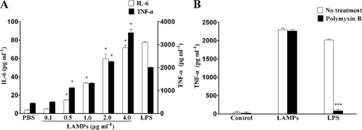
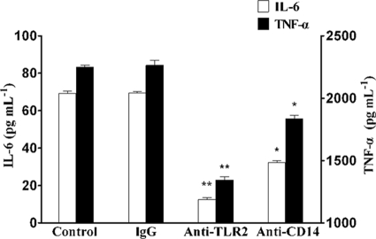
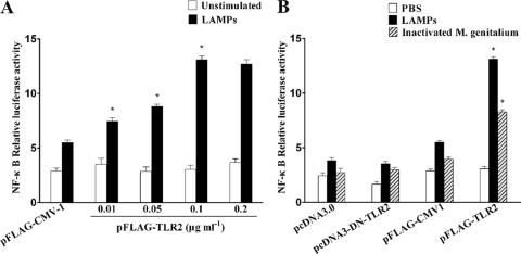
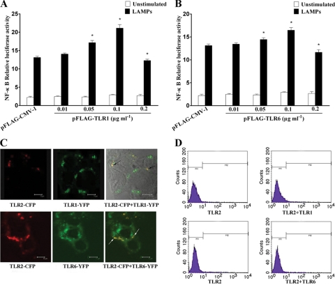
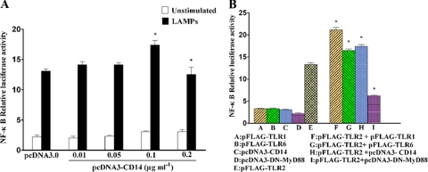
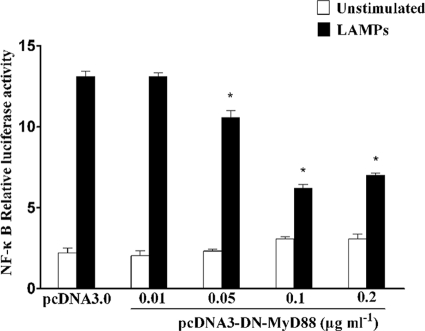
Mycoplasma genitalium is a leading pathogen of nongonoccocal chlamydia-negative urethritis, which has been implicated directly in numerous other genitourinary and extragenitourinary tract pathologies. The pathogenesis of infection is attributed in part to excessive immune responses. M. genitalium-derived lipid-associated membrane proteins (LAMPs) are a mixture of bacterial lipoproteins, exposed at the surface of mycoplasma, that are potent inducers of the host innate immune system. However, the interaction of M. genitalium-derived LAMPs as pathogenic agents with Toll-like receptors (TLRs) and the signaling pathways responsible for active inflammation and NF-kappaB activation have not been fully elucidated. In this study, LAMPs induced the production of tumor necrosis factor alpha (TNF-alpha) and interleukin-6 (IL-6) in a dose-dependent manner. Blocking assays showed that TLR2- and CD14-neutralizing antibodies reduced the expression of TNF-alpha and IL-6 in THP-1 cells. Furthermore, LAMP-induced NF-kappaB activation was increased in 293T cells transfected with TLR2 plasmid. The activity of NF-kappaB was synergically augmented by cotransfected TLR1, TLR6, and CD14. Additionally, LAMPs were shown to inhibit NF-kappaB expression by cotransfection with dominant-negative MyD88 and TLR2 plasmids. These results suggest that M. genitalium-derived LAMPs activate NF-kappaB via TLR1, TLR2, TLR6, and CD14 in a MyD88-dependent pathway.


Figures

FIG. 1.
FIG. 1.
Production of TNF-α and IL-6 by LAMP-induced THP-1 cells. (A) THP-1 cells were cultured in serum medium for 24 h in 24-well tissue culture plates, and the indicated concentrations of LAMPs were added to the medium (PBS or 100 ng ml−1 of LPS was used as the negative or positive control, respectively). After being treated for 8 h, THP-1 cells were lysed, and the culture supernatants were assayed for the proinflammatory cytokines by ELISA (IL-6, left y axis; TNF-α, right y axis). Values represent the means ± standard deviations from three independent experiments assayed in duplicate. P < 0.05 (*) was considered significant. (B) LAMPs (2.0 μg ml−1) or 100 ng ml−1 LPS was added to the medium. After 8 h of culture, supernatant was collected and the concentration of TNF-α in the medium was determined by ELISA as described in Materials and Methods. Values represent the means ± standard deviations from three independent experiments assayed in duplicate.
FIG. 2.
FIG. 2.
Inhibitory effect of anti-TLR2 and anti-CD14 MAb on cytokine production. THP-1 cells were pretreated with 10 μg ml-1 mouse IgG1, anti-TLR2 MAb (IgG1), or anti-CD14 MAb (IgG1) for 30 min and then stimulated with 2.0 μg ml−1 LAMPs. After being treated for 8 h, THP-1 cells were lysed and the culture supernatant was assayed for the proinflammatory cytokines by ELISA (IL-6, left y axis; TNF-α, right y axis). Values represent the means ± standard deviations from three independent experiments assayed in duplicate.
FIG. 3.
FIG. 3.
TLR2 is required for NF-κB activation by LAMPs. (A) HEKHKE293T cells were transiently cotransfected with the indicated concentrations of pFLAG-TLR2, 0.1 μg ml−1 pNF-κB-luc, and 0.01 μg ml−1 pRL-TK. After 24 h of incubation, the cells were stimulated for 8 h with 2.0 μg ml−1 LAMPs. All values represent the means and standard deviations from three assays. P < 0.05 (*) was considered significant. (B) HEK293T cells were transiently cotransfected with the indicated constructs, 0.01 μg ml−1 pNF-κB-luc, and 0.01 μg ml−1 pRL-TK. The total amount of cDNA transfected was kept constant with the level of control construct. After 24 h of incubation, the cells were stimulated for 8 h with 2.0 μg ml−1 LAMPs or inactivated M. genitalium. The cells then were lysed and assayed for luciferase reporter activity. Values represent the means ± standard deviations from three independent experiments assayed in duplicate.
FIG. 4.
FIG. 4.
TLR1 and TLR6 enhanced TLR2-mediated NF-κB activation. (A) HEK293T cells were transiently cotransfected with the indicated concentrations of pFLAG-TLR1, 0.1 μg ml−1 pFLAG-TLR2, 0.1 μg ml−1 pNF-κB-luc, and 0.01 μg ml−1 pRL-TK. After 24 h of incubation, the cells were stimulated for 8 h with 2.0 μg ml−1 LAMPs. The cells then were lysed and assayed for luciferase reporter activity. All values represent the means and standard deviations from three assays. P < 0.05 (*) was considered significant. (B) HEK293T cells were transiently cotransfected with the indicated concentrations of pFLAG-TLR6, 0.1 μg ml−1 pFLAG-TLR2, 0.1 μg ml−1 pNF-κB-luc, and 0.01 μg ml−1 pRL-TK. After 24 h of incubation, the cells were stimulated for 8 h with 2.0 μg ml−1 LAMPs. The cells then were lysed and assayed for luciferase reporter activity. All values represent the means and standard deviations from three assays. P < 0.05 (*) was considered significant. (C) HEK293T cells were transiently cotransfected with TLR2-CFP, and TLR1-YFP or TLR6-YFP was grown on glass-bottomed tissue culture dishes. After 24 h of incubation, the cells were stimulated for 8 h with 2.0 μg ml−1 LAMPs. The living cells then were analyzed by confocal microscopy as described in the text. To the left is the distribution of TLR2; in the center is the localization of TLR1 or TLR6; to the right is the colocalization of TLR2 with TLR1 or TLR6 (black arrows or white arrows). Representative confocal sections of cells are shown. (D) HEK293T cells were transiently cotransfected with the indicated constructs. After 24 h of incubation, the cells were stimulated for 8 h with 2.0 μg ml−1 LAMPs. The cells then were incubated with anti-TLR2 MAb and FITC-labeled secondary Ab. The cell surface expression of TLR2 was analyzed by flow cytometry. Representative confocal sections of cells are shown.
FIG. 5.
FIG. 5.
CD14 enhanced TLR2-mediated NF-κB activation. (A) HEK293T cells were transiently cotransfected with the indicated concentrations of pcDNA3-CD14, 0.1 μg ml−1 pFLAG-TLR2, 0.1 μg ml−1 pNF-κB-luc, and 0.01 μg ml−1 pRL-TK. After 24 h of incubation, the cells were stimulated for 8 h with 2.0 μg ml−1 LAMPs. The cells then were lysed and assayed for luciferase reporter activity. All values represent the means and standard deviations from three assays. P < 0.05 (*) was considered significant. (B) HEK293T cells were transiently cotransfected with the indicated plasmids (the concentration of each plasmid was 0.1 μg ml−1). After 24 h of incubation, the cells were stimulated for 8 h with 2.0 μg ml−1 LAMPs. The cells then were lysed and assayed for luciferase reporter activity. All values represent the means and standard deviations from three assays.
FIG. 6.
FIG. 6.
Activation of NF-κB by LAMPs was TLR2 and MyD88 dependent. HEK93T cells were transiently cotransfected with the indicated concentrations of pcDNA3-DN-MyD88, 0.1 μg ml−1 pFLAG-TLR2, 0.1 μg ml−1 pNF-κB-luc, and 0.01 μg ml−1 pRL-TK. After 24 h of incubation, the cells were stimulated for 8 h with 2.0 μg ml−1 LAMPs. The cells then were lysed and assayed for luciferase reporter activity. All values represent the means and standard deviations from three assays. P < 0.05 (*) was considered significant.

Similar articles

Cited by

References

    1. Akira, S., K. Takeda, and T. Kaisho. 2001. Toll-like receptors: critical proteins linking innate and acquired immunity. Nat. Immunol. 2:675-680. - PubMed
    1. Akira, S., K. Takeda, and T. Kaisho. 2004. Toll-like receptor signaling. Nat. Rev. Immunol. 4:499-511. - PubMed
    1. Barton, G. M., and R. Medzhitov. 2003. Toll-like receptor signaling pathways. Science 300:1524-1525. - PubMed
    1. Baseman, J. B., M. Cagle, J. E. Korte, C. Herrera, W. G. Rasmussen, J. G. Baseman, R. Shain, and J. M. Piper. 2004. Diagnostic assessment of Mycoplasma genitalium in culture-positive women. J. Clin. Microbiol. 42:203-211. - PMC - PubMed
    1. Beutler, B. 2004. Inferences, questions and possibilities in Toll-like receptor signalling. Nature 430:257-263. - PubMed

Publication types

MeSH terms