. 2010 Jan;91(Pt 1):122-32.
doi: 10.1099/vir.0.015909-0. Epub 2009 Sep 30.

Requirement of cellular DDX3 for hepatitis C virus replication is unrelated to its interaction with the viral core protein

Affiliations

Requirement of cellular DDX3 for hepatitis C virus replication is unrelated to its interaction with the viral core protein

Allan G N Angus et al. J Gen Virol. 2010 Jan.

Abstract

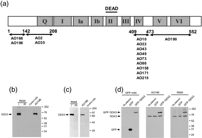
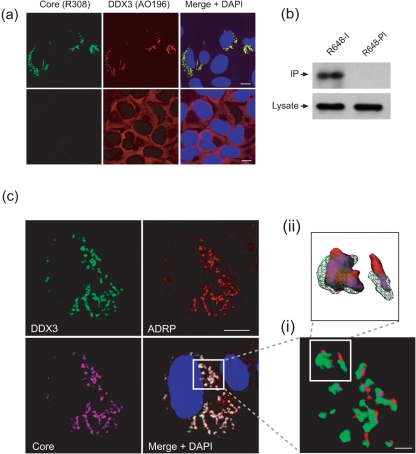
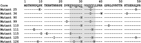
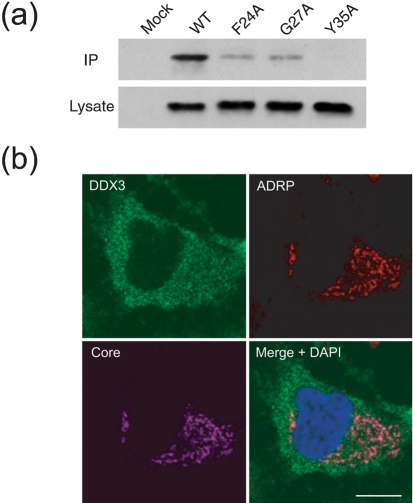
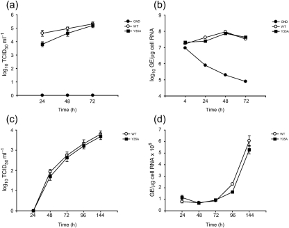
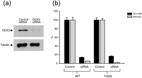
The cellular DEAD-box protein DDX3 was recently shown to be essential for hepatitis C virus (HCV) replication. Prior to that, we had reported that HCV core binds to DDX3 in yeast-two hybrid and transient transfection assays. Here, we confirm by co-immunoprecipitation that this interaction occurs in cells replicating the JFH1 virus. Consistent with this result, immunofluorescence staining of infected cells revealed a dramatic redistribution of cytoplasmic DDX3 by core protein to the virus assembly sites around lipid droplets. Given this close association of DDX3 with core and lipid droplets, and its involvement in virus replication, we investigated the importance of this host factor in the virus life cycle. Mutagenesis studies located a single amino acid in the N-terminal domain of JFH1 core that when changed to alanine significantly abrogated this interaction. Surprisingly, this mutation did not alter infectious virus production and RNA replication, indicating that the core-DDX3 interaction is dispensable in the HCV life cycle. Consistent with previous studies, siRNA-led knockdown of DDX3 lowered virus production and RNA replication levels of both WT JFH1 and the mutant virus unable to bind DDX3. Thus, our study shows for the first time that the requirement of DDX3 for HCV replication is unrelated to its interaction with the viral core protein.


Figures

Fig. 1.
Fig. 1.
Generation of anti-DDX3 antibodies. (a) Schematic representation of DDX3 protein structure showing the nine conserved motifs, including motif II (or the DEAD motif). The arrows mark the epitopes recognized by the mouse mAbs. (b) Characterization of anti-DDX3 antibodies. HuH-7 cell lysates were immunoprecipitated with the immune (I) and pre-immune (PI) antiserum R648, or mAb AO196 or an isotype control mAb. The immune complexes were analysed by Western blotting using biotinylated R648. Positions of molecular mass markers are shown (in kDa). (c) Western blotting of HuH-7 cell lysates with R648-I, -PI, mAb AO196 and isotype control mAb. (d) Western blotting of lysates of HEK cells transfected with a plasmid expressing EGFP alone or an EGFP–DDX3 fusion protein using anti-GFP mAb, AO196 and R648.
Fig. 2.
Fig. 2.
Sequestration of DDX3 by HCV core. (a) DDX3 co-localizes with core in JFH1-infected cells. HuH-7 cells were either mock-infected (lower panel) or infected with JFH1 (top panel). At 3 days post-infection, cells were fixed and probed with HCV core and DDX3 using antibodies R308 and A0196, respectively. Cell nuclei were detected by counterstaining with DAPI. (b) Co-immunoprecipitation of core by anti-DDX3 antiserum. HuH-7 cells electroporated with viral RNA were lysed at 72 h post-incubation, and the lysate immunoprecipitated with anti-DDX3 R648 immune (I) or pre-immune (PI) serum. The resulting precipitates were examined by immunoblotting using anti-core mAb c7-50 (top panel). One twentieth of the cell lysate used in the co-immunoprecipitation assay was immunoblotted for core with mAb c7-50 as the input control (bottom panel). (c) Localization of DDX3 and core on LDs. HuH-7 cells electroporated with the JFH1WT RNA were fixed at 72 h post-transfection and probed using antibodies to core (R308), DDX3 (AO196) and ADRP. Z-stack analysis of all three proteins was performed by recording a series of approximately 20 images. 3D reconstructions of the boxed areas are shown (i) and a selected area is shown in greater detail (ii) where DDX3 is depicted as a wire frame to reveal the core–ADRP association. Bars, 10 μm for confocal images and 2 μm for 3D image.
Fig. 3.
Fig. 3.
Identification of core residues critical for its interaction with DDX3. Amino acid substitutions in residues 1–59 of core in nine mutants unable to interact with DDX3. Residues in the region 24–36 (shaded box) were targeted for alanine-scanning mutagenesis, which identified residues at positions 24, 27, 30, 33, 34 and 35 (underlined in the sequence at the top) that were critical for DDX3 interaction.
Fig. 4.
Fig. 4.
Analysis of JFH1 core mutant viruses. HuH-7 cells electroporated with viral RNAs as shown were subjected to Western immunoblotting at 72 h post-incubation using anti-NS5a mAb 9E10, anti-core antibodies mAb c7-50 or R308, anti-DDX3 mAb AO196 and an anti-tubulin antibody.
Fig. 5.
Fig. 5.
Analysis of the interaction of DDX3 with core mutants. (a) HuH-7 cells were electroporated with different viral RNAs and HCV core co-immunoprecipitated at 72 h post-incubation using the anti-DDX3 serum R648 as described in the legend to Fig. 2b. (b) HuH-7 cells electroporated with JFH1Y35A were fixed at 72 h post-incubation and analysed by confocal microscopy for the intracellular distribution of core, DDX3 and ADRP using appropriate antibodies. Bar, 10 μm.
Fig. 6.
Fig. 6.
Analysis of the core mutant viruses. (a, b) Determination of virus yield and intracellular RNA levels following transfection. HuH-7 cells were electroporated with RNA derived from JFH1WT, JFH1Y35A or JFH1GND cDNA. At the indicated time points (a) the released virus titres and (b) the intracellular HCV RNA levels were quantified by TCID50 and RT-qPCR, respectively. (c and d) Determination of virus yield and intracellular RNA levels following infection. Naïve HuH-7 cells were infected with JFH1WT or JFH1Y35A (obtained from the electroporated cells above) at an m.o.i. of 0.02. The (c) titres of infectious virus released into the medium and (d) intracellular viral RNA levels were measured over 6 days as described above. Means and error ranges from duplicate electroporations and infections are shown.
Fig. 7.
Fig. 7.
Infection of DDX3 knockdown cells. (a) HuH-7 cells were transfected with siRNA duplexes as indicated. Cells were harvested after 2 days and examined by Western blotting using the anti-DDX3 mAb AO196. (b) Naïve and DDX3-deficient HuH-7 cells were infected with WT JFH1WT or JFH1Y35A virus. At 2 days post-infection the levels of released virus and intracellular HCV RNA were measured by TCID50 assay and RT-qPCR, respectively. Means and error ranges from duplicate infections are shown.

Similar articles

Cited by

References

    1. Ariumi, Y., Kuroki, M., Abe, K., Dansako, H., Ikeda, M., Wakita, T. & Kato, N. (2007). DDX3 DEAD-box RNA helicase is required for hepatitis C virus RNA replication. J Virol 81, 13922–13926. - PMC - PubMed
    1. Barba, G., Harper, F., Harada, T., Kohara, M., Goulinet, S., Matsuura, Y., Eder, G., Schaff, Z., Chapman, M. J. & other authors (1997). Hepatitis C virus core protein shows a cytoplasmic localization and associates to cellular lipid storage droplets. Proc Natl Acad Sci U S A 94, 1200–1205. - PMC - PubMed
    1. Beckham, C., Hilliker, A., Cziko, A. M., Noueiry, A., Ramaswami, M. & Parker, R. (2008). The DEAD-Box RNA helicase Ded1p affects and accumulates in Saccharomyces cerevisiae P-bodies. Mol Biol Cell 19, 984–993. - PMC - PubMed
    1. Blight, K. J., Kolykhalov, A. A. & Rice, C. M. (2000). Efficient initiation of HCV RNA replication in cell culture. Science 290, 1972–1974. - PubMed
    1. Botlagunta, M., Vesuna, F., Mironchik, Y., Raman, A., Lisok, A., Winnard, P., Jr, Mukadam, S., Van Diest, P., Chen, J. H. & other authors (2008). Oncogenic role of DDX3 in breast cancer biogenesis. Oncogene 27, 3912–3922. - PMC - PubMed

Publication types

Substances

LinkOut - more resources