. 2009 Nov;20(22):4777-89.
doi: 10.1091/mbc.e09-07-0643. Epub 2009 Sep 30.

The M phase kinase Greatwall (Gwl) promotes inactivation of PP2A/B55delta, a phosphatase directed against CDK phosphosites

Affiliations

The M phase kinase Greatwall (Gwl) promotes inactivation of PP2A/B55delta, a phosphatase directed against CDK phosphosites

Priscila V Castilho et al. Mol Biol Cell. 2009 Nov.

Abstract

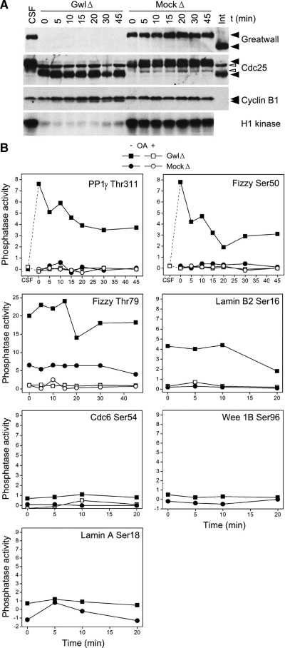
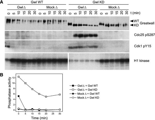
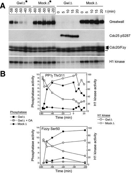
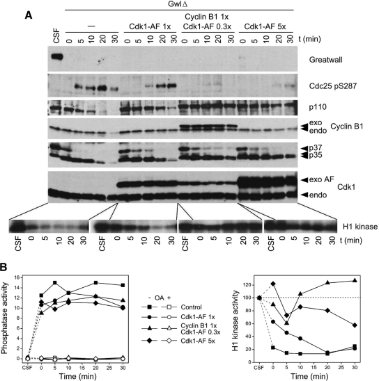
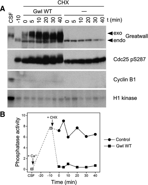
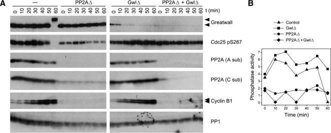
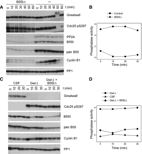
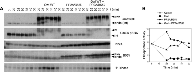
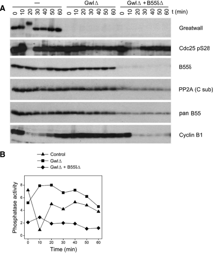
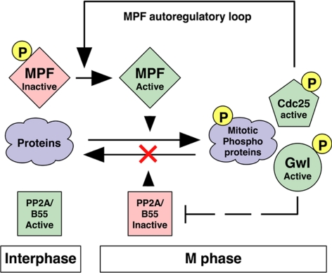
We have previously shown that Greatwall kinase (Gwl) is required for M phase entry and maintenance in Xenopus egg extracts. Here, we demonstrate that Gwl plays a crucial role in a novel biochemical pathway that inactivates, specifically during M phase, "antimitotic" phosphatases directed against phosphorylations catalyzed by cyclin-dependent kinases (CDKs). A major component of this phosphatase activity is heterotrimeric PP2A containing the B55delta regulatory subunit. Gwl is activated during M phase by Cdk1/cyclin B (MPF), but once activated, Gwl promotes PP2A/B55delta inhibition with no further requirement for MPF. In the absence of Gwl, PP2A/B55delta remains active even when MPF levels are high. The removal of PP2A/B55delta corrects the inability of Gwl-depleted extracts to enter M phase. These findings support the hypothesis that M phase requires not only high levels of MPF function, but also the suppression, through a Gwl-dependent mechanism, of phosphatase(s) that would otherwise remove MPF-driven phosphorylations.


Figures

Figure 1.
Figure 1.
Gwl depletion from CSF extracts induces OA-sensitive phosphatase(s) directed against CDK phosphosites. (A) Depletion of Gwl from CSF extracts causes pseudomitotic exit. Immunodepletion was performed as described (Yu et al., 2006) by incubating protein A beads coated with affinity-purified anti-Gwl for 1 h at 4°C and then removing the beads by centrifugation. The supernatant was then incubated at 22°C starting at t = 0. Mock-depleted (MockΔ) control extracts were treated with protein A beads alone. MockΔ and CSF extracts remain in M phase as demonstrated by M phase-specific phosphorylations of Gwl and Cdc25, as well as high histone H1 kinase activity (measuring CDK function). In contrast, Gwl-depleted (GwlΔ) extracts have low H1 kinase, and Cdc25 migrates at its interphase (Int) position. During this pseudomitotic exit, cyclin B1 remains undegraded, whereas inhibitory phosphorylations accumulate on Thr14 and Tyr15 of Cdk1 (Yu et al., 2006; see also Figure 2 below). The white arrowheads point to nonspecific background bands. (B) Induction of anti-CDK phosphatase activity after Gwl depletion. Three-microliter aliquots of the extracts shown in A were assayed for phosphatase activity as described (Mochida and Hunt, 2007), with or without the addition of 2.5 μM okadaic acid (OA). The substrates for this assay were MBP fusion proteins labeled in vitro with [γ-32P]ATP by Cdk2/cyclin A; the fusion proteins contained ∼25 residue regions surrounding known CDK targets (for details, see Mochida and Hunt, 2007). The y-axes of the graphs show the percentage of radioactive phosphate released from the input substrate. Dephosphorylations of substrates containing the sites Thr311 of the γ isoform of PP1, Ser50 and Thr79 of Fizzy/Cdc20, and Ser16 of Lamin B2 are strongly induced by Gwl depletion and are OA-sensitive. As previously noted (Mochida and Hunt, 2007), the Fizzy Thr79 phosphosite is dephosphorylated at a low rate during M phase (as seen in mock-depleted extracts), whereas dephosphorylation of the other three substrates is undetectable during M phase. The decay over time in the phosphatase activity of GwlΔ likely reflects the slow reacquisition of the extract's M phase characteristics due to the continued synthesis of Gwl and cyclins from endogenous mRNAs (Yu et al., 2006). The other assayed substrates shown in the figure (Ser54 of Cdc6, Ser96 of Wee1B, and Ser18 of Lamin A) are not obviously targeted by the phosphatase induced by Gwl depletion.
Figure 2.
Figure 2.
Rescue of Gwl depletion from CSF extracts with exogenous Gwl protein. (A) Pseudomitotic exit caused by Gwl depletion (GwlΔ) of CSF extracts can be reversed by the addition of active wild-type (WT) but not kinase-dead (KD) Gwl protein, as judged by the interphase-specific phosphorylations of Cdc25 Ser287 (Stanford and Ruderman, 2005) and Cdk1 Tyr15 (Yu et al., 2006; Zhao et al., 2008), and the M phase-specific phosphorylation of histone H1 by CDKs. Mock-depleted (MockΔ) extracts remain in M phase regardless of the nature of the exogenously added protein. The exogenous proteins were purified from OA-treated Sf9 insect cells expressing baculovirus constructs (Zhao et al., 2008), and were added in threefold excess with respect to endogenous Gwl. Gwl proteins were added immediately after depletion at 4°C (t = 0), and the samples were then incubated at 22°C. (B) Phosphatase assays of the same samples shown in A, using the substrate containing the Thr311 PP1γ phosphosite. Phosphatase activity was consistently very low during M phase, but elevated during interphase (that is, in the GwlΔ samples supplemented with Gwl KD). The intermediate level of phosphatase activity observed in the GwlΔ sample supplemented with Gwl WT at t = 0 suggests that the exogenous kinase has not yet restored the extracts to M phase, in agreement with the extent of Cdc25 Ser287 and Cdk1 Tyr15 phosphorylations seen in A at t = 0.
Figure 3.
Figure 3.
Gwl addition to interphase cycling extracts inhibits anti-CDK phosphatase activity. (A) Cycling extracts at interphase were prepared as previously described (Yu et al., 2006; Zhao et al., 2008). At t = 0, the extracts were supplemented with active wild-type (Gwl WT) or kinase-dead Gwl (Gwl KD) and then incubated at 22°C. The series at the left is a control cycling extract without added Gwl. The exogenous proteins (tagged with the immunoglobulin Z domain, whose interactions with secondary antibodies account for ∼50% of the total signal on Western blots) were purified from OA-treated Sf9 insect cells expressing baculovirus constructs and were added in threefold excess with respect to endogenous (endo) Gwl, because Gwl WT prepared in this way has only about one-third the specific activity of the enzyme in M phase extracts (Zhao et al., 2008). Immunoblots were analyzed for Gwl, phosphorylation of Ser287 of Cdc25 (a marker for interphase (Int); Stanford and Ruderman, 2005), and cyclin B1. Gwl WT acquires more phosphorylations (including autophosphorylations) than Gwl KD (right and middle, respectively) in the OA-treated insect cells and in M phase Xenopus egg extracts; in the control, endo Gwl migrates during M phase roughly at the position of Gwl KD (arrowhead). Autoradiographs below show histone H1 kinase activity in the same samples. Addition of WT, but not KD, Gwl causes immediate M phase entry. (B) Phosphatase activity of the samples shown in A, measured using the PP1γ Thr311 substrate. Phosphatase levels are consistently greatly reduced during mitosis in the control and Gwl KD samples; this is also true of the precocious M phase caused by Gwl WT. Phosphatase levels during the first 10 min of incubation with Gwl WT are slightly elevated over M phase levels, probably because the exogenous Gwl and the interphase anti-CDK phosphatase compete to inactivate each other.
Figure 4.
Figure 4.
When Gwl is depleted from CSF extracts, phosphatase activity is induced before MPF activity is lost. (A) Protein A beads coated with purified anti-Gwl antibodies, or control protein A beads, were added at t = −60 min to CSF extracts. To investigate the state of the extracts during the course of the immunodepletion, aliquots were taken at the indicated times (GwlΔ* and MockΔ*). At t = 0, extracts that had been immunodepleted for 1 h at 4°C were then incubated at 22°C and processed as in Figure 1 (i.e., GwlΔ* and GwlΔ indicate successive examinations of the same sample, the former during the immunodepletion at 4°C and the latter after transfer of the depleted samples to 22°C). (B) The samples in A were also analyzed for phosphatase activity against the CDK phosphosites at Thr311 of PP1γ (top) and Ser50 of Cdc20/Fizzy (bottom). The results are plotted on the same graph with quantitative data from phosphorimager scans of the H1 kinase assays shown in A. Note that the GwlΔ* samples from t = −55 min and t = −50 min reveal the induction of considerable phosphatase activity even though H1 kinase (i.e., CDK) activity remains at high, M phase-like levels similar to those in the mock depletion.
Figure 5.
Figure 5.
Gwl depletion induces anti-CDK phosphatase activity even in the presence of constitutively active MPF. (A) CSF extracts were immunodepleted for Gwl as in Figure 1. At t = 0, the indicated proteins were added, and the extracts incubated at 22°C. The added proteins were synthesized from baculovirus constructs in Sf9 insect tissue culture cells treated with OA (Zhao et al., 2008). Cdk1-AF refers to constitutively active Cdk1 kinase with S14A and Y15F mutations that block inactivating phosphorylations at these sites; 0.3×, 1×, and 5×, to levels of exogenous protein compared with those of the corresponding endogenous protein. A mixture of cyclin B1 and Cdk1-AF is more effective than Cdk1-AF alone in maintaining M phase characteristics (such as the lack of phosphorylation of Ser287 on Cdc25 and high histone H1 kinase activity), likely because endogenous cyclin B is already bound to CDKs and exchanges only very slowly with exogenous Cdk1-AF (Kobayashi et al., 1994). p35, p37, and p110 indicate unknown proteins of these molecular weights revealed with an antibody directed against phospho-serine within (K/R)pSPX(K/R) motifs (Cell Signaling Technology; no. 2324). Constitutively active MPF or Cdk1 help retain M phase specific phosphorylations on p35 and p110, but not on p37, demonstrating differential sensitivities of these pS/TP phosphosites to variations in the ratio of CDKs and the anti-CDK phosphatase(s) that are induced when Gwl is depleted. (B) Graphs of the phosphatase activity (left; directed against Thr311 of PP1γ) and H1 kinase activity (right) of the samples shown in A. Note that in the samples supplemented with Cdk1-AF and cyclin B1 (i.e., constitutively active MPF) that Gwl depletion induces phosphatase activity even though H1 kinase levels are often even higher than those in CSF extracts.
Figure 6.
Figure 6.
Gwl inhibits anti-CDK phosphatase activity even in the near absence of CDK activity. (A) A CSF extract was treated with 0.3 mM Ca2+ and incubated for 20 min at 22°C to cause cyclin degradation and M phase exit. At that time (t = −10 min), 100 ng/μl cycloheximide (CHX) was added, and samples were incubated for 10 min at 22°C to prevent further protein synthesis. CDK (H1 kinase) activity is very low at this and subsequent times because of the lack of cyclins. At t = 0, the extract was split in two, and Gwl WT was added to one sample at about five times the concentration of the endogenous Gwl. (Our anti-Gwl antibody recognizes the most highly phosphorylated forms of Gwl less efficiently than other forms; see control extract in Figure 3.) Cdc25 in the Gwl-supplemented samples gradually acquires some M phase-like phosphorylations (compare with Figure 3) in spite of the lack of CDK activity. These phosphorylations are likely due to kinases like MAPK with CDK-like target specificities (Sheridan et al., 2008), coupled with the low phosphatase activity against these Cdc25 phosphosites. (B) Phosphatase assays of the samples shown in A, using substrate containing Thr311 of PP1γ. The addition of Gwl WT inhibits anti-CDK phosphatase either directly or indirectly.
Figure 7.
Figure 7.
Immunodepletion of PP2A removes most of the phosphatase activity directed against CDK sites. (A) Interphase cycling extracts were immunodepleted at 4°C for Gwl, PP2A, or both enzymes as indicated. PP2A immunodepletion was performed with the 6F9 mAb against the structural A subunit (Kremmer et al., 1997; Covance; no. MRT-204R) coupled to protein G-Sepharose (Zymed, South San Francisco, CA). The control extract was mock-depleted for both Gwl and PP2A, the GwlΔ extract was mock-depleted for PP2A, and the PP2AΔ extract was mock-depleted for Gwl. The extracts were then incubated at 22°C, and samples were removed for immunoblotting at the indicated times. Antibodies used for the Western blot included the 6F9 antibody for the PP2A A subunit and antibodies against the catalytic (C) subunits of PP1 and PP2A previously described by (Maton et al., 2005); these blots show the specificity of the immunodepletion for PP2A but not PP1. All samples depleted for PP2A remain in interphase because cyclin B1 is degraded and fails to accumulate for unknown reasons (see text). (B) Phosphatase assays of the samples shown in A, using substrate containing Thr311 of PP1γ. Although the PP2A depleted samples are clearly in interphase, they have on average <30% of the phosphatase activity of the corresponding nonPP2A-depleted controls.
Figure 8.
Figure 8.
PP2A/B55δ is a major constituent of the phosphatase directed against CDK phosphosites. (A) Cycling extracts were either mock-depleted (−) or depleted for B55δ (B55δΔ) for 1 h at 4°C and then incubated at 22°C starting at t = 0. The B55δ-depleted extracts were unable to enter M phase because cyclins were immediately degraded (see Discussion). (B) Phosphatase assays of the first four timed aliquots of the samples shown in A, using substrate containing Thr311 of PP1γ. Note that although both the control and B55δ-depleted extracts were in interphase during these times, the large majority of measured phosphatase activity was removed in the latter samples. (C) CSF extracts were either successively depleted for Gwl and B55δ (GwlΔ + B55δΔ), depleted for Gwl and then mock-depleted for B55δ (GwlΔ) or double mock-depleted (CSF). (D) Phosphatase assays of the samples shown in C, using substrate containing Thr311 of PP1γ. The phosphatase activity induced by Gwl depletion was substantially removed by subsequent B55δ immunodepletion. As judged by comparing the Western blot signals obtained with B55δ and pan B55 antibodies from the B55δ-depleted samples, B55δ accounted for more than 80% of the total B55 population in the extracts used in A and B and ∼60% of the B55 molecules in the extracts used in C and D.
Figure 9.
Figure 9.
Gwl blocks the activity of PP2A/B55δ. (A) Interphase cycling extracts were incubated for 20 min at 22°C and then supplemented with a fivefold excess of Gwl WT, a twofold excess of heterotrimeric PP2A containing the B55δ regulatory subunit, or both. Samples were processed for immunoblotting and H1 kinase assays. PP2A was detected with mAb against the A subunit and antibody against B55δ. (B) Phosphatase assays of the samples shown in A. In vitro experiments (not shown) indicate that the exogenous PP2A/B55δ contributes a level of anti-CDK phosphosite activity roughly equivalent to that of the endogenous phosphatase in the extract, consistent with the idea that the large majority of phosphatase assayed in extracts is contributed by PP2A associated with B55-type regulatory subunits. Note that Gwl WT suppresses the activity of both exogenous and endogenous phosphatases. The substrate used is that containing Thr311 of PP1γ.
Figure 10.
Figure 10.
B55δ removal rescues M phase entry defects in Gwl-depleted cycling extracts. (A) Cycling extracts were prepared as in Figures 3 and 8, except that the eggs were incubated an additional 30 min after calcium ionophore treatment but before crushing. Extracts were Gwl-depleted as indicated and then immediately afterward were either B55δ-depleted by three successive 30-min incubations at 4°C with protein G beads coupled to anti-B55δ (GwlΔ + B55δΔ) or mock-depleted with beads without the B55δ antibody (GwlΔ). Control extracts (−) were double mock-depleted. At t = 0, the extracts were incubated at 22°C. B55δ accounted for ∼70% of the B55-type subunits in these extracts, as estimated by comparing immunoblot signals with the B55δ antibody and a pan-B55 antibody. As judged by loss of the interphase-specific phosphorylation on Ser287 of Cdc25 and the degradation of cyclin B, control extracts and extracts depleted for Gwl and B55δ enter M phase at ∼20 min, whereas extracts depleted for Gwl alone do not enter M phase during the incubation. (B) Phosphatase assays of the samples shown in A; the substrate used is that containing Thr311 of PP1γ. Note that the phosphatase activity decreases in the control extract during M phase, whereas the activity is much reduced in the B55δ-depleted sample.
Figure 11.
Figure 11.
A model for Gwl function in M phase. MPF phosphorylates many target proteins, including but not limited to Gwl and MPF autoregulatory loop components such as Cdc25 and (kinases [not shown] such as Plx1 and Myt1/Wee1). Phosphorylated, active Greatwall directly, or more likely indirectly, inactivates PP2A/B55δ, thus protecting MPF substrates from dephosphorylation. In this way, Greatwall can simultaneously influence the autoregulatory loop and also function outside of the loop. This scheme implies that M phase (both in mitosis and meiosis) requires feedback mechanisms that not only positively regulate MPF kinase, but also negatively regulate countering “antimitotic” phosphatases including PP2A/B55δ.

References

    1. Agostinis P., Derua R., Sarno S., Goris J., Merlevede W. Specificity of the polycation-stimulated (type-2A) and ATP, Mg-dependent (type-1) protein phosphatases toward substrates phosphorylated by p34Cdc2 kinase. Eur. J. Biochem. 1992;205:241–248. - PubMed
    1. Che S., Wu W., Nelman-Gonzalez M., Stukenberg T., Clark R., Kuang J. A phosphatase activity in Xenopus oocyte extracts preferentially dephosphorylates the MPM-2 epitope. FEBS Lett. 1998;424:225–233. - PubMed
    1. Clarke P. R., Hoffmann I., Draetta G., Karsenti E. Dephosphorylation of Cdc25-C by a type-2A protein phosphatase: specific regulation during the cell cycle in Xenopus egg extracts. Mol. Biol. Cell. 1993;4:397–411. - PMC - PubMed
    1. Dephoure N., Zhou C., Villen J., Beausoleil S. A., Bakalarski C. E., Elledge S. J., Gygi S. P. A quantitative atlas of mitotic phosphorylation. Proc. Natl. Acad. Sci. USA. 2008;105:10762–10767. - PMC - PubMed
    1. Eto M., Karginov A., Brautigan D. L. A novel phosphoprotein inhibitor of protein type-1 phosphatase holoenzymes. Biochemistry. 1999;38:16952–16957. - PubMed

Publication types

MeSH terms

LinkOut - more resources