Comparative Study
. 2009 Sep 30;29(39):12045-58.
doi: 10.1523/JNEUROSCI.1365-09.2009.

Control of NMDA receptor function by the NR2 subunit amino-terminal domain

Affiliations
Comparative Study

Control of NMDA receptor function by the NR2 subunit amino-terminal domain

Hongjie Yuan et al. J Neurosci. .

Abstract

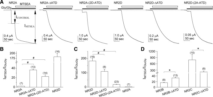
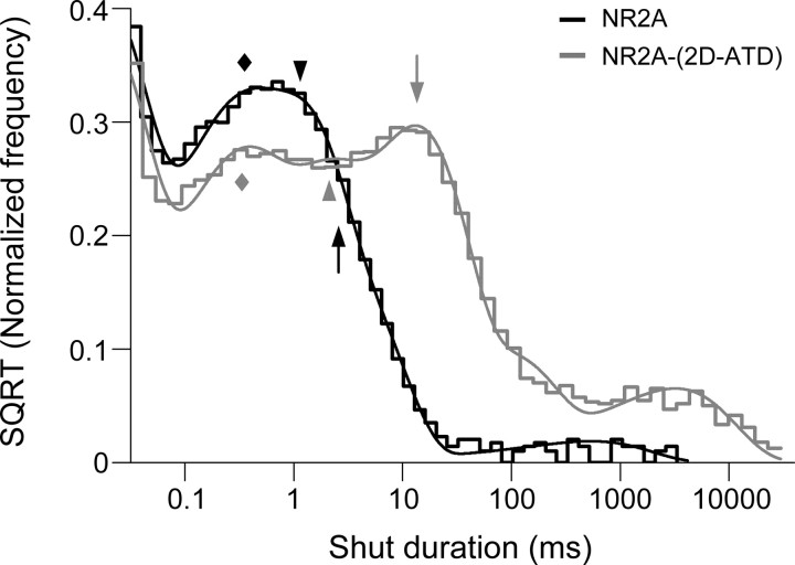
NMDA receptors comprised of different NR2 subunits exhibit strikingly unique biophysical and pharmacological properties. Here, we report that the extracellular amino-terminal domain (ATD) of the NR2 subunit controls pharmacological and kinetic properties of recombinant NMDA receptors, such as agonist potency, deactivation time course, open probability (P(OPEN)), and mean open/shut duration. Using ATD deletion mutants of NR2A, NR2B, NR2C, NR2D, and chimeras of NR2A and NR2D with interchanged ATD [NR2A-(2D-ATD) and NR2D-(2A-ATD)], we show that the ATD contributes to the low glutamate potency of NR2A-containing NMDA receptors and the high glutamate potency of NR2D-containing receptors. The ATD influences the deactivation time courses of NMDA receptors, as removal of the ATD from NR2A slows the deactivation rate, while removal of the ATD from NR2B, NR2C and NR2D accelerates the deactivation rate. Open probability also is influenced by the ATD. Removal of the ATD from NR2A or replacement of the NR2A-ATD with that of NR2D decreases P(OPEN) in single-channel recordings from outside-out patches of HEK 293 cells. In contrast, deletion of the ATD from NR2D or replacement of the NR2D ATD with that of NR2A increases P(OPEN) and mean open duration. These data demonstrate the modular nature of NMDA receptors, and show that the ATD of the different NR2 subunits plays an important role in fine-tuning the functional properties of the individual NMDA receptor subtypes.


Figures

Figure 1.
Figure 1.
Schematic representation of the NR2 ATD deletion constructs and NR2A-NR2D chimeras. A, Hypothetical arrangement of two of the four subunits that comprise NMDA receptors (left). The diagram on the right illustrates the key features of a single subunit. The ATD shown in green was generated from a homology model based on the mGluR1 crystal structure (Yuan et al., 2009), the agonist-binding domains shown in blue are the crystal structure of the NR1-NR2A heterodimer (PDB code 2A5T) (Furukawa et al., 2005), and the transmembrane domains shown in orange are the M1, M2, and M3 regions aligned with the structure of the P-loop region of KcsA (PDB code 1BL8). The C-terminal domain and the transmembrane helix M4 from NR1 and NR2A is omitted from the hypothetical arrangement (left). B, Linear schematic organization of NR2 subunits. Each subunit consists of the ATD, segments S1 and S2, three transmembrane helices (M1, M3, and M4), one cytoplasmic re-entrant pore loop (M2), and an intracellular C-terminal domain (CTD). Together, the S1 and S2 segments form the agonist-binding domain. C, The ATD deletion constructs of the NR2 subunits were developed from wild-type NR2A, NR2B, NR2C, NR2D, and NR2B-(ΔS28-M394). All ATD deletion constructs encode the first 28 residues of NR2B, including the signal peptide. The residue following Ser 28 of NR2B was His 405 of NR2A, His 405 of NR2B, His 415 of NR2C, and His 428 of NR2D. D, Constructs of NR2A-NR2D chimeras were developed by interchanging the ATD of NR2A and NR2D. The residue following Gln 427 of NR2D was His 405 of NR2A, and the residue following Asn 404 of NR2A was His 428 of NR2D. Amino acid numbering is done according to full-length protein, including the signal peptide.
Figure 2.
Figure 2.
The NR2 ATD controls the EC50 values for glutamate and glycine on recombinant NMDA receptors expressed in oocytes. A, ATD deletion in the NR2A, NR2B, NR2C, and NR2D subunits causes minimal change in glutamate EC50 values (in the presence of 100 μm glycine) compared with the corresponding wild type. However, the glutamate EC50 of NR2A-(2D-ATD) is similar to that of wild-type NR2D, and the EC50 of NR2D-(2A-ATD) is similar to that of wild-type NR2A. B, Glycine EC50 (in the presence of 100 μm glutamate) was increased following ATD deletion in NR2B, NR2C, and NR2D subunits.
Figure 3.
Figure 3.
The NR2 ATD controls the deactivation time course in NMDA receptors. A, Representative whole-cell currents elicited by glycine and glutamate (50 μm each, black bar) are shown in whole-cell voltage-clamp (VHOLD = −60 mV) current recordings from HEK 293 cells expressing recombinant NR1 with NR2A or NR2A-ΔATD. Glycine was present in all solutions. Right panel shows superimposed traces (black: NR2A; gray: NR2A-ΔATD). B, Representative traces of currents from NR1 coexpressed with NR2D or NR2D-ΔATD. C, Representative traces of currents from NR1 coexpressed with NR2A-(2D-ATD) or NR2D-(2A-ATD). D, Representative traces of currents from NR1 coexpressed with NR2B or NR2B-ΔATD. Right panel shows superimposed traces (black: NR2B; gray: NR2B-ΔATD). E, Representative traces of currents from NR1 with NR2C or NR2C-ΔATD. Right panel shows superimposed traces (black: NR2C; gray: NR2C-ΔATD). F, Summary of the mean deactivation time constants. #p < 0.01, one-way ANOVA with Bonferroni's post hoc test, compared with the corresponding wild type. *p < 0.01, unpaired t test, compared with the corresponding wild type.
Figure 4.
Figure 4.
NR2 ATD control of open probability evaluated by the degree of MTSEA potentiation. A, Representative two-electrode voltage-clamp current recordings obtained from oocytes coexpressing recombinant NR1-A652C with NR2A, NR2A-ΔATD, NR2A-(2D-ATD), NR2D, NR2D-ΔATD, or NR2D-(2A-ATD) to evaluate the degree of potentiation by 0.2 mm MTSEA after the receptors are activated by glutamate and glycine (100 μm). Application of MTSEA to agonist-bound NR1-A652C-containing channels irreversibly locks open the pore following covalent modification. MTSEA-modified receptors have an open probability (POPEN) of ∼1.0, which renders the degree of MTSEA potentiation of the maximal agonist response inversely related to the open probability of the channel. B–D, Summary of the degree of MTSEA potentiation. Open probability estimated from these data are given in Table 3; #p < 0.01, one-way ANOVA with Bonferroni's post hoc test, compared with the corresponding wild type. *p < 0.01, unpaired t test, compared with the corresponding wild type.
Figure 5.
Figure 5.
ATD deletion from NR2A decreases the single-channel open probabilities in outside-out patch-clamp recordings from transiently transfected HEK 293 cells. Aa1, Bb1, Steady-state recordings of NR1/NR2A (Aa1) and NR1/NR2A-ΔATD (Bb1) unitary currents in an outside-out patch that contained one active channel activated by 50 μm glycine and 1 mm glutamate are shown at different time scales. Aa2 and Bb2 show the open period histogram, shut duration histogram, amplitude histogram, and the stability plot of the amplitude for the channel in this patch.
Figure 6.
Figure 6.
ATD deletion from NR2D increases the single-channel open probability and mean open duration. Aa1, Bb1, Steady-state recordings of NR1 with NR2D (Aa1) or NR2D-ΔATD (Bb1) unitary currents in an outside-out patch that contained one active channel are shown at different time scales. Aa2 and Bb2 show the open period histogram, shut duration histogram, amplitude histogram, and the stability plot of the amplitude for the channel in this patch.
Figure 7.
Figure 7.
Chimeric NR2A-(2D-ATD) shows lower open probability and briefer open periods than wild-type NR2A. A, Steady-state recordings of NR1/NR2A-(2D-ATD) unitary currents in an outside-out patch that contained one active channel are shown at different time scales. B shows the open period histogram and amplitude histogram.
Figure 8.
Figure 8.
Chimeric NR2D-(2A-ATD) shows an increased open probability and longer open periods than NR2D. A, Steady-state recordings of NR1/NR2D-(2A-ATD) unitary currents in an outside-out patch that contained one active channel are shown at different time scales. B shows the open period histogram and amplitude histogram.
Figure 9.
Figure 9.
NR2D-ATD controls τ3 from the exponential components fitted to the shut duration histogram. Overlay of the normalized composite fitted histograms of shut durations from wild-type NR2A (black line) and NR2A-(2D-ATD) (gray line). Each histogram was normalized by dividing the frequency of each dwell time (i.e., each bin) with the total number of dwell times used to generate the histogram. τ3 (arrow) of NR2A-(2D-ATD) was right-shifted 4.6-fold compared with the wild-type NR2A, while τ1 (diamond) and τ2 (arrowhead) show either no shift and shifted to right side only by 1.4-fold, respectively.
Figure 10.
Figure 10.
The linker between the ATD and the agonist binding domain influences receptor open probability. A, Amino acid alignment of the linker region between the ATD and the agonist binding domain of NR2A and NR2D. The chimeras NR2A-(2D-linker) and NR2D-(2A-linker) were generated by interchanging the linker between the ATD and S1 (16 aa) from NR2A (V389-N404) and NR2D (L413-Q427). B, Summary of the degree of MTSEA (200 μm) potentiation of responses to glutamate and glycine (100 μm) from oocytes coexpressing NR1-A652C with NR2A-(2D-linker) or NR2D-(2A-linker). C, Summary of the mean deactivation time constants of responses to 50 μm glutamate obtained from whole-cell voltage-clamp (VHOLD = −60 mV) current recordings from HEK 293 cells transfected with NR1 and NR2A-(2D-linker) or NR2D-(2A-linker); 50 μm glycine was present in all solutions. The number of oocytes and HEK 293 cells is indicated in parenthesis above each bar. #p < 0.01, one-way ANOVA with Bonferroni's post hoc test, compared with the corresponding wild type.

References

    1. Béhé P, Colquhoun D, Wyllie DJ. Activation of Single AMPA- and NMDA-Type Glutamate-Receptor Channels. In: Jonas P, Monyer H, editors. Ionotropic glutamate receptors in the CNS. Springer; 1999. pp. 175–218.
    1. Chen PE, Geballe MT, Katz E, Erreger K, Livesey MR, O'Toole KK, Le P, Lee CJ, Snyder JP, Traynelis SF, Wyllie DJ. Modulation of glycine potency in rat recombinant NMDA receptors containing chimeric NR2A/2D subunits expressed in Xenopus laevis oocytes. J Physiol. 2008;586:227–245. - PMC - PubMed
    1. Choi Y, Chen HV, Lipton SA. Three pairs of cysteine residues mediate both redox and Zn2+ modulation of the NMDA receptor. J Neurosci. 2001;21:392–400. - PMC - PubMed
    1. Choi YB, Lipton SA. Identification and mechanism of action of two histidine residues underlying high-affinity Zn2+ inhibition of the NMDA receptor. Neuron. 1999;23:171–180. - PubMed
    1. Colquhoun D, Hawkes AG. Stochastic properties of ion channel openings and bursts in a membrane patch that contains two channels: evidence concerning the number of channels present when a record containing only single openings is observed. Proc R Soc Lond B Biol Sci. 1990;240:453–477. - PubMed

Publication types