. 2009 Dec;297(6):F1566-74.
doi: 10.1152/ajprenal.00214.2009. Epub 2009 Sep 30.

A new function for parietal epithelial cells: a second glomerular barrier

Affiliations

A new function for parietal epithelial cells: a second glomerular barrier

Takamoto Ohse et al. Am J Physiol Renal Physiol. 2009 Dec.

Abstract

The functional role of glomerular parietal epithelial cells (PECs) remains poorly understood. To test the hypothesis that PECs form an impermeable barrier to filtered protein through the formation of tight junctions (TJ), studies were performed in normal animals and in the anti-glomerular basement membrane (GBM) model of crescentic nephritis. Electron microscopy showed well-defined TJ between PECs in normal mice, which no longer could be identified when these cells became extensively damaged or detached from their underlying Bowman's basement membrane. The TJ proteins claudin-1, zonula occludens-1, and occludin stained positive in PECs; however, staining decreased in anti-GBM disease. To show that these events were associated with increased permeability across the PEC-Bowman's basement membrane barrier, control and diseased animals were injected intravenously with either Texas red-conjugated dextran (3 kDa) or ovalbumin (45 kDa) tracers. As expected, both tracers were readily filtered across the glomerular filtration barrier and taken up by proximal tubular cells. However, when the glomerular filtration barrier was injured in anti-GBM disease, tracers were taken up by podocytes and PECs. Moreover, tracers were also detected between PECs and the underlying Bowman's basement membrane, and in many instances were detected in the extraglomerular space. We propose that together with its underlying Bowman's basement membrane, the TJ of PECs serve as a second barrier to protein. When disturbed following PEC injury, the increase in permeability of this layer to filtered protein is a mechanism underlying periglomerular inflammation characteristic of anti-GBM disease.


Figures

Fig. 1.
Fig. 1.
Tight junction proteins expression in developing mouse kidney. Staining for claudin-1, zonula occludin-1 (ZO-1), and PAX8 is shown from the comma stage through the capillary loop stages in the developing kidney. AE: claudin-1 staining in epithelial cell layers in comma- and early S-shaped body stages. E: high magnification of the inset in D. Examples of positive stains are shown by arrows. FJ: staining for ZO-1 in different stages of glomerulogenesis. ZO-1 staining is not detected until late S-shaped body and capillary loop stages in immature parietal epithelial cells (PECs) and immature podocytes. Examples are represented by arrows. KN: PAX8 is also shown to indicate the locations of PECs and podocytes in the fetal kidney. By the S-shaped body stage, both PECs and podocytes stain positive for PAX8, whereas PECs stain positive and podocytes lose expression of PAX8 by the capillary loop stage.
Fig. 2.
Fig. 2.
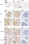
Tight junction protein staining in normal adult mouse, rat, and human glomeruli. AC: claudin-1 staining was confined specifically to PECs in normal mouse, rat, and human glomeruli. DF: ZO-1 staining was present in both podocytes and PECs in normal glomeruli from each species. High-power magnification of the insets are shown in GI, and examples are illustrated by arrows. JL: occludin staining was detected in PECs and podocytes in normal mouse, rat, and human glomeruli. High-power magnification of the insets are shown in MO, and examples are illustrated by arrows.
Fig. 3.
Fig. 3.
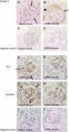
Tight junction protein staining in experimental anti-glomerular basement membrane (GBM) disease. A: claudin-1 staining is detected in PECs in control mice (arrows). B: staining was decreased in segments of the glomerulus in mice with anti-GBM disease (arrowhead). C and D: staining was not detected in negative controls when the primary antibody was omitted in control mice and mice with anti-GBM disease, respectively. E: ZO-1 staining in PECs in control mice (arrows). F: ZO-1 staining decreased in PECs in anti-GBM disease. G: occludin staining was detected in normal PECs (arrows). H: there was a substantial decrease in occludin staining in PECs in anti-GBM disease. No staining was detected in the negative controls in control (I) and diseased (J) mice.
Fig. 4.
Fig. 4.
Electron microscopy showing tight junctions between adjacent PECs. Electron microscopic study was performed to examine the tight junction structure in PECs. A: example of a tight junction is shown on the apical side of adjacent PECs in a control mouse (arrow). B: in anti-GBM disease, one of the tight junctions is intact (arrow), but others are less apparent as PECs are injured or become detached from their underlying basement membranes. The distance between adjacent PECs is wider in anti-GBM disease compared with control mice. US, urinary space; BBM, Bowman's basement membrane.
Fig. 5.
Fig. 5.
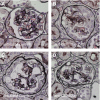
Silver staining of anti-GBM nephritis mouse. Silver staining was performed to visualize any defects in BBM in the mouse anti-GBM nephritis model. Compared with control mice (A), the pattern of silver staining of the BBM was indistinguishable from anti-GBM disease at days 7, 9, and 14 (BD). In D, the BBM was duplicated in some segments of glomeruli at day 14, “engulfing” PECs.
Fig. 6.
Fig. 6.
Tracer study in anti-GBM nephritis mouse. Texas red staining was performed to detect the glomerular localization of intravenously injected Texas red-conjugated dextran (3 kDa) and ovalbumin (45 kDa). Texas red staining for dextran was seen within capillary loops and in the brush border of proximal tubules in control animals, which received only normal sheep IgG (A and D). Texas red staining was observed in 5 stages in mice with anti-GBM nephritis: stage 1, within glomerular capillary loop; stage 2, within podocyte cell body; stage 3, within PEC cell body; stage 4, between PEC and BBM; stage 5, between cell layers in crescents; and stage 6, in the extraglomerular space (B and C). Texas red staining for ovalbumin showed limited distribution compared with dextran (E). Images of the various staining controls are shown (F). Several images focusing on the tracers distributed in the extraglomerular region are shown. G and H: images from control mouse. IN: images from diseased mouse. Corresponding high-magnification images are on the right of each image of the whole glomerulus. Arrows indicate the positive signals of leaked tracers. Note that the white arrowhead in H indicates the tracers on the brush border of proximal tubules.
Fig. 7.
Fig. 7.
Double staining of tracer and claudin-1 in anti-GBM nephritis mouse. Double staining was performed to show the relationship between the extraglomerular distribution of the dextran tracer and changes in claudin-1 staining. A: in the control mice, claudin-1 (brown) was expressed in PECs in a linear pattern, and no extraglomerular dextran was observed (blue). B: in mice with anti-GBM nephritis, less claudin-1 staining was observed (brown) near areas with extraglomerular staining for dextran (blue, arrows).
Fig. 8.
Fig. 8.
Schema. PECs work as a second barrier of glomerular filtrate with their tight junctions. Details are described in the text.

References

    1. Akilesh S, Huber TB, Wu H, Wang G, Hartleben B, Kopp JB, Miner JH, Roopenian DC, Unanue ER, Shaw AS. Podocytes use FcRn to clear IgG from the glomerular basement membrane. Proc Natl Acad Sci USA 105: 967–972, 2008 - PMC - PubMed
    1. Atkins RC, Nikolic-Paterson DJ, Song Q, Lan HY. Modulators of crescentic glomerulonephritis. J Am Soc Nephrol 7: 2271–2278, 1996 - PubMed
    1. Barisoni L, Nelson PJ. Collapsing glomerulopathy: an inflammatory podocytopathy? Curr Opin Nephrol Hypertens 16: 192–195, 2007 - PubMed
    1. Gonzalez-Mariscal L, Betanzos A, Nava P, Jaramillo BE. Tight junction proteins. Prog Biophys Mol Biol 81: 1–44, 2003 - PubMed
    1. Griffin SV, Krofft RD, Pippin JW, Shankland SJ. Limitation of podocyte proliferation improves renal function in experimental crescentic glomerulonephritis. Kidney Int 67: 977–986, 2005 - PubMed

Publication types

MeSH terms