. 2009 Sep;21(9):2859-77.
doi: 10.1105/tpc.109.066118. Epub 2009 Sep 30.

A tripartite SNARE-K+ channel complex mediates in channel-dependent K+ nutrition in Arabidopsis

Affiliations

A tripartite SNARE-K+ channel complex mediates in channel-dependent K+ nutrition in Arabidopsis

Annegret Honsbein et al. Plant Cell. 2009 Sep.

Abstract

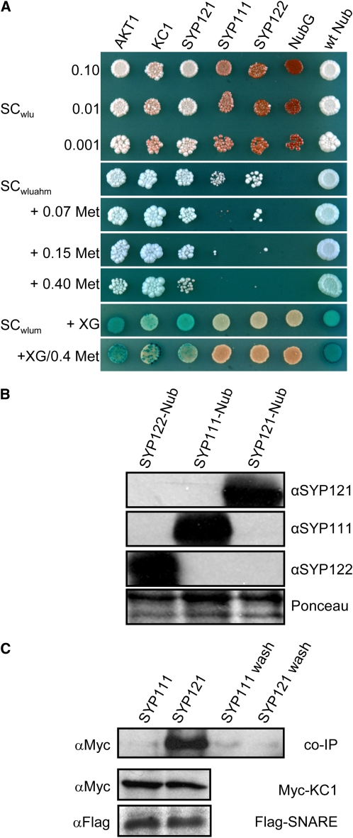
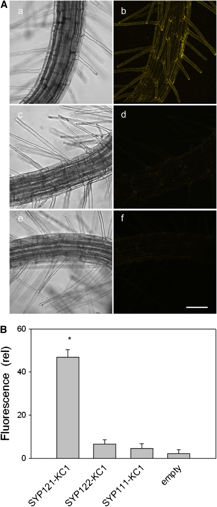
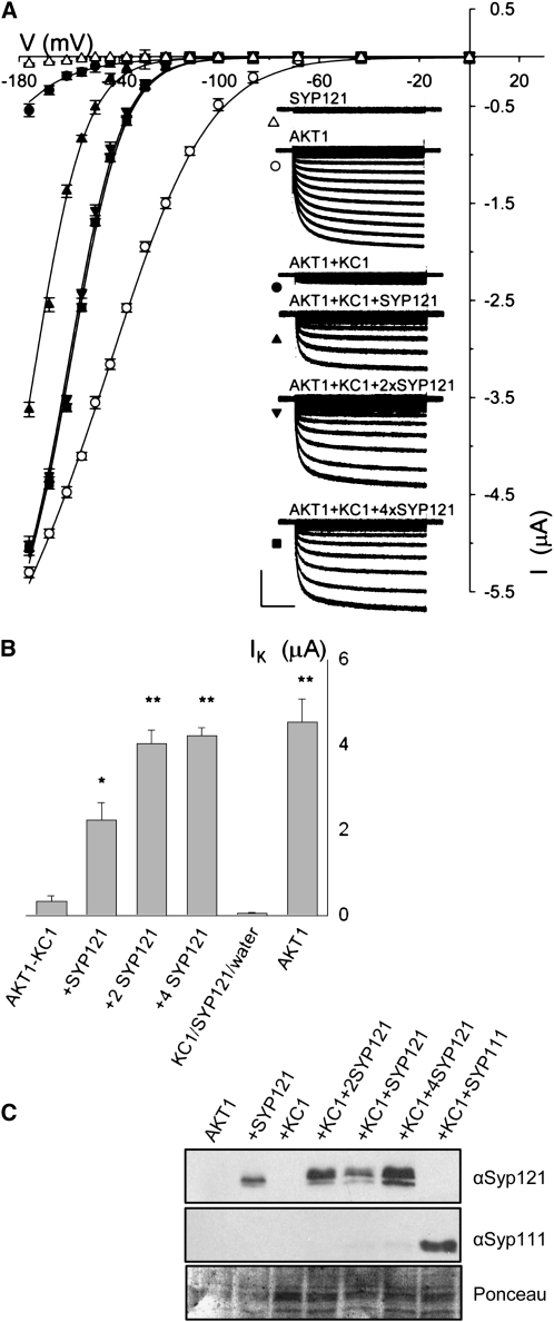
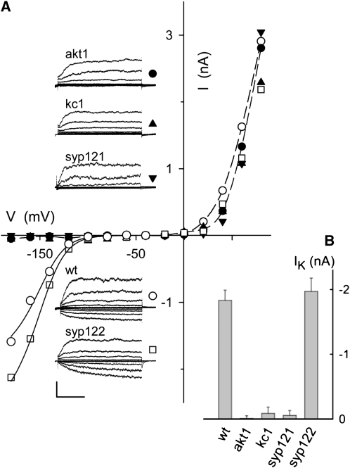
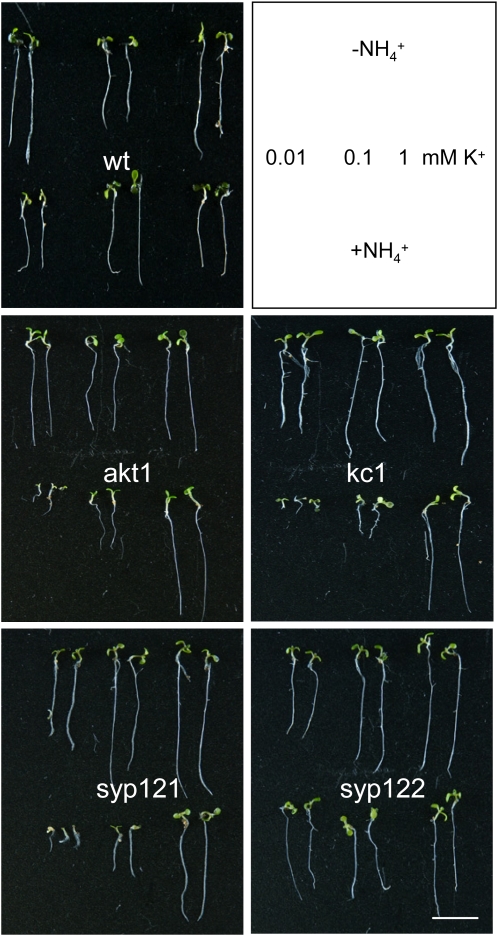
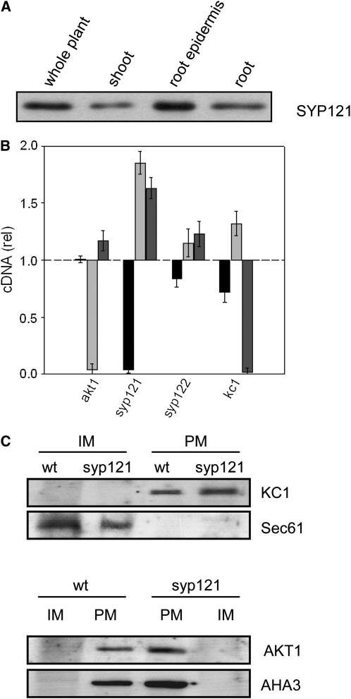
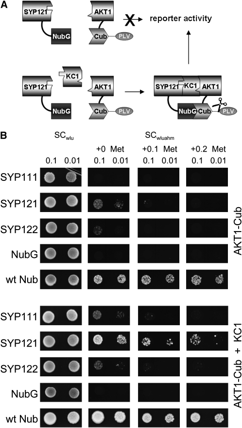
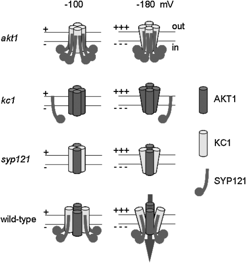
A few membrane vesicle trafficking (SNARE) proteins in plants are associated with signaling and transmembrane ion transport, including control of plasma membrane ion channels. Vesicle traffic contributes to the population of ion channels at the plasma membrane. Nonetheless, it is unclear whether these SNAREs also interact directly to affect channel gating and, if so, what functional impact this might have on the plant. Here, we report that the Arabidopsis thaliana SNARE SYP121 binds to KC1, a regulatory K(+) channel subunit that assembles with different inward-rectifying K(+) channels to affect their activities. We demonstrate that SYP121 interacts preferentially with KC1 over other Kv-like K(+) channel subunits and that KC1 interacts specifically with SYP121 but not with its closest structural and functional homolog SYP122 nor with another related SNARE SYP111. SYP121 promoted gating of the inward-rectifying K(+) channel AKT1 but only when heterologously coexpressed with KC1. Mutation in any one of the three genes, SYP121, KC1, and AKT1, selectively suppressed the inward-rectifying K(+) current in Arabidopsis root epidermal protoplasts as well as K(+) acquisition and growth in seedlings when channel-mediated K(+) uptake was limiting. That SYP121 should be important for gating of a K(+) channel and its role in inorganic mineral nutrition demonstrates an unexpected role for SNARE-ion channel interactions, apparently divorced from signaling and vesicle traffic. Instead, it suggests a role in regulating K(+) uptake coordinately with membrane expansion for cell growth.


Figures

Figure 1.
Figure 1.
The Silent K+ Channel KC1 Interacts with the SNARE SYP121 in Arabidopsis. (A) Yeast mating-based split-ubiquitin assay for interaction with KC1-Cub. Yeast diploids created with Nub-X constructs of AKT1, KC1, SYP121, SYP111, SYP122, and controls (negative, NubG; positive, wild-type Nub) spotted (left to right) on synthetic complete (SC) medium without Trp, Leu, and Ura (SCwlu) to verify crossing and test for adenine synthesis (white colonies, top panel). SC without Trp, Leu, Ura, Ade, His, and Met (SCwluahm) used to verify Ade- and His-independent growth (second panel), and the addition of 0.07, 0.15, and 0.40 mM Met (next three panels) used to verify interaction at lower KC1-Cub expression levels. SC medium without Trp, Leu, Ura, and Met (SCwlum; last two panels) alone and with addition of 0.4 mM Met used with an overlay of X-Gal–containing agarose to assay β-galactosidase activity (Obrdlik et al., 2004). Serial dilutions 0.1, 0.01, and 0.001 of diploid cultures as indicated for spots on SCwlu. Otherwise, only 0.1 dilutions are shown. Note KC1 interaction with AKT1, itself, and SYP121 and the absence of specific interaction with SYP111 and SYP122. (B) Verification of prey protein expression in diploid yeasts carrying KC1-Cub and Nub-SNAREs. Protein gel blot analysis of total protein extracted from yeast diploids expressing KC1-Cub with Nub-SYP122, Nub-SYP111, and Nub-SYP121 using antibodies specific for SYP122, SYP111, or SYP121 (top panels). Ponceau S stain was used as loading control (bottom panel). (C) Coimmunoprecipitation of Myc-tagged KC1 by retention with Flag-tagged SYP121 on anti-Flag-coupled Sepharose. Protein gel blot analysis (top, left to right) of eluates coimmunoprecipitating with Flag-tagged SYP111, with SYP121, and the precipitated terminal washes prior to elution from each, respectively, probed with anti-Myc antibody. Control protein gel blots (bottom) show equivalent levels of expression for KC1 and for the SNAREs in each solubilized fraction after coexpression in Sf9 insect cells.
Figure 2.
Figure 2.
Selective BiFC of KC1 with SYP121 Expressed in Arabidopsis Root Epidermis as Fusion Constructs with the N- and C-Terminal Halves of YFP (nYFP and cYFP), Respectively. (A) Three-dimensional reconstructions from confocal fluorescence image stacks of seedlings expressing (bright-field, left; fluorescence, right) SYP121-nYFP with KC1-cYFP ([a] and [b]), SYP122-nYFP with KC1-cYFP ([c] and [d]), and with the empty (untransformed) Agrobacterium ([e] and [f]). Bar = 50 μm. Images with SYP111-nYFP paired with KC1-cYFP were similar to those of (c) and (d). (B) Mean fluorescence intensities (arbitrary units) ± se after correction for background of (nontransfected) control measurements (n > 6 independent experiments in each case; * indicates significant difference from the empty control at P < 0.01). See also Supplemental Figure 2 and Supplemental Movie 1 online.
Figure 3.
Figure 3.
Coexpression with SYP121 Selectively Rescues AKT1-KC1 K+ Current in Xenopus Oocytes. (A) Current traces and steady state current voltage curves recorded under voltage clamp in 96 mM K+ from oocytes expressing SYP121 alone (triangles), AKT1 alone (open circles), AKT1 with KC1 (closed circles∂, molar ratio 1:1), and AKT1 with KC1 and SYP121 (SYP121:KC1 cRNA molar ratios: 1:1, upright triangle; 2:1, downward triangle; 4:1, square). Clamp cycles: holding voltage, −50 mV; voltage steps, 0 to −180 mV. Insets: Corresponding whole-cell currents cross-referenced by symbol. Scale: 2 μA and 1 s. Currents from oocytes injected with water and with KC1 cRNA only gave results similar to those for SYP121, and currents from oocytes injected with cRNAs for AKT1 together with KC1 and SYP111 or SYP122 were indistinguishable from those injected with cRNAs for AKT1 plus KC1 alone. Currents from oocytes injected with cRNAs for AKT1 and SYP121 showed gating characteristics similar to those for AKT1 alone and expressing KC1 alone or with SYP121 failed to yield a measureable current (data not shown). All measurements performed as coexpressions with CBL1 and CIPK23 essential for AKT1 function in oocytes in 1:1:1 molar ratios with AKT1 (Xu et al., 2006), and no current was observed in the absence of CBL1/CIPK23 expression (data not shown). Final cRNA volume for each oocyte was equal. Similar results were obtained with the same construct combinations when expressed in Sf9 insect cells (see Supplemental Figure 3 online) without the additional expression load of CBL1/CIPK23. Solid curves are the results of joint, nonlinear least squares fitting of the K+ currents (IK) to a Boltzmann function IK = gmax(V − EK)/(1 + eδ(V−V1/2)/RT), where gmax is the conductance maximum, and V, EK, R, and T have their usual meanings. The characteristic voltage dependence (V1/2) indicates the midpoint of the voltage range for gating, and the apparent gating charge (δ) is an unique property of the gate, its sensitivity to voltage changes and the associated conformations. Best fittings were obtained with gmax held in common and with separate, joint values for δ with and without SYP121 expression. Similar results were obtained in each of eight separate analyses. (see Table 1). (B) Summary of K+ current amplitudes recorded from oocytes expressing AKT with KC1 in combinations with SYP121 (cRNA injection ratios indicated) and expressing AKT1 alone. Data are means ±se obtained at -160 mV from >9 independent measurements in each case (significant difference from AKT1+KC1 ** at P < 0.01 and * at P < 0.05). (C) Verification of SNARE protein expression in oocytes. Oocytes were injected with AKT1, CIPK23, CBL1 cRNA (AKT1, molar ratios 1:1:1), and with SYP121 (+SYP121, molar ratios 1:1:1:1), KC1 (+KC1, molar ratios 1:1:1:1), KC1 and Syp121 (+KC1+SYP121, molar ratios 1:1:1:1:1; +KC1+2SYP121, molar ratios 1:1:1:1:2; +KC1+4SYP121, molar ratios 1:1:1:1:4), and KC1 and SYP111 (+KC1+SYP111, molar ratios 1:1:1:1:1). Protein gel blot analysis of total membrane protein extracted from oocytes collected after electrical recordings detected with antibodies specific to SYP121 and SYP111. Ponceau S stain was used to normalize SYP121 expression levels for lanes with KC1 and yielded ratios of 1: 2.04:3.4.
Figure 4.
Figure 4.
syp121 and kc1 Mutations Phenocopy the akt1 Mutant in Suppressing Inward-Rectifying K+ Current. (A) Whole-cell currents (insets) and steady state current-voltage curves from representative Arabidopsis root epidermal protoplasts in 100 mM K+ under voltage clamp, cross-referenced by symbol. Scale bar for current traces (below): 2 nA and 1 s. Clamp cycles: holding voltage, −50 mV; voltage steps, +80 to −180 mV, tailing voltage, −50 mV. Dashed curves in the current voltage plots are empirical polynomial fittings for outward-rectifying K+ currents from wild-type and akt1 mutant protoplasts and are included for visual guidance only. Solid curves are results of joint fittings of inward-rectifying K+ currents to a Boltzmann function (see Figure 3 legend) with V1/2 and δ held in common between wild-type and syp122 mutant protoplasts. The parameter values, δ, −1.97 ± 0.13 and V1/2, −142 ± 5 mV, were statistically equivalent to similar fittings for K+ currents on heterologous expression of AKT1 with KC1 and SYP121 (Figure 3; see also Supplemental Figure 3 online). Note that protoplasts from the akt1, kc1, and syp121 mutants, but not from wild-type and syp122 mutant plants, all showed a loss in K+ current at voltages negative of −100 mV. Records from all protoplasts retain the outward-rectifying K+ current that is nominally dependent on the GORK K+ channel. (B) Summary of inward-rectifying K+ current amplitudes recorded from wild-type and mutant Arabidopsis root epidermal protoplasts. Data are mean steady state amplitudes ± se obtained at −160 mV from five to seven independent experiments in each case. Means for akt1, kc1, and syp121 mutants are significantly different from the wild type at P < 0.01.
Figure 5.
Figure 5.
syp121 and kc1 Mutations Phenocopy the akt1 Mutant in Suppressing NH4+-Sensitive Growth at Submillimolar [K+]. Wild-type and mutant Arabidopsis seedlings germinated and grown in modified Murashige and Skoog (MS) with 0.01, 0.1, and 1.0 mM K+ with and without 2 mM NH4+ for 10 d. Reference to growth conditions indicated in frame (top right). Bar = 1 cm. Statistical analysis of root length and K+ content are summarized in Supplemental Figure 4 online.
Figure 6.
Figure 6.
SNARE and K+ Channel Transcription and Expression in Arabidopsis. (A) The SNARE SYP121 is expressed strongly in the Arabidopsis root and root epidermis. Protein gel blot analysis of total proteins (10 μg/lane) extracted from whole Arabidopsis, shoot, root, and root epidermis and probed with anti-SYP121 primary antibody. All four lanes yielded a single band close to 37 kD, corresponding to SYP121 (Tyrrell et al., 2007) and consistent with the expression patterns for AKT1 and KC1 (Birnbaum et al., 2003). (B) Quantification of SYP121 and K+ channel transcripts in the wild type and mutant Arabidopsis. Real-time PCR of SYP121 (black bars), AKT1 (light-gray bars), and KC1 (dark-gray bars) transcript levels in each of the mutant lines syp121-1, syp122-1, akt1-1, and kc1-1 after standardization on ACT2 transcript levels. Data normalized to the corresponding amplification yields in the wild type and are means ± se from three independent experiments. No appreciable decrease was evident for the K+ channel subunits in either of the SNARE mutant or the complementary K+ channel mutant lines. A significant increase (P < 0.05) in relative transcript level was evident in the kc1 and akt1 mutants for AKT1 and KC1 genes, respectively. (C) KC1 K+ channel localization to the plasma membrane is unaffected in syp121-1 mutant Arabidopsis. Protein gel blot analysis of plasma membrane (PM) and inner membrane (IM) fractions separated by two-phase partitioning of microsomal membranes isolated from roots of wild-type and syp121-1 mutant plants. Parallel SDS-PAGE was run of all fractions (1.3 μg protein/lane), and PVDF membranes were probed with polyclonal antibodies to KC1 and AKT1 (see Supplemental Figure 6 online) before stripping and reprobing with antibodies to the endoplasmic reticulum Sec61 (Yuasa et al., 2005) as a marker for inner membranes and with polyclonal antibody to the H+-ATPase AHA3 (Pardo and Serrano, 1989) as a marker for the plasma membrane. Protein gel blots were visualized by 125I radiotracer phosphor imaging.
Figure 7.
Figure 7.
The syp121-1 Mutation Does Not Affect K+ Channel Expression at the Root Epidermal Plasma Membrane. (A) Longitudinal optical section (top three panels: bright-field, composite, YFP fluorescence) and three-dimensional transect analysis (bottom six panels) of KC1-YFP distribution in an Arabidopsis syp121-1 mutant root hair. Transects taken at positions (in μm) from the apex as indicated by the dashed lines above. Note the peripheral distribution of the fluorescence and its virtual absence from the dense cytosol and tonoplast boundary behind the tip (top, labeled schematic illustration drawn to scale for reference). Similar results were obtained in each of nine separate experiments with the syp121 mutant and in five experiments with expression in wild-type seedlings (data not shown). Bar = 5 μm. (B) Mean fluorescence intensities ± se (arbitrary units) of root hairs expressing AKT1-GFP after correction for background of control measurements transfected with nontransformed Agrobacterium (n > 12 independent experiments in each case; means differ significantly at P < 0.05).
Figure 8.
Figure 8.
The syp121 Mutation Does Not Affect K+ Channel Mobility at the Cell Periphery Assessed by FRAP and FLIP in Arabidopsis syp121 Mutant Root Hairs. (A) and (B) AKT1-GFP FLIP in an Arabidopsis syp121-1 mutant root hair. (A) Bright-field and fluorescence image taken before (top two frames) and at times during repetitive photobleaching (start of photobleach cycles, relative t = 0) at the distal end of the root hair (photobleach area indicated in the first fluorescence image). See Supplemental Movie 3 online for full image sequence. (B) FLIP analysis of the fluorescence signals taken from the regions indicated (inset, green and red boxes) and corrected for background fluorescence decay shows a limited loss of signal outside the area of photobleaching (photobleach periods indicated by gray bars). The solid curve is the result of nonlinear least squares fitting of the postbleach fluorescence signal to a single exponential function yielding an immobile fraction of 0.84. Similar results were obtained in each of four separate experiments with the syp121 mutant and in four experiments with wild-type seedlings and KC1-YFP in place of AKT1-GFP. Bar = 10 μm. (C) and (D) AKT-GFP FRAP in an Arabidopsis syp121-1 mutant root hair. (C) Bright-field and fluorescence image taken before (top two frames) and after photobleaching (end of photobleach, relative t = 0) the distal end of the root hair (photobleach areas indicated in the first fluorescence image). See Supplemental Movie 2 online for full image sequence. (D) FRAP analysis of the fluorescence signals taken from the regions indicated (inset, green and red boxes) and corrected for background fluorescence decay shows the loss of signal after photobleaching and its limited recovery. Photobleach time indicated by gray bar. The solid curve is the result of nonlinear least squares fitting of the postbleach fluorescence signal to a single exponential function. Similar results were obtained in each of eight separate experiments with the syp121 mutant and in seven experiments with wild-type seedlings, yielding immobile fractions of 0.86 ± 0.04 and 0.84 ± 0.03, respectively. Bar = 10 μm.
Figure 9.
Figure 9.
Tripartite Assembly of SYP121 with AKT1 Requires the KC1 Subunit as a Bridge. (A) Concept of the yeast SUB assay for formation of a tripartite protein assembly. By analogy with the mating-based split-ubiquitin approach, the SUB assay depends on protein interaction to bring together the two halves of the ubiquitin protein, which leads to cleavage of the VP16 transactivator (PLV) and activation of the reporter gene. In this case, however, assembly of the proteins carrying the split-ubiquitin moieties depends on inclusion of the third protein (KC1) component. (B) Yeast split-ubiquitin assay for interaction after transformation with AKT1-Cub, the Nub-X fusion constructs of SYP121, SYP122, and SYP111, with (bottom) and without (top) coexpression of KC1. Controls (negative, NubG; positive, wtNub) are included and transformed yeast are spotted (top to bottom) on SC medium without Trp, Leu, and Ura (SCwlu) to verify crossing. Yeast growth on SC without Trp, Leu, Ura, Ade, His, and Met (SCwluahm) was used to verify Ade- and His-independent growth (second panel), and the addition of 0.1 and 0.2 mM Met was used to verify interaction at lower AKT1-Cub expression levels. Serial dilutions (0.1 and 0.01) as indicated for each frame. Note yeast growth on Met only with KC1 coexpression. Protein expression was verified by protein gel blot analysis in each case (data not shown).
Figure 10.
Figure 10.
Tripartite Assembly with SYP121 Determines the Gating of Heteromeric AKT1-KC1 K+ Channels. Model of channel subunit assembly and gating in the plant based on channel gating characteristics on heterologous expression and in the plant (Figures 3 and 4; see Supplemental Figure 3 online). Eliminating any one of the three proteins AKT1 (akt1), KC1 (kc1) or SYP121 (syp121) prohibits normal gating and K+ flux, either by preventing the assembly of the necessary heteromeric core of AKT1 and KC1 subunits (akt1 and kc1) or by preventing association of SYP121 with the channel core through its binding with KC1 (kc1 and syp121). We assume that AKT1 and KC1 form the core of the channel and its pore, consistent with their structural homologies to other Kv-like K+ channel subunits and the observation that heterologous expression of AKT1 alone and with KC1 yields a current on heterologous expression. The approximate twofold change in the apparent gating charge on coexpression with SYP121 (Figure 3; see also Supplemental Figure 3 online) points to profound changes in the conformation of the channel. Thus, assembly of the heteromeric AKT1-KC1 channel core and binding of at least two SYP121 proteins to each KC1 subunit (wild type) gives a channel gate with native characteristics, underpinning its physiological voltage dependence and K+ flux (arrow).

Similar articles

Cited by

References

    1. Alexandersson, E., Saalbach, G., Larsson, C., and Kjellbom, P. (2004). Arabidopsis plasma membrane proteomics identifies components of transport, signal transduction and membrane trafficking. Plant Cell Physiol. 45 1543–1556. - PubMed
    1. Amtmann, A., and Blatt, M.R. (2007). Regulation of ion transporters. In Plant Solute Transport, A.R. Yeo and T. Flowers, eds (Oxford, UK: Blackwell), pp. 99–132.
    1. Amtmann, A., and Blatt, M.R. (2009). Regulation of macronutrient transport. New Phytol. 181 35–52. - PubMed
    1. Ashley, M.K., Grant, M., and Grabov, A. (2006). Plant responses to potassium deficiencies: A role for potassium transport proteins. J. Exp. Bot. 57 425–436. - PubMed
    1. Assaad, F.F., Qiu, J.L., Youngs, H., Ehrhardt, D., Zimmerli, L., Kalde, M., Wanner, G., Peck, S.C., Edwards, H., Ramonell, K., Somerville, C.R., and Thordal-Christensen, H. (2004). The PEN1 syntaxin defines a novel cellular compartment upon fungal attack and is required for the timely assembly of papillae. Mol. Biol. Cell 15 5118–5129. - PMC - PubMed

Publication types

MeSH terms