. 2010 Feb;30(2):311-22.
doi: 10.1038/jcbfm.2009.211. Epub 2009 Sep 30.

Pharmacological uncoupling of activation induced increases in CBF and CMRO2

Affiliations

Pharmacological uncoupling of activation induced increases in CBF and CMRO2

Christoph Leithner et al. J Cereb Blood Flow Metab. 2010 Feb.

Abstract

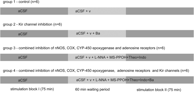
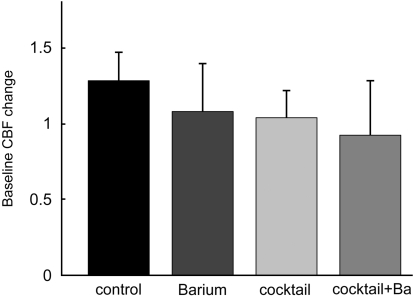
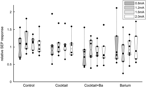
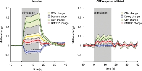
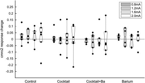
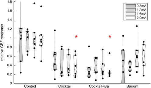
Neurovascular coupling provides the basis for many functional neuroimaging techniques. Nitric oxide (NO), adenosine, cyclooxygenase, CYP450 epoxygenase, and potassium are involved in dilating arterioles during neuronal activation. We combined inhibition of NO synthase, cyclooxygenase, adenosine receptors, CYP450 epoxygenase, and inward rectifier potassium (Kir) channels to test whether these pathways could explain the blood flow response to neuronal activation. Cerebral blood flow (CBF) and cerebral metabolic rate of oxygen (CMRO(2)) of the somatosensory cortex were measured during forepaw stimulation in 24 rats using a laser Doppler/spectroscopy probe through a cranial window. Combined inhibition reduced CBF responses by two-thirds, somatosensory evoked potentials and activation-induced CMRO(2) increases remained unchanged, and deoxy-hemoglobin (deoxy-Hb) response was abrogated. This shows that in the rat somatosensory cortex, one-third of the physiological blood flow increase is sufficient to prevent microcirculatory increase of deoxy-Hb concentration during neuronal activity. The large physiological CBF response is not necessary to support small changes in CMRO(2). We speculate that the CBF response safeguards substrate delivery during functional activation with a considerable 'safety factor'. Reduction of the CBF response in pathological states may abolish the BOLD-fMRI signal, without affecting underlying neuronal activity.


Figures

Figure 1
Figure 1
The experimental paradigm. A stimulation block with randomly distributed stimulations at 0.4 to 2.0 mA was followed by a 60-min waiting period (wash in of vehicle/inhibitors), followed by a second stimulation block analogous to the first one. Each experimental group consisted of six animals. The first group served as the time control series. After the first block, superfusion was switched to aCSF+vehicle (v, ethanol 0.5%). In a second, Kir channel inhibition group, superfusion was switched to aCSF plus vehicle containing 500 μmol/L BaCl2. In the third group, combined inhibition of nNOS, COX, CYP450 epoxygenase, and adenosine receptors was achieved with superfusion of aCSF containing vehicle, L-NNA (1 mmol/L), indomethacin (500 μmol/L), MS-PPOH (20 μmol/L), and theophylline (50 μmol/L). In the fourth group, BaCl2 (500 μmol/L) was added on top of the inhibitory cocktail of group 3.
Figure 2
Figure 2
Baseline CBF changes. The relative changes in baseline CBF were calculated as the ratio of the mean of all CBF data of the first block (superfusion of aCSF in all groups) to the mean of all CBF data of the second block (superfusion of aCSF+vehicle in the control group, superfusion aCSF+inhibitors for the other groups). Baseline CBF increased by 28%±19% in the control group. In comparison with the control group, baseline CBF was reduced by approximately 20% in all other groups, with a nonsignificant trend towards stronger reduction in the cocktail plus BaCl2 group. Error bars denote 95% confidence intervals.
Figure 3
Figure 3
SEP responses. The SEP amplitudes were calculated as amplitude between the first negative and positive peak (N1P1). Average amplitudes of the first stimulation block were divided by average amplitudes of the second block (after superfusion of vehicle/inhibitors) for each stimulation intensity. The figure shows the relative changes in SEP amplitude after superfusion of vehicle/inhibitors; black dots denote individual animals and boxes contain the second and third quartiles. There is no significant change in the SEP amplitudes, although a trend towards small reduction can be seen with the cocktail+BaCl2 and BaCl2 groups.
Figure 4
Figure 4
Major CBF-response reduction without CMRO2 impairment. Time courses of CBF, CBV, CMRO2, and deoxy-Hb during baseline conditions with superfusion of aCSF (no additional vehicle or inhibitor, n=24) and superfusion of inhibitory cocktails (CBF response inhibition, n=12) for a stimulation intensity of 2.0 mA. Shaded areas denote 95% confidence intervals. At baseline, CBF increases by ∼50%, CBV by ∼20% (in conformity with Grubbs relationship), CMRO2 by ∼10%, and deoxy-Hb level decreases by ∼15%. The inhibitory cocktail reduces CBF responses by two-thirds, leaving CMRO2 unchanged. No change in deoxy-Hb level can be detected under these conditions (‘BOLD-blind' coupling). Shaded areas denote 95% confidence intervals.
Figure 5
Figure 5
CMRO2 responses unaltered. Mean CMRO2 change before minus the mean CMRO2 change after superfusion of vehicle/inhibitors (ΔCMRO2 after−ΔCMRO2 before). Subtraction rather than division was used because CMRO2 changes were small and close to zero for some animals. Black dots denote individual animals and boxes contain the second and third quartiles. Superfusion of inhibitors did not lead to a significant change in CMRO2, despite a relevant reduction in CBF (see Figure 6).
Figure 6
Figure 6
CBF responses pharmacologically reduced. The relative changes in activity-induced increase in CBF were calculated by individually dividing the average CBF increases of all animals of each group before by the average CBF increase after superfusion of vehicle/inhibitors. Black dots denote individual animals and boxes contain the second and third quartiles. CBF responses were largely reduced to 30% to 40% of the baseline response in the cocktail and cocktail+BaCl2 group. BaCl2 alone led to less pronounced reductions in CBF response. CBF response reduction was statistically tested for the 2.0-mA stimulation intensity. The red stars denote significant differences compared with the control group.

References

    1. Bakalova R, Matsuura T, Kanno I. The cyclooxygenase inhibitors indomethacin and rofecoxib reduce regional cerebral blood flow evoked by somatosensory stimulation in rats. Exp Med Biol (Maywood) 2002;227:465–473. - PubMed
    1. Burke M, Bührle C.2006BOLD response during uncoupling of neuronal activity and CBF Neuroimage 321–8.e-pub 2006 May 3 - PubMed
    1. Buxton RB, Frank LR. A model for the coupling between cerebral blood flow and oxygen metabolism during neural activation. J Cereb Blood Flow Metab. 1997;17:64–72. - PubMed
    1. Diamond J. Quantitative evolutionary design. J Physiol. 2002;542 (Pt 2:337–345. - PMC - PubMed
    1. Dirnagl U, Niwa K, Lindauer U, Villringer A. Coupling of cerebral blood flow to neuronal activation: role of adenosine and nitric oxide. Am J Physiol. 1994;267 (1 Pt 2:H296–H301. - PubMed

Publication types

MeSH terms

Substances

LinkOut - more resources