

Fluid dynamics in developmental biology: moving fluids that shape ontogeny
- PMID: 19794816
- PMCID: PMC2707792
- DOI: 10.2976/1.3043738
Fluid dynamics in developmental biology: moving fluids that shape ontogeny
Abstract
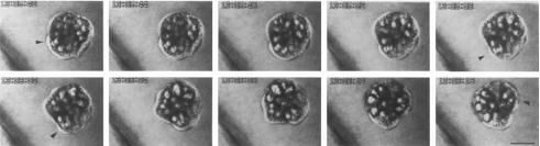
Human conception, indeed fertilization in general, takes place in a fluid, but what role does fluid dynamics have during the subsequent development of an organism? It is becoming increasingly clear that the number of genes in the genome of a typical organism is not sufficient to specify the minutiae of all features of its ontogeny. Instead, genetics often acts as a choreographer, guiding development but leaving some aspects to be controlled by physical and chemical means. Fluids are ubiquitous in biological systems, so it is not surprising that fluid dynamics should play an important role in the physical and chemical processes shaping ontogeny. However, only in a few cases have the strands been teased apart to see exactly how fluid forces operate to guide development. Here, we review instances in which the hand of fluid dynamics in developmental biology is acknowledged, both in human development and within a wider biological context, together with some in which fluid dynamics is notable but whose workings have yet to be understood, and we provide a fluid dynamicist's perspective on possible avenues for future research.
Figures
Similar articles
-
Early object relations into new objects.Psychoanal Study Child. 2001;56:39-67; discussion 68-75. doi: 10.1080/00797308.2001.11800664. Psychoanal Study Child. 2001. PMID: 12102023
-
Physical Biology : challenges for our second decade.Phys Biol. 2014 Jun;11(3):030201. doi: 10.1088/1478-3975/11/3/030201. Epub 2014 Apr 15. Phys Biol. 2014. PMID: 24732666
-
Chronoastrobiology: proposal, nine conferences, heliogeomagnetics, transyears, near-weeks, near-decades, phylogenetic and ontogenetic memories.Biomed Pharmacother. 2004 Oct;58 Suppl 1:S150-87. doi: 10.1016/s0753-3322(04)80025-8. Biomed Pharmacother. 2004. PMID: 15754855
-
Fluid dynamics of heart development.Cell Biochem Biophys. 2011 Sep;61(1):1-22. doi: 10.1007/s12013-011-9158-8. Cell Biochem Biophys. 2011. PMID: 21327946 Review.
-
Theoretical aspects of Systems Biology.Prog Biophys Mol Biol. 2013 May;112(1-2):33-43. doi: 10.1016/j.pbiomolbio.2013.03.019. Epub 2013 Apr 3. Prog Biophys Mol Biol. 2013. PMID: 23562476 Review.
Cited by
-
Coupling between hydrodynamic forces and planar cell polarity orients mammalian motile cilia.Nat Cell Biol. 2010 Apr;12(4):341-50. doi: 10.1038/ncb2040. Epub 2010 Mar 21. Nat Cell Biol. 2010. PMID: 20305650
-
The use of biophysical approaches to understand ciliary beating.Biochem Soc Trans. 2020 Feb 28;48(1):221-229. doi: 10.1042/BST20190571. Biochem Soc Trans. 2020. PMID: 31922188 Free PMC article. Review.
-
Basal Xenobot transcriptomics reveals changes and novel control modality in cells freed from organismal influence.Commun Biol. 2025 Apr 22;8(1):646. doi: 10.1038/s42003-025-08086-9. Commun Biol. 2025. PMID: 40263484 Free PMC article.
-
Simulation of microtubule-cytoplasm interaction revealed the importance of fluid dynamics in determining the organization of microtubules.Plant Direct. 2023 Jul 25;7(7):e505. doi: 10.1002/pld3.505. eCollection 2023 Jul. Plant Direct. 2023. PMID: 37502315 Free PMC article.
-
Mechanistic basis of otolith formation during teleost inner ear development.Dev Cell. 2011 Feb 15;20(2):271-8. doi: 10.1016/j.devcel.2010.12.006. Dev Cell. 2011. PMID: 21316594 Free PMC article.
References
-
- Abraham, E R (1998). “The generation of plankton patchiness by turbulent stirring.” Nature (London) NATUAS10.1038/35361 391, 577–580. - DOI
-
- Alward, W LM (2003). “A new angle on ocular development.” Science SCIEAS 299, 1578–1581. - PubMed
-
- Arnold, J M, and Williams-Arnold, L D (1980). “Development of the ciliature pattern on the embryo of the squid Loligo pealei: a scanning electron microscope study.” Biol. Bull. ZZZZZZ 159, 102–116.
-
- Assheton, R (1896). “Notes on the ciliation of the ectoderm of the amphibian embryo.” J. Cell. Sci. JNCSAI 38, 465–484.
-
- Bagnat, M, Cheung, I D, Mostov, K E, and Stainier, D YR (2007). “Genetic control of single lumen formation in the zebrafish gut.” Nat. Cell Biol. NCBIFN 9, 954–960. - PubMed
LinkOut - more resources
Full Text Sources