. 2009:2009:147946.
doi: 10.1155/2009/147946. Epub 2009 Sep 23.

Ischemia alters the expression of connexins in the aged human brain

Affiliations

Ischemia alters the expression of connexins in the aged human brain

Taizen Nakase et al. J Biomed Biotechnol. 2009.

Abstract

Although the function of astrocytic gap junctions under ischemia is still under debate, increased expression of connexin 43 (Cx43) has been observed in ischemic brain lesions, suggesting that astrocytic gap junctions could provide neuronal protection against ischemic insult. Moreover, different connexin subtypes may play different roles in pathological conditions. We used immunohistochemical analysis to investigate alterations in the expression of connexin subtypes in human stroke brains. Seven samples, sectioned after brain embolic stroke, were used for the analysis. Data, evaluated semiquantitatively by computer-assisted densitometry, was compared between the intact hemisphere and ischemic lesions. The results showed that the coexpression of Cx32 and Cx45 with neuronal markers was significantly increased in ischemic lesions. Cx43 expression was significantly increased in the colocalization with astrocytes and relatively increased in the colocalization with neuronal marker in ischemic lesions. Therefore, Cx32, Cx43, and Cx45 may respond differently to ischemic insult in terms of neuroprotection.


Figures

Figure 1
Figure 1
Representative picture of brain slice stained with H&E. Pale colored area shows the infarct lesion. Rectangle lined areas indicate the selected regions of interest (ischemic cortex (a) and intact cortex (b)).
Figure 2
Figure 2
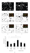
Cx32 immunofluorescent stainings indicate amplified expression in the ischemic lesion (b) compared to the intact area (a). Inset pictures of (c) and (d) represent double immunofluorescent staining with Cx32 (green) and MAP2 (red). Representative pictures in second row show the extracted area as yellow region of the double immunofluorescent staining from intact (c) and ischemic area (d). The yellow region which stands for Cx32 immunopositive area colocalized with MAP2 shows increased expression in the ischemic periphery. Inset pictures of (e) and (f) represent double immunofluorescent staining with Cx32 (green) and GFAP (red). Representative pictures in third row show the extracted area as yellow region of the double immunofluorescent staining from intact (e) and ischemic area (f). The yellow region which stands for Cx32 immunopositive area colocalized with GFAP seems similar level of the expression between normal and ischemic area. Black bars in graph (g) indicate the average counts of protein expressions. The Cx32 expression was relatively increased in the ischemic area compared to the intact area (†: P = .0510). The coexpression of Cx32 and MAP2 was significantly increased in the ischemic lesion as compared to the intact area (**: P < .01). However, there was no statistical difference in Cx32 and GFAP coexpression between intact and ischemic areas. Scale bar indicates 50 μm.
Figure 3
Figure 3
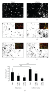
Cx45 immunofluorescent stainings indicate amplified expression in the ischemic lesion (b) compared to the intact area (a). Inset pictures of (c) and (d) represent double immunofluorescent staining with Cx45 (green) and MAP2 (red). Representative pictures in second row show the extracted area as yellow region of the double immunofluorescent staining from intact (c) and ischemic area (d). The yellow region which stands for Cx45 immunopositive area colocalized with MAP2 shows increased expression in the ischemic periphery. Inset pictures of (e) and (f) represent double immunofluorescent staining with Cx45 (green) and GFAP (red). Representative pictures in third row show the extracted area as yellow region of the double immunofluorescent staining from intact (e) and ischemic area (f). The yellow region which stands for Cx45 immunopositive area colocalized with GFAP shows no difference of expression between intact and ischemic area. Black bars in graph (g) indicate the average counts of protein expressions. The Cx45 expression was significantly increased in the ischemic area compared to the intact area. The coexpression of Cx45 and MAP2 was significantly increased in the ischemic lesion as compared to the intact area. However, no statistical alteration was observed in the amount of coexpression of Cx45 and GFAP between intact and ischemic regions. Scale bar indicates 50 μm. **: P < .01.
Figure 4
Figure 4
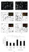
Cx43 immunofluorescent stainings indicate amplified expression in the ischemic lesion (b) compared to the intact area (a). Inset pictures of (c) and (d) represent double immunofluorescent staining with Cx43 (green) and MAP2 (red). Representative pictures in second row show the extracted area as yellow region of the double immunofluorescent staining from intact (c) and ischemic area (d). The yellow region which stands for Cx43 immunopositive area colocalized with MAP2 shows amplified expression in the ischemic periphery. Inset pictures of (e) and (f) represent double immunofluorescent staining with Cx43 (green) and GFAP (red). Representative pictures in third row show the extracted area as yellow region of the double immunofluorescent staining from intact (e) and ischemic area (f). The yellow region which stands for Cx43 immunopositive area colocalized with GFAP also shows increased expression level in ischemic lesion as compared to intact area. Black bars in graph (g) indicate the average counts of protein expressions. The Cx43 expression alone and the amount of coexpression with Cx43 or GFAP were significantly increased in the ischemic lesion compared to the intact area. The coexpression of Cx43 and MAP2 was increased in ischemic lesion compared to intact area (†: P = .0599). Scale bar indicates 50 μm. **: P < .01, *: P < .05.
Figure 5
Figure 5
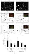
Cx26 immunofluorescent stainings show similar finding between intact area (a) and ischemic lesion (b). Inset pictures represent double immunofluorescent staining with Cx26 (green) and MAP2 (red) in (c) and (d) and with Cx26 (green) and GFAP (red) in (e) and (f). Representative pictures in second row show the extracted area as yellow region of the double immunofluorescent staining from intact (c) and ischemic area (d). The yellow region which stands for Cx26 immunopositive area colocalized with MAP2 shows the same amount of expression. The yellow region which stands for Cx26 immunopositive area colocalized with GFAP shows no difference of expression between intact and ischemic area. Black bars in graph (g) indicate the average counts of protein expressions. The Cx26 expression was not changed in the ischemic lesion as compared to the intact area. No statistical alteration was observed in the amount of coexpression of Cx26 and MAP2 nor Cx26 and GFAP between intact and ischemic regions. Scale bar indicates 50 μm.

Similar articles

Cited by

References

    1. Araque A, Parpura V, Sanzgiri RP, Haydon PG. Tripartite synapses: glia, the unacknowledged partner. Trends in Neurosciences. 1999;22(5):208–215. - PubMed
    1. Dermietzel R, Spray DC. From neuro-glue (‘Nervenkitt’) to glia: a prologue. Glia. 1998;24(1):1–7. - PubMed
    1. Giaume C, Fromaget C, El Aoumari A, Cordier J, Glowinski J, Gros D. Gap junctions in cultured astrocytes: single-channel currents and characterization of channel-forming protein. Neuron. 1991;6(1):133–143. - PubMed
    1. Naus CCG, Bechberger JF, Caveney S, Wilson JX. Expression of gap junction genes in astrocytes and C6 glioma cells. Neuroscience Letters. 1991;126(1):33–36. - PubMed
    1. Siushansian R, Bechberger JF, Cechetto DF, Hachinski VC, Naus CCG. Connexin43 null mutation increases infarct size after stroke. Journal of Comparative Neurology. 2001;440(4):387–394. - PubMed