. 2009 Apr;10(2):133-42.
doi: 10.2174/138920209787847014.

The arrestin fold: variations on a theme

Affiliations

The arrestin fold: variations on a theme

Laurence Aubry et al. Curr Genomics. 2009 Apr.

Abstract

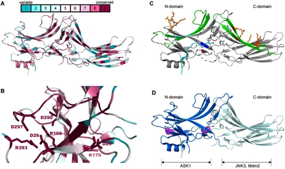
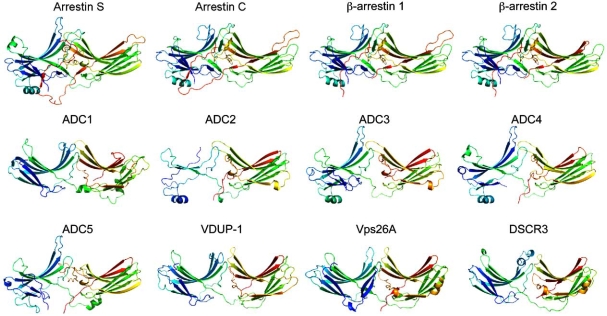
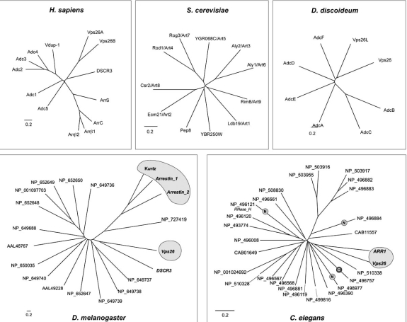
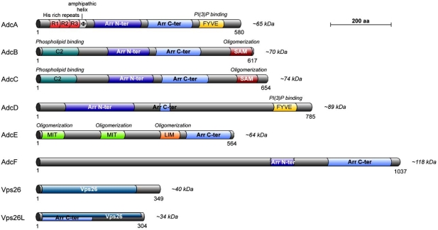
Endocytosis of ligand-activated plasma membrane receptors has been shown to contribute to the regulation of their downstream signaling. beta-arrestins interact with the phosphorylated tail of activated receptors and act as scaffolds for the recruitment of adaptor proteins and clathrin, that constitute the machinery used for receptor endocytosis. Visual- and beta-arrestins have a two-lobe, immunoglobulin-like, beta-strand sandwich structure. The recent resolution of the crystal structure of VPS26, one of the retromer subunits, unexpectedly evidences an arrestin fold in this protein, which is otherwise unrelated to arrestins. From a functional point of view, VPS26 is involved in the retrograde transport of the mannose 6-P receptor from the endosomes to the trans-Golgi network. In addition to the group of genuine arrestins and Vps26, mammalian cells harbor a vast repertoire of proteins that are related to arrestins on the basis of their PFAM Nter and Cter arrestin- domains, which are named Arrestin Domain- Containing proteins (ADCs). The biological role of ADC proteins is still poorly understood. The three subfamilies have been merged into an arrestin-related protein clan.This paper provides an overall analysis of arrestin clan proteins. The structures and functions of members of the subfamilies are reviewed in mammals and model organisms such as Drosophila, Caenorhabditis, Saccharomyces and Dictyostelium.

Keywords: Arrestins; GPCR; Vps26; endocytosis.; retromer; trafficking.


Figures

Fig. (1). Residue conservation among members of the arrestin family. Localization of binding sites on β -arrestins.
A. Amino acid conservation in human rod and cone arrestins and β-arrestins 1 and 2 was calculated with the online Consurf software (http://consurf.tau.ac.il) and represented with PyMol using Consurf coloring (see scale) on β-arrestin 1 template (1ZSH). B. Blowup of the polar core showing the high conservation of the residues important for the polar core highlighted as sticks. Electrostatic bonds between core residues are indicated as dotted lines. C. The structure of β-arrestin 1 (PDB 1ZSH) is represented with the PyMol software. The localization of the polar core is indicated by a dashed ellipse with positive residues in blue and negative residues in red. Receptor binding β-strands are indicated in green. The adaptin AP-2 binding site is indicated in cyan. Low- and high-affinity binding sites for InsP6 in the N- and C-domains, respectively are indicated as orange sticks. D. The N- and C-domains of β-arrestin 2 (PDB 1JSY) are indicated in dark and light blue, respectively. Prolines involved in Src binding appear as magenta spheres. The binding domains of ASK1, JNK3 and Mdm2 are indicated.
Fig. (2). 3D-structure of human arrestin clan proteins.
The structure of human arrestin clan proteins was modeled with the Phyre software (http://www.sbg.bio.ic.ac.uk ~phyre/) and visualized with the Pymol software (http://www.pymol.org). The PDB templates used for the modeling were 1CF1 for arrestin S, ADC2, ADC3, ADC4 and ADC5, 1JSY for ß-arrestin 1, ß-arrestin 2 and arrestin C, 2FAU for ADC1, VDUP, Vps26A and DSCR3.
Fig. (3). Diversity of arrestin clan proteins in human, fruit fly, nematode, yeast and amoeba.
Sequences of human arrestin clan proteins were aligned with ClustalW software [102]. An unrooted phylogenetic tree was drawn with Tree-Dyn for each species (http://www.phylogeny.fr) [103, 104]. Labels in the nematode tree indicate genes in which only the arrestin N- or C-domain was found. A grey cloud highlights proteins in nematode and fruit fly for which literature is available.
Fig. (4). Domain organization of Dictyostelium arrestin clan proteins.
Dictyostelium arrestin-domain proteins (Adcs), Vps26 and Vps26L/DSCR3 are represented by their colored domains as indicated. Number of amino acids and molecular masses of each protein are indicated. The functions of each domain are indicated on top.

Similar articles

Cited by

References

    1. Zuckerman R, Buzdygon B, Philp N, Liebman P, Sitara-mayya A. Arrestin: an ATP/ADP exchange protein that regulates cGMP phosphodiesterase activity in retinal rod disk membranes (RDM) Biophys. J. 1985;47:37a. - PubMed
    1. Zuckerman R, Cheasty JE. A 48 kDa protein arrests cGMP phosphodiesterase activation in retinal rod disk membranes. FEBS Lett. 1986;207:35–41. - PubMed
    1. DeWire SM, Ahn S, Lefkowitz RJ, Shenoy SK. Beta-arrestins and cell signaling. Annu. Rev. Physiol. 2007;69:483–510. - PubMed
    1. Gurevich EV, Gurevich VV. Arrestins: ubiquitous regulators of cellular signaling pathways. Genome Biol. 2006;7:236. - PMC - PubMed
    1. Moore CA, Milano SK, Benovic JL. Regulation of receptor trafficking by GRKs and arrestins. Annu. Rev. Physiol. 2007;69:451–482. - PubMed

LinkOut - more resources