

Three-dimensional culture models of mammary gland
- PMID: 19794898
- PMCID: PMC2710524
- DOI: 10.4161/org.5.2.8321
Three-dimensional culture models of mammary gland
Abstract
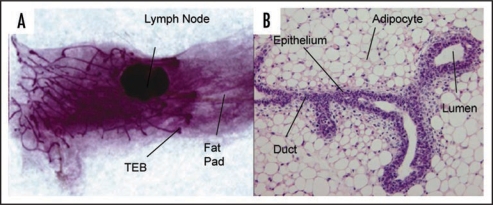
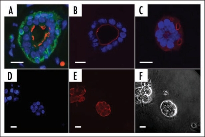
The mammary gland is a complex tissue comprised of a branching network of ducts embedded within an adipocyte-rich stroma. The ductal epithelium is a bi-layer of luminal and myoepithelial cells, the latter being in contact with a basement membrane. During pregnancy, tertiary branching occurs and lobuloalveolar structures, which produce milk during lactation, form in response to hormonal and cytokine signals. Postlactational regression is characterized by extensive cell death and tissue remodeling. These complex developmental events have been difficult to mimic in cell culture although many useful culture models exist. Recently, considerable advances in three-dimensional modelling of the mammary gland have been made with the use of collagen and other biomaterials for the study of branching morphogenesis and tumorigenesis, techniques which may enable rapid advances in our understanding of both basic biology and the study of cancer therapeutics.
Keywords: adipocytes; cell culture; cell-lines; epithelium; extracellular matrix; mammary gland; models; scaffolds; tissue engineering.
Figures
Similar articles
-
Evaluation of mammary gland development and function in mouse models.J Vis Exp. 2011 Jul 21;(53):2828. doi: 10.3791/2828. J Vis Exp. 2011. PMID: 21808224 Free PMC article.
-
[Mammary gland development: Role of basal myoepithelial cells].J Soc Biol. 2006;200(2):193-8. doi: 10.1051/jbio:2006021. J Soc Biol. 2006. PMID: 17151555 Review. French.
-
Mammary ductal elongation and myoepithelial migration are regulated by the composition of the extracellular matrix.J Microsc. 2013 Sep;251(3):212-23. doi: 10.1111/jmi.12017. Epub 2013 Feb 22. J Microsc. 2013. PMID: 23432616 Free PMC article.
-
Milk protein expression and ductal morphogenesis in the mammary gland in vitro: hormone-dependent and -independent phases of adipocyte-mammary epithelial cell interaction.Dev Biol. 1987 Mar;120(1):245-58. doi: 10.1016/0012-1606(87)90122-9. Dev Biol. 1987. PMID: 3817293
-
Application of the D492 Cell Lines to Explore Breast Morphogenesis, EMT and Cancer Progression in 3D Culture.J Mammary Gland Biol Neoplasia. 2019 Jun;24(2):139-147. doi: 10.1007/s10911-018-09424-w. Epub 2019 Jan 25. J Mammary Gland Biol Neoplasia. 2019. PMID: 30684066 Review.
Cited by
-
Mammary gland 3D cell culture systems in farm animals.Vet Res. 2021 Jun 2;52(1):78. doi: 10.1186/s13567-021-00947-5. Vet Res. 2021. PMID: 34078471 Free PMC article. Review.
-
Adipose-derived mesenchymal stem cells formed acinar-like structure when stimulated with breast epithelial cells in three-dimensional culture.PLoS One. 2018 Oct 18;13(10):e0204077. doi: 10.1371/journal.pone.0204077. eCollection 2018. PLoS One. 2018. PMID: 30335754 Free PMC article.
-
3D culture models for studying branching morphogenesis in the mammary gland and mammalian lung.Biomaterials. 2019 Apr;198:135-145. doi: 10.1016/j.biomaterials.2018.08.043. Epub 2018 Aug 23. Biomaterials. 2019. PMID: 30174198 Free PMC article. Review.
-
Advances in 3D cell culture technologies enabling tissue-like structures to be created in vitro.J Anat. 2015 Dec;227(6):746-56. doi: 10.1111/joa.12257. Epub 2014 Nov 20. J Anat. 2015. PMID: 25411113 Free PMC article. Review.
-
PtdIns(3,4,5)P3-dependent Rac exchanger 1 (P-Rex1) promotes mammary tumor initiation and metastasis.Proc Natl Acad Sci U S A. 2020 Nov 10;117(45):28056-28067. doi: 10.1073/pnas.2006445117. Epub 2020 Oct 23. Proc Natl Acad Sci U S A. 2020. PMID: 33097662 Free PMC article.
References
-
- Senger DR, Van de Water L, Brown LF, Nagy JA, Yeo KT, Yeo TK, et al. Vascular permeability factor (VPF, VEGF) in tumor biology. Cancer Metastasis Rev. 1993;12:303–324. - PubMed
-
- Brennan KR, Brown AMC. Wnt Proteins in Mammary development and cancer. J Mammary Gland Biol Neoplasia. 2004;9:119–131. - PubMed
-
- Trichopoulos D, Adami HO, Ekbom A. Early life events and conditions and breast cancer risk: From epidemiology to etiology. Int J Cancer. 2008;122:481–485. - PubMed
-
- Robinson GW. Cooperation of signalling pathways in embryonic mammary gland development. Nat Rev Genet. 2007;8:963–972. - PubMed
Grants and funding
LinkOut - more resources
Full Text Sources