. 2009 Apr;5(2):50-6.
doi: 10.4161/org.5.2.8347.

Adipose tissue-organotypic culture system as a promising model for studying adipose tissue biology and regeneration

Affiliations

Adipose tissue-organotypic culture system as a promising model for studying adipose tissue biology and regeneration

Shuji Toda et al. Organogenesis. 2009 Apr.

Abstract

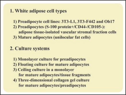
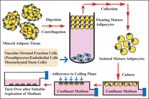
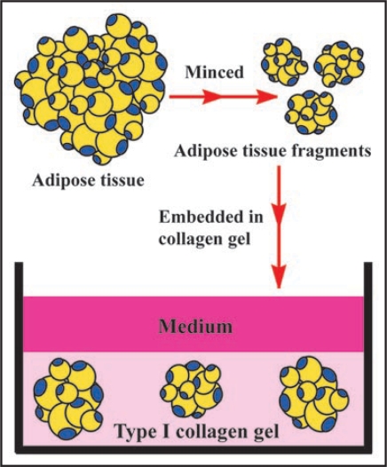
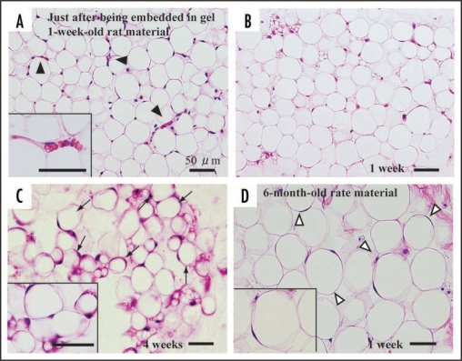
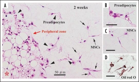
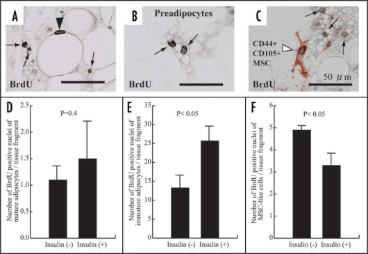
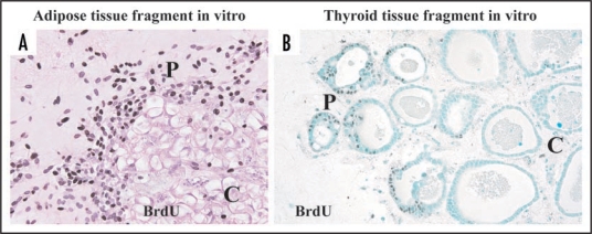
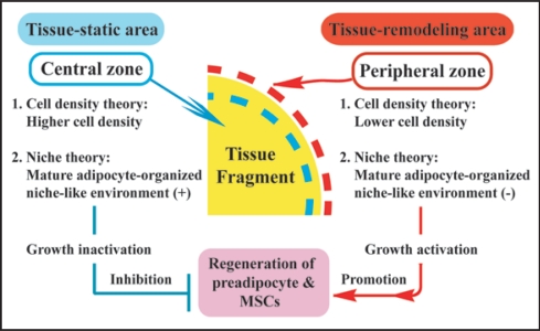
Adipose tissue consists of mature adipocytes, preadipocytes and mesenchymal stem cells (MSCs), but a culture system for analyzing their cell types within the tissue has not been established. We have recently developed "adipose tissue-organotypic culture system" that maintains unilocular structure, proliferative ability and functions of mature adipocytes for a long term, using three-dimensional collagen gel culture of the tissue fragments. In this system, both preadipocytes and MSCs regenerate actively at the peripheral zone of the fragments. Our method will open up a new way for studying both multiple cell types within adipose tissue and the cell-based mechanisms of obesity and metabolic syndrome. Thus, it seems to be a promising model for investigating adipose tissue biology and regeneration. In this article, we introduce adipose tissue-organotypic culture, and propose two theories regarding the mechanism of tissue regeneration that occurs specifically at peripheral zone of tissue fragments in vitro.

Keywords: adipokines; adipose tissue-organotypic culture; central zone; mature adipocytes; mesenchymal stem cells; peripheral zone; preadipocytes (immature adipocytes); three-dimensional; tissue fragments; tissue regeneration.


Figures

Figure 1
Figure 1
Adipose cell types and their suitable culture systems. As preadipocyte cell lines, 3T3-L1 and 3T3-F442 cells are used more frequently than Ob17 cells. Primary preadipocytes (S-100 protein+/CD44−/CD105−) and mature adipocytes that are all isolated from adipose tissue are also utilized. Preadipocyte types are easily monolayer-cultured. Floating culture of mature adipocytes is used, but it maintains viable mature adipocytes only for a short term. In contrast, ceiling culture maintains viable mature adipocytes for a long term and is useful for studying their growth and differentiation. Ceiling culture is able to be applied to culture of tissue fragments in monolayer. Three-dimensional collagen gel culture is useful for both mature adipocytes and preadipocytes.
Figure 2
Figure 2
Ceiling culture method. Minced adipose tissue is digested by collagenase and then centrifuged. After centrifugation, mature adipocyte layer floats at top of test tube, while vascular stromal fraction cell types that consist of preadipocytes, endothelial cells and MSCs precipitate at bottom. After isolated mature adipocytes are collected from mature adipocyte layer, they are pored into culture bottle filled with medium. The bottle is placed in upside-down fashion in culture incubator. After mature adipocytes adhere to ceiling plane, medium is aspirated and the bottle is placed in upside-down fashion. In this way, ceiling culture of mature adipocytes is set up.
Figure 3
Figure 3
Adipose tissue-organotypic culture and its organization procedure. After rinsing adipose tissue, it was minced in about 0.5 mm in diameter. The minced tissue fragments are embedded in type I collagen gel. After the gel is fully firm, the gel is covered with medium and cultured.
Figure 4
Figure 4
Histology of adipose tissue in adipose tissue-organotypic cultures of 1-week-old (A, B and C) and 6-month-old rat materials (D). (A) Adipose tissue fragment just after being embedded in collagen gel has viable mature adipocytes with a large single lipid droplet and a peripherally located nucleus, hence have been called unilocular fat cells. Capillary network (arrowheads and inset) is seen among mature adipocytes. Note that erythrocytes are seen in capillaries. (B) At 1 week in culture, viable mature adipocytes are maintained at the center of the tissue fragment and they have no drastic morphological change. However, capillary network disappears within the tissue fragments. Spindle-shaped cells do not develop at the central part of the fragment. (C) Even at 4 weeks in culture, mature adipocytes are well retained within the fragment, and spindle-shaped cells are also not observed at the center. (D) Mature adipocytes within the fragments derived from 6-month-old rats are also well retained at 1 week in culture. Notice that the size of 6-month-old rat-derived mature adipocytes is about 1.5 to 2 times that of 1-week-old rat-derived mature adipocytes (B). A, B, C and D, H–E staining.
Figure 5
Figure 5
Development of preadipocytes and MSCs at the peripheral zone of adipose tissue fragments derived from 1-week-old rats. (A) At 2 weeks in culture, preadipocytes and MSCs appear actively at the peripheral part of the fragment. Preadipocytes (arrowheds) have fine lipid droplets in the cytoplasm. MSCs without lipid droplets (C) are also seen. (D) Lipid droplets (red in color) are confirmed by oil red O staining. (A, B and C), H-E staining. *central zone of tissue fragment.
Figure 6
Figure 6
Proliferation of mature adipocytes (A and D), preadipocytes (B and E) and MSCs (C and F) at 1 week in cultures of 1-week-old rat materials with or without insulin. In cultures with insulin, its stimulation was carried out at culture day 2, 4 and 6. Cell growth is assessed by immunohistochemistry with BrdU. A mature adipocyte shows intranuclear BrdU uptake in black (arrowhead in panel A). Preadipocytes show intranuclear BrdU uptake (allows in panels A, B and C). CD44+/CD105+ MSCs in brownish red (C) has intranuclear BrdU intake (arrowhead in C). (D) There is no significant difference of number of BrdU-positive mature adipocytes between cultures with and without insulin, although insulin tends to enhance BrdU uptake of mature adipocytes. (E and F) Insulin enhances BrdU uptake of preadipocytes (E), while it inhibits BrdU uptake of MSCs (F), indicating their significant difference between that of immature adipocytes or MSC-like cells with and without insulin.
Figure 7
Figure 7
Immunohistochemistry with BrdU in adipose (A) and thyroid (B) tissue-organotypic culture after 48 hour incubation with 30 µg/ml BrdU. Intranuclear BrdU uptakes of adipose cell types and thyrocytes at peripheral zones of adipose (A) and thyroid tissue fragments (B) are extensively greater than those at the central zones. P, peripheral zone. C, central zone.
Figure 8
Figure 8
Two theories regarding the mechanism of preadipocyte and MSC regeneration that occurs specifically at peripheral zone of the tissue fragments in vitro. In regard to “cell density theory,” central zone of adipose tissue fragments is characterized by higher cell density, whereas the peripheral zone is characterized by lower cell density. In general, increased cell density in a microenvironment inhibits the regeneration and growth of cells that are subjected to contact inhibition of cell growth. Namely, central zone is tissue-static area with cell growth inactivation, while peripheral zone is tissue-remodeling area with cell growth activation. Thus, lower cell density of the peripheral zone may contribute to active development of preadipocytes and MSCs. In regard to “niche theory,” central zone concentrated by mature adipocytes may be subjected to mature adipocyte-organized niche-like environment, whereas peripheral zone with sparse population of mature adipocytes may lose the environment. In general, a niche environment for stem cell types maintains their resting state. Thus, the niche-like environment formed by mature adipocytes may inhibit regeneration of preadipocytes and MSCs at the center, while its loss at the peripheral zone may contribute to their regeneration.

Similar articles

Cited by

References

    1. Zuk PA, Zhu M, Ashjian P, De Ugarte DA, Huang JI, Mizuno H, et al. Human adipose tissue is a source of multipotent stem cells. Mol Biol Cell. 2002;13:4279–4295. - PMC - PubMed
    1. Jensen MD. Role of body fat distribution and the metabolic complications of obesity. J Clin Endocrinol Metab. 2008;93:S57–S63. - PMC - PubMed
    1. Anghel SI, Wahli W. Fat poetry: a kingdom for PPAR gamma. Cell Res. 2007;17:486–511. - PubMed
    1. Wozniak SE, Gee LL, Wachtel MS, Frezza EE. Adipose Tissue: The new endocrine organ? A Review Article. Dig Dis Sci. 2008 - PubMed
    1. Rasouli N, Kern PA. Adipocytokines and the metabolic complications of obesity. J Clin Endocrinol Metab. 2008;93:S64–S73. - PMC - PubMed

LinkOut - more resources