. 2009 Oct 1;4(10):e0007243.
doi: 10.1371/journal.pone.0007243.

Histone H1 subtypes differentially modulate chromatin condensation without preventing ATP-dependent remodeling by SWI/SNF or NURF

Affiliations

Histone H1 subtypes differentially modulate chromatin condensation without preventing ATP-dependent remodeling by SWI/SNF or NURF

Jaime Clausell et al. PLoS One. .

Abstract

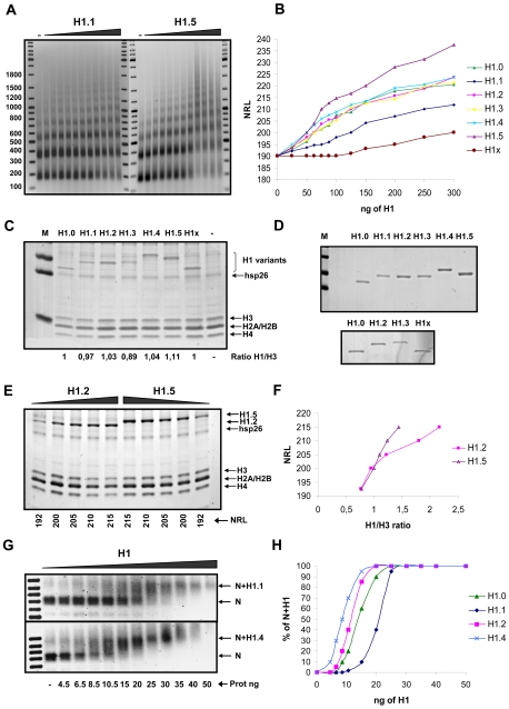
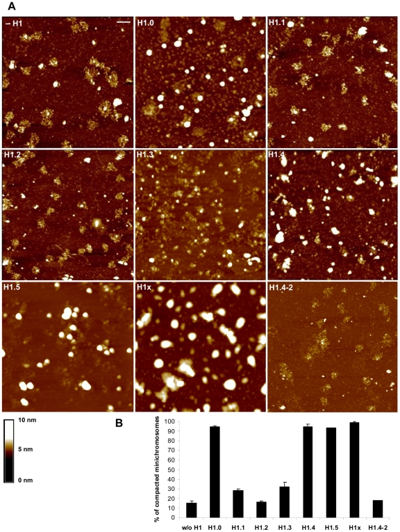
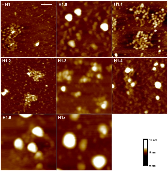
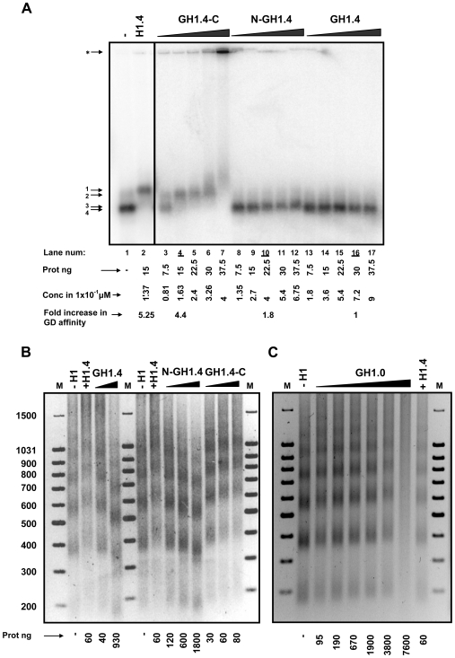
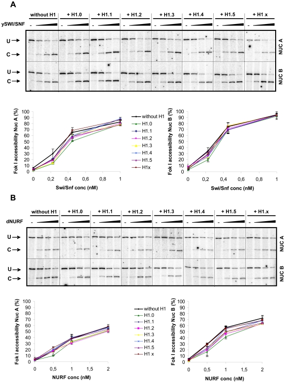
Although ubiquitously present in chromatin, the function of the linker histone subtypes is partly unknown and contradictory studies on their properties have been published. To explore whether the various H1 subtypes have a differential role in the organization and dynamics of chromatin we have incorporated all of the somatic human H1 subtypes into minichromosomes and compared their influence on nucleosome spacing, chromatin compaction and ATP-dependent remodeling. H1 subtypes exhibit different affinities for chromatin and different abilities to promote chromatin condensation, as studied with the Atomic Force Microscope. According to this criterion, H1 subtypes can be classified as weak condensers (H1.1 and H1.2), intermediate condensers (H1.3) and strong condensers (H1.0, H1.4, H1.5 and H1x). The variable C-terminal domain is required for nucleosome spacing by H1.4 and is likely responsible for the chromatin condensation properties of the various subtypes, as shown using chimeras between H1.4 and H1.2. In contrast to previous reports with isolated nucleosomes or linear nucleosomal arrays, linker histones at a ratio of one per nucleosome do not preclude remodeling of minichromosomes by yeast SWI/SNF or Drosophila NURF. We hypothesize that the linker histone subtypes are differential organizers of chromatin, rather than general repressors.


Conflict of interest statement

Competing Interests: The authors have declared that no competing interests exist.

Figures

Figure 1
Figure 1. Affinity of histone H1 subtypes for chromatin.
(A) Nucleosome ladders obtained after micrococcal nuclease (MNase) digestion of minichromosomes assembled in preblastodermic Drosophila embryo extracts (DREX) with increasing amounts of histone H1 subtypes. Results with H1.1 and H1.5 are shown as an example. DNA size markers are shown on both sides of the MNase digested samples. Numbers on the left indicate fragment length in base pairs. (B) Graphic representation of the Nucleosomal Repeat Length (NRL) calculated from experiments similar to that shown in (A) with the amount of each H1 subtype indicated in nanograms. (C) Minichromosomes, assembled with each H1 subtype to yield a NRL of 200 bp, were precipitated with buffer containing 20 mM MgCl2, and their proteins were electrophoresed on a 16% SDS-Polyacrylamide gel (SDS-PAGE), and visualized by Coomassie G-250 staining. The bands were quantified using Quantity One software (Bio-Rad) and the ratio between each H1 and H3 is shown below the corresponding gel lane. H1.4 runs slower than the other H1 subtypes as it contains a FLAG tag. Abbreviation and Symbol: M, Marker; -, without H1. (D) Staining of 2 µg of each purified H1 subtypes (H1.0 to H1.5) with Coomassie G-250. The lower panel is from a separate experiment that included H1x and a subset of the other subtypes for comparison. (E) Minichromosomes assembled with increasing amounts of H1.2 and H1.5 corresponding to the NRL indicated below, were purified as in (C). Proteins were separated by electrophoresis and stained with Coomassie G-250. (F) The intensity of the bands corresponding to H3, H1.2 and H1.5 in (E) were quantitated, normalised according to their different staining ability shown in (D), and the NRL plotted against the H1/H3 ratio. (G) Mononucleosomes assembled by salt dialysis on 100 ng of a 220 bp DNA fragment were incubated for 20 minutes with increasing amounts of H1.1 and H1.4 and analysed on a 0.7% agarose gel. The position of the mononucleosome without H1 (N) and with H1 is indicated on the right margin. (H) Graphic representation of the extent of H1.0, H1.1, H1.2 and H1.4 binding, calculated from the band shift experiments as shown in (G).
Figure 2
Figure 2. Influence of H1 subtypes and the H1.4–2 chimera on chromatin compaction.
(A) Minichromosomes were assembled without H1 or with the indicated H1 subtype or the chimeric protein H1.4–2, all at concentrations yielding a NRL of 200 bp. Minichromosomes were purified through a sucrose gradient and a sucrose cushion, resuspended in 20 mM KCl containing buffer, immobilized on an APTES mica surface and visualised using TMAFM (Tapping Mode Atomic Force Microscopy). The horizontal bar represents 200 nm and the vertical scale (in nm) is shown below; the grid size is 2 µm. Nucleosomes appear as balls of 11 nm and compacted minichromosomes as globules of 50–60 nm in diameter. Single nucleosomes are lost from the minichromosomes during the fixation process and appear as individual balls of approximately 11 nm. (B) The percentage of compacted minichromosomes was calculated from at least two independent experiments and is shown in the bottom diagram, with the error bars corresponding to S.E.M.
Figure 3
Figure 3. 500 nm magnifications of minichromosomes assembled without or with each somatic H1.
The same techniques as in Figure 2 were used. The horizontal bar corresponds to 100 nm and the grid size is 500 nm. The vertical scale is shown.
Figure 4
Figure 4. Role of H1 domains in chromatin affinity and nucleosome spacing.
(A) Centrally positioned mononucleosomes were assembled on a 220 bp DNA fragment corresponding to the nucleosome B in the MMTV promoter. After a glycerol gradient purification, mononucleosome particles were incubated with increasing amounts of the following H1.4 domain mutants: the globular domain (GH1.4), the N-terminal plus the globular domain (N-GH1.4), and the globular plus the C-terminal domain (GH1.4-C), and analyzed on a 0.7% (w/v) agarose gel. A lane with wild type H1.4 is shown as control. The different particles are numbered on the left margin: 1) Mononucleosome + H1.4; 2) Mononucleosome + GH1.4-C; 3) Mononucleosome + N-GH1.4; 4) Mononucleosome + GH1.4. The symbol ‘*’ indicates the appearance of aggregates. The amount of each protein is indicated at the base of the gel (lanes 2–17). Protein concentrations are also expressed in 1×10⁻¹ µM units. (B) MNase digestion of minichromosomes assembled with the indicated linker histone. The amount of linker histone added (in nanograms per 200 ng of DNA) is indicated bellow each lane. Lettering is as in (A). Numbers on the left margin refer to the size of the markers (lane M) in base pairs. (C) MNase digestions of minichromosomes with increasing amounts of GH1.0. Also shown is the digestion in the absence of linker histone (−H1) and in the presence of H1.4 (+H1.4). Markers and the amount of protein added are indicated as in (B).
Figure 5
Figure 5. Effect of histone H1 subtypes on ATP-dependent chromatin remodeling.
MMTV minichromosomes were reconstituted in the presence of the histone H1 subtypes, as indicated, purified, resuspended in a 60 mM KCl and 5 mM MgCl2 containing buffer, and incubated with Fok I that cleaves in nucleosome A and B of the MMTV promoter. Increasing nM concentrations of SWI/SNF (A) and NURF (B) complexes were added and incubated for 30 minutes at 30°C. The reaction was stopped and a linear extension PCR with a radiolabeled oligonucleotide was performed. The cleavage products were visualised in a 10% polyacrylamide denaturing gel and quantified using Image Quant software (Amersham). Minichromosomes of two independent purifications were tested and the results represented with the corresponding S.E.M in the graphs bellow. Abbreviations: Nuc A, nucleosome A; Nuc B, nucleosome B; U, uncut material; C, cut material.

Similar articles

Cited by

References

    1. Luger K, Mader AW, Richmond RK, Sargent DF, Richmond TJ. Crystal structure of the nucleosome core particle at 2.8 A resolution. Nature. 1997;389:251–260. - PubMed
    1. Ramakrishnan V. Histone structure and the organization of the nucleosome. Annu Rev Biophys Biomol Struct. 1997;26:83–112. - PubMed
    1. Thoma F, Koller T, Klug A. Involvement of histone H1 in the organization of the nucleosome and of the salt-dependent superstructures of chromatin. J Cell Biol. 1979;83:403–427. - PMC - PubMed
    1. Huynh VA, Robinson PJ, Rhodes D. A method for the in vitro reconstitution of a defined “30 nm” chromatin fibre containing stoichiometric amounts of the linker histone. J Mol Biol. 2005;345:957–968. - PubMed
    1. Butler PJ, Thomas JO. Changes in chromatin folding in solution. J Mol Biol. 1980;140:505–529. - PubMed

Publication types

MeSH terms

LinkOut - more resources